. 2019 Apr 23;87(5):e00119-19.
doi: 10.1128/IAI.00119-19. Print 2019 Mar.

Alternative Enzyme Protection Assay To Overcome the Drawbacks of the Gentamicin Protection Assay for Measuring Entry and Intracellular Survival of Staphylococci

Affiliations

Alternative Enzyme Protection Assay To Overcome the Drawbacks of the Gentamicin Protection Assay for Measuring Entry and Intracellular Survival of Staphylococci

Jin-Hahn Kim et al. Infect Immun. .

Abstract

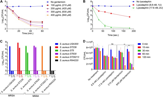
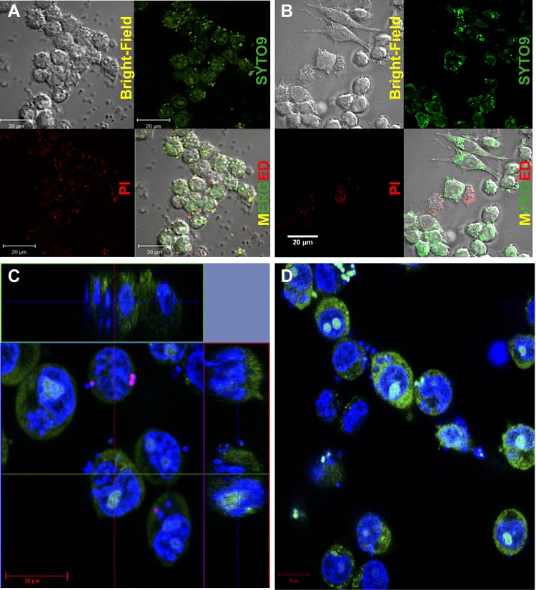
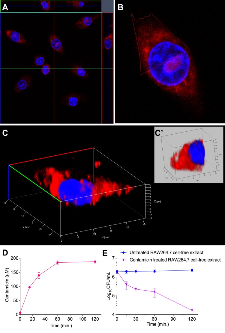
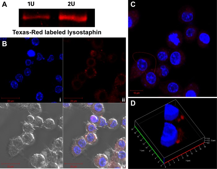
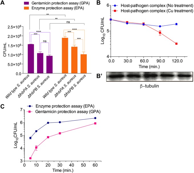
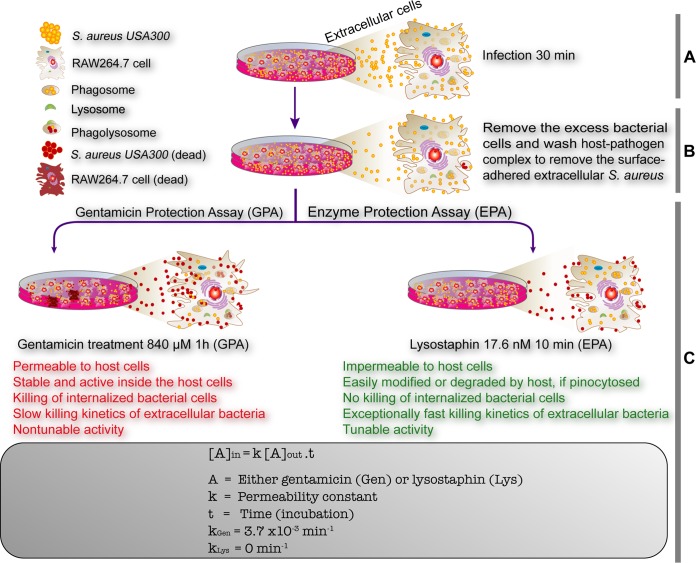
Precise enumeration of living intracellular bacteria is the key step to estimate the invasion potential of pathogens and host immune responses to understand the mechanism and kinetics of bacterial pathogenesis. Therefore, quantitative assessment of host-pathogen interactions is essential for development of novel antibacterial therapeutics for infectious disease. The gentamicin protection assay (GPA) is the most widely used method for these estimations by counting the CFU of intracellular living pathogens. Here, we assess the longstanding drawbacks of the GPA by employing an antistaphylococcal endopeptidase as a bactericidal agent to kill extracellular Staphylococcus aureus We found that the difference between the two methods for the recovery of intracellular CFU of S. aureus was about 5 times. We prove that the accurate number of intracellular CFU could not be precisely determined by the GPA due to the internalization of gentamicin into host cells during extracellular bacterial killing. We further demonstrate that lysostaphin-mediated extracellular bacterial clearance has advantages for measuring the kinetics of bacterial internalization on a minute time scale due to the fast and tunable activity and the inability of protein to permeate the host cell membrane. From these results, we propose that accurate quantification of intracellular bacteria and measurement of internalization kinetics can be achieved by employing enzyme-mediated killing of extracellular bacteria (enzyme protection assay [EPA]) rather than the host-permeative drug gentamicin, which is known to alter host physiology.

Keywords: Staphylococcus aureus; bacteria; enzyme protection assay; gentamicin protection assay; host; lysostaphin.


Figures

FIG 1
FIG 1
Comparison between the gentamicin protection assay (GPA) and the lysostaphin-mediated enzyme protection assay (EPA). (A) Quantitative assessment of gentamicin concentration and time-dependent killing efficiency for S. aureus cells at various concentrations from 100 μg/ml (210 μM) to 400 μg/ml (840 μM). Gentamicin at a concentration of 400 μg/ml (840 μM) could kill 99.999% of S. aureus cells within 60 min. (B) Concentration and time-dependent killing of S. aureus cells using lysostaphin. One unit (8.8 nM) and 2 U (17.6 nM) of lysostaphin were incubated with 1 × 107 S. aureus cells in 1 ml DMEM containing 10% FBS in a 5% CO2 environment at 37°C. Nearly all (99.999%) the S. aureus cells were killed within 180 s with 2 U lysostaphin. (C) Killing efficiency of lysostaphin against various methicillin-sensitive S. aureus (MSSA) (ST630, RN4220, and MSSA29213) and methicillin-resistant S. aureus (MRSA) (ST5, ST239, and USA300) strains. Two units of lysostaphin eradicated all the tested clinical methicillin-sensitive and methicillin-resistant strains of S. aureus. C, control. (D) Concentration and time-dependent killing of S. aureus in host cells. After infection of RAW264.7 cells (1 × 106) by S. aureus cells (1 × 107) for 30 min, gentamicin (210 μM and 840 μM) or lysostaphin (1 and 2 U) was applied to the host-pathogen mixture for 15, 30, 60, and 120 min. After rigorous washing using PBS to remove gentamicin or EDTA-quenched lysostaphin in the infection medium, the host cells were lysed to release internalized bacteria, followed by serial dilution and plating to count CFU of intracellular S. aureus. The GPA killed a significant number of intracellular S. aureus bacteria compared to the EPA. The intracellular killing of S. aureus during the GPA versus the EPA was analyzed using Student’s t tests. ns, nonsignificant; **, P < 0.01.
FIG 2
FIG 2
Visualization of intracellular killing of S. aureus during the GPA and EPA. The Live/Dead BacLight bacterial viability kit (catalog no. L7007) was used for the qualitative assessment of dead bacteria with SYTO9 (green fluorescence) and propidium iodide (PI) (red fluorescence), which stain all cells and membrane-damaged cells, respectively, and thus, all bacteria are shown in green, while only membrane-damaged bacterial cells are indicated by red dots. (A and B) Confocal images of RAW264.7 cells infected with S. aureus after GPA-mediated (A) and EPA-mediated (B) eradication of extracellular S. aureus showing that red dots are not found in lysostaphin-treated cells (B) but are found in gentamicin-treated cells (A), indicating the presence of intracellular killing of S. aureus during the GPA. (C and D) Central z-stack images of the host-pathogen complex (RAW264.7 cells and S. aureus) with gentamicin killing for 2 h (C) and the without-gentamicin-killing control (D) showing that red cells are found only under conditions of gentamicin treatment, while green cells are found under both conditions. These images indicate that gentamicin causes intracellular killing of S. aureus.
FIG 3
FIG 3
Qualitative and quantitative assessment of internalization of gentamicin. (A) Central z-stack image of RAW264.7 cells indicating the internalization of gentamicin conjugated with Texas Red (GTTR). RAW264.7 cells seeded for 24 h were stained with 20 μg/ml Hoechst 33258 (blue) and 50 μg/ml GTTR (red) at 37°C for 1 h. (B) Enlarged single image of the image in panel A. (C) Three-dimensional image of the cell in panel B. (C′) Dissected section of the single cell marked in panel B. (D) Time-dependent internalization of gentamicin in host cells. RAW264.7 cells were treated with gentamicin at a concentration of 840 μM for various time periods (0, 15, 30, 60, and 120 min) in six-well microtiter plates. The host cells were then harvested and intensively washed to avoid gentamicin contamination. The concentration of intracellular gentamicin was calculated at each time point and plotted. (E) Antibacterial activity of gentamicin-treated host cells. RAW264.7 cells were treated with gentamicin (840 μM) for various time periods. The host cells were harvested and intensively washed to avoid gentamicin contamination. The host cell lysates were applied to S. aureus cells to investigate the antibacterial activity of internalized gentamicin. RAW264.7 cells treated with water at an equivalent volume were used as a control for each time point. CFU count is decreased up to 100-fold in gentamicin-treated host cell lysate compared to its corresponding control.
FIG 4
FIG 4
Assessment of lysostaphin internalization into the host cell. (A) Red fluorescence signals (excitation/emission wavelength of 588/615 nm) of 1 U (5 μl; 8.8 nM) and 2 U (10 μl; 17.6 nM) lysostaphin-TR on SDS-PAGE gels. (B) RAW264.7 cells imaged by confocal microscopy shown in split channels, with (i) host cell nucleus stained with 20 μg/ml Hoechst 33258 (blue), (ii) host cells stained with 2 U of lysostaphin-TR (red), (iii) bright-field images, and (iv) merged image of bright-field, blue, and red signals. (C and D) Image in panel B enlarged in a z-stack (C) and analyzed in a 3D dissected section (D), confirming that the red signal from lysostaphin-TR was located on the cell surface and not inside the host cells.
FIG 5
FIG 5
Applications of the lysostaphin-mediated enzymatic protection assay. (A) Assessment of the internalizing potential of wild-type S. aureus and its isogenic mutants lacking functional FnbPA and FnbPA (fnbPAΩEmr and fnbPBΩEmr). Wild-type, fnbPAΩEmr, and fnbPBΩEmr S. aureus strains were added to nonphagocytic HEK293 cells for 30 min in a 5% CO2 environment at 37°C. After killing of the extracellular bacteria with gentamicin (300 μg/ml for 1 h and 100 μg/ml) and lysostaphin (2 U for 5 min), the internalized S. aureus cells were assessed by counting CFU. Both fnbP mutant strains showed differential reductions in internalization potential, demonstrating that the EPA can precisely discern a phenotypic change between mutant and WT bacteria. Internalization potentials of S. aureus strains in nonphagocytic HEK293 cells were compared by one-way ANOVA with Bonferroni’s multiple-comparison test. ns, nonsignificant; **, P < 0.01; ***, P < 0.001; ****, P < 0.0001. (B) Measurement of the bactericidal activity of RAW264.7 cells by the EPA. After 30 min of infection of RAW264.7 cells with S. aureus, the extracellular bacteria were killed by lysostaphin. The infected RAW264.7 cells were treated with 100 μM Cu2+ for various time periods (0, 30, 60, 90, and 120 min) in Hanks’ balanced salt solution (HBSS). The number of intracellular S. aureus cells was monitored by measuring CFU. As a control, β-tubulin was monitored by Western blotting. The time-dependent reduction of intracellular CFU suggests that internalized bacteria were killed by a copper-mediated defense mechanism of the host. This experiment demonstrates that the EPA can be applied to monitor changes in intracellular bacteria. (C) Internalization kinetics of S. aureus entering RAW264.7 cells using the GPA and the EPA. RAW264.7 cells were infected with S. aureus for various time periods (5, 10, 20, 30, and 60 min). At each time point, the extracellular bacteria were killed by gentamicin (400 μg/ml for 1 h of incubation, followed by gentamicin removal by washing) or lysostaphin (2 U for 10 min of incubation, followed by EDTA quenching and removal by washing), and measurement of cell lysate CFU was performed. The internalization kinetics measured by applying the GPA were significantly lower than those measured via the EPA.
FIG 6
FIG 6
Schematic diagrams showing the summary of drawbacks of the gentamicin protection assay and its comparison with the newly established enzyme protection assay. The GPA is one of the key techniques in infectious disease biology to assess the invasion/infection potential of bacterial pathogens and the efficacy of newly identified antibiotics. The major drawbacks of the GPA (text in red font), which kills internalized bacterial cells, are recognized. The EPA has been devised to alleviate the drawbacks of the GPA with added advantages of measuring the internalization/invasion kinetics due to its exceptionally fast and tunable enzymatic killing efficiency (text in green font). (A and B) Both the GPA and EPA share the first two steps: infection (A) and removal of excess bacterial cells (B). (C) The killing of extracellular and surface-bound bacterial pathogens is the key step wherein gentamicin or lysostaphin was used, based on the results that the killing agent cannot enter mammalian host cells. Gentamicin seeps into the cells (kGen = 3.7 × 10−3 min−1) and kills the internalized bacteria, resulting in the misleading infection potential of the bacterial pathogen and precise drug efficacy. In the EPA, lysostaphin efficiently kills host cell surface-bound bacterial cells and cannot enter the host cells (kLys = 0), which avoids any misleading calculation and interpretation of both the invasion potential and antibiotic drug efficacy.

References

    1. Ribet D, Cossart P. 2015. How bacterial pathogens colonize their hosts and invade deeper tissues. Microbes Infect 17:173–183. doi: 10.1016/j.micinf.2015.01.004. - DOI - PubMed
    1. Thammavongsa V, Kim HK, Missiakas D, Schneewind O. 2015. Staphylococcal manipulation of host immune responses. Nat Rev Microbiol 13:529–543. doi: 10.1038/nrmicro3521. - DOI - PMC - PubMed
    1. van Kessel KP, Bestebroer J, van Strijp JA. 2014. Neutrophil-mediated phagocytosis of Staphylococcus aureus. Front Immunol 5:467. doi: 10.3389/fimmu.2014.00467. - DOI - PMC - PubMed
    1. Cossart P, Sansonetti PJ. 2004. Bacterial invasion: the paradigms of enteroinvasive pathogens. Science 304:242–248. doi: 10.1126/science.1090124. - DOI - PubMed
    1. Monack DM, Mueller A, Falkow S. 2004. Persistent bacterial infections: the interface of the pathogen and the host immune system. Nat Rev Microbiol 2:747–765. doi: 10.1038/nrmicro955. - DOI - PubMed

Publication types

MeSH terms