. 2019 Feb 19;9(1):2262.
doi: 10.1038/s41598-018-38458-7.

High-throughput, Efficient, and Unbiased Capture of Small RNAs from Low-input Samples for Sequencing

Affiliations

High-throughput, Efficient, and Unbiased Capture of Small RNAs from Low-input Samples for Sequencing

Cassandra D Belair et al. Sci Rep. .

Abstract

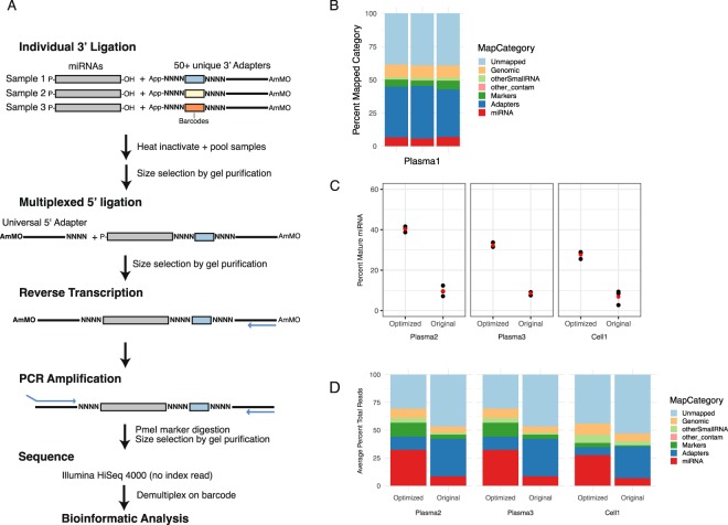
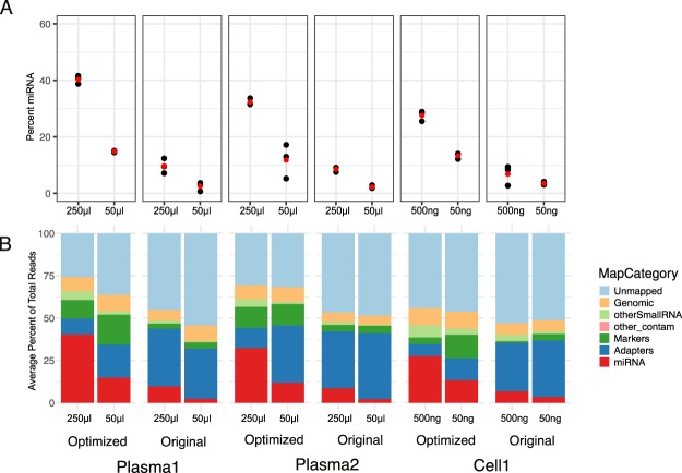
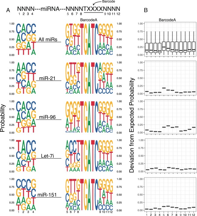
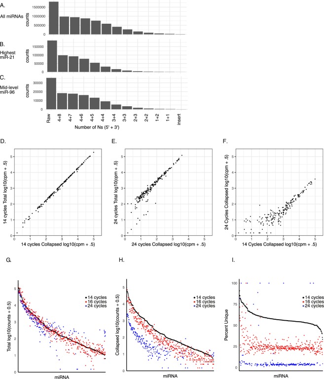
MicroRNAs hold great promise as biomarkers of disease. However, there are few efficient and robust methods for measuring microRNAs from low input samples. Here, we develop a high-throughput sequencing protocol that efficiently captures small RNAs while minimizing inherent biases associated with library production. The protocol is based on early barcoding such that all downstream manipulations can be performed on a pool of many samples thereby reducing reagent usage and workload. We show that the optimization of adapter concentrations along with the addition of nucleotide modifications and random nucleotides increases the efficiency of small RNA capture. We further show, using unique molecular identifiers, that stochastic capture of low input RNA rather than PCR amplification influences the biased quantitation of intermediately and lowly expressed microRNAs. Our improved method allows the processing of tens to hundreds of samples simultaneously while retaining high efficiency quantitation of microRNAs in low input samples from tissues or bodily fluids.


Conflict of interest statement

The authors declare no competing interests.

Figures

Figure 1
Figure 1
Modifications to high-throughput sequencing method improves capture of miRNAs. (A) Schematic of protocol to prepare miRNA libraries for sequencing. Modifications from original protocol noted in bold. (B) Percentage of different classes of RNAs captured from a plasma sample using the original conditions (0.85 μM 3′ adapter, 3.3 μM unmodified 5′ adapter). Ligations were performed in triplicate from the same RNA. Each replicate is shown as an individual bar. Note the low percentage of reads mapping to miRNAs (red). (C) Percentage of mature miRNAs captured using the optimized conditions (0.05 μΜ 3′ adapter, 0.33 μΜ amino-modified 5′ adapter) were compared to original conditions in three independent biological samples. The ligation reactions were performed in triplicate for each sample and protocol. Each replicate is shown as a black dot. Red dot represents average. (D) The average percentage of reads mapping to different classes of RNA for each sample and condition shown in C.
Figure 2
Figure 2
Improved miRNA capture also seen at low concentrations of input RNA. (A) The percentage of reads mapping to miRNAs at the indicated input following either the optimized (0.05 μΜ 3′ adapter, 0.33 μΜ amino-modified 5′adapter) or the original (0.85 μM 3′ adapter, 3.3 μM unmodified 5′ adapter) protocol. Black dots represent the three replicates from each input RNA for each sample, protocol, and concentration. Red dots represent average. (B) The average percentage of reads mapping to different RNA classes for each sample, protocol, and concentration.
Figure 3
Figure 3
Nucleotide biases seen at ligation sites and varies between individual miRNAs. (A) SeqLogo representation of the base composition of the degenerate Ns over all miRNAs and select miRs representing high (miR-21) mid (Let-7i, miR-96) and low (miR-151) expression. The height of the letter representing the base is proportional to its probability. Bases 1–4 are at the 3′ end of 5′ adapter. Bases 5–8 are at the 5′ end of 3′ adapter. Bases 9–12 follow barcode in 3′ adapter. (B) Cumulative divergence from expected probability of nucleotide composition at each base across all miRNAs, and the same select miRs as (A). Data shown is from a 500 ng cellular RNA input sample.
Figure 4
Figure 4
Unique molecular identifiers (UMIs) collapse duplicate reads and reveal linear relationship at low PCR cycles between total and collapsed counts, but drop out at high PCR cycles. (A-C) Effect of increasing length of UMI on number of miRNA counts following collapsing of miRNA + UMI, compared to total “raw” count. Number to left of + sign represents Ns on the 5′ adapter while numbers to right represent 3′ adapter. Insert represent collapse on miRNAs alone (i.e. without and UMI). Raw represents uncollapsed read count. Analysis shown for all miRNAs (A), a highly expressed miRNA (miR-21, B) and intermediately expressed miRNA (miR-96, C). (D) Correlation plot of log10 counts per million for each miRNA comparing collapsed versus uncollapsed (total) reads for a library amplified for 14 cycles. All 12 random nucleotides were used for collapsing. (E) Same as D, but amplified for 24 cycles showing reduction in correlation for low expressed miRNAs. (F) Same as D, but comparing only collapsed reads between library amplified for 14 versus 24 cycles, showing much poorer correlation for low to intermediate expressed miRNAs. (G-I) Direct comparison of read counts for each miRNA from libraries differing in the number of PCR amplification cycles. miRNAs are ordered from high to low expression in 14 cycle library. (G) Uncollapsed (total) counts per million. (H) Collapsed counts per million. (I) Percent unique reads (i.e. collapsed counts/total counts *100). Note noise created by high PCR cycle number on the lowly to intermediately expressed miRNAs. All libraries were made from one cellular input RNA.

References

    1. Xu C, et al. Characterization of microRNAs in serum: a novel class of biomarkers for diagnosis of cancer and other diseases. Cell Res. 2008;18:cr2008282. - PubMed
    1. Gilad S, et al. Serum MicroRNAs Are Promising Novel Biomarkers. Plos One. 2008;3:e3148. doi: 10.1371/journal.pone.0003148. - DOI - PMC - PubMed
    1. Calin, G. A., Schwarzenbach, H., Pantel, K. & Nishida, N. Clinical relevance of circulating cell-free microRNAs in cancer. Nat. Rev. Clin. Oncol. 11, nrclinonc.2014.5 (2014). - PubMed
    1. Turchinovich A, Weiz L, Langheinz A, Burwinkel B. Characterization of extracellular circulating microRNA. Nucleic Acids Res. 2011;39:7223–7233. doi: 10.1093/nar/gkr254. - DOI - PMC - PubMed
    1. Zhang Z, Lee JE, Riemondy K, Anderson EM, Yi R. High-efficiency RNA cloning enables accurate quantification of miRNA expression by deep sequencing. Genome Biol. 2013;14:R109. doi: 10.1186/gb-2013-14-10-r109. - DOI - PMC - PubMed

Publication types

MeSH terms