

Neuroprotective effect of solanesol against 3-nitropropionic acid-induced Huntington's disease-like behavioral, biochemical, and cellular alterations: Restoration of coenzyme-Q10-mediated mitochondrial dysfunction
- PMID: 30783323
- PMCID: PMC6364342
- DOI: 10.4103/ijp.IJP_11_18
Neuroprotective effect of solanesol against 3-nitropropionic acid-induced Huntington's disease-like behavioral, biochemical, and cellular alterations: Restoration of coenzyme-Q10-mediated mitochondrial dysfunction
Abstract
Objective: The aim of the present study was to evaluate the solanesol (SNL)-mediated coenzyme-Q10 restoration to ameliorate 3-nitropropionic (3-NP)-induced behavioral, biochemical, and histological changes which resemble Huntington's disease (HD)-like symptoms in men.
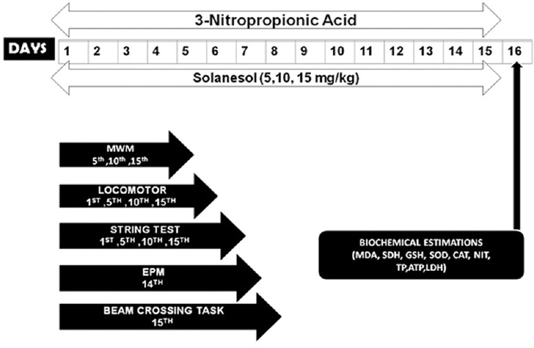
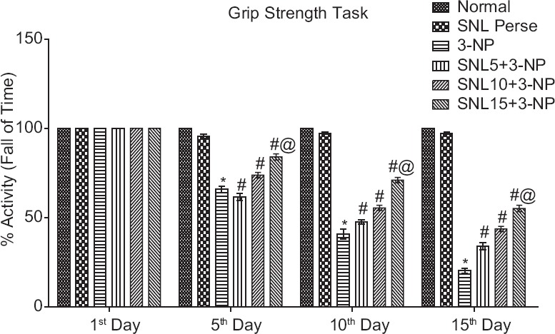
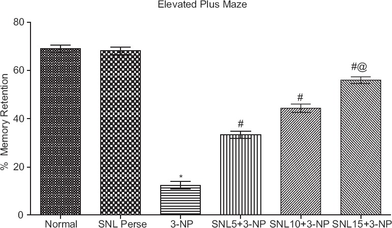
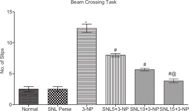
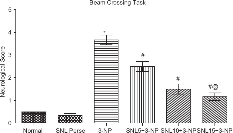
Materials and methods: Various behavioral and biochemical parameters were carried out to evaluate the activity of SNL on 3-NP-treated rats. To determine the therapeutic significance of SNL on HD, different behavioral tests such as memory task, locomotor activity, grip strength, and beam cross and some biochemical test along with histopathological findings were done.
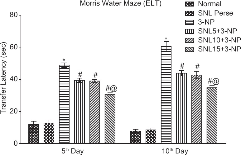
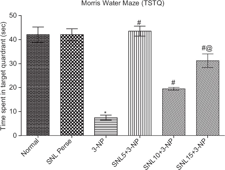
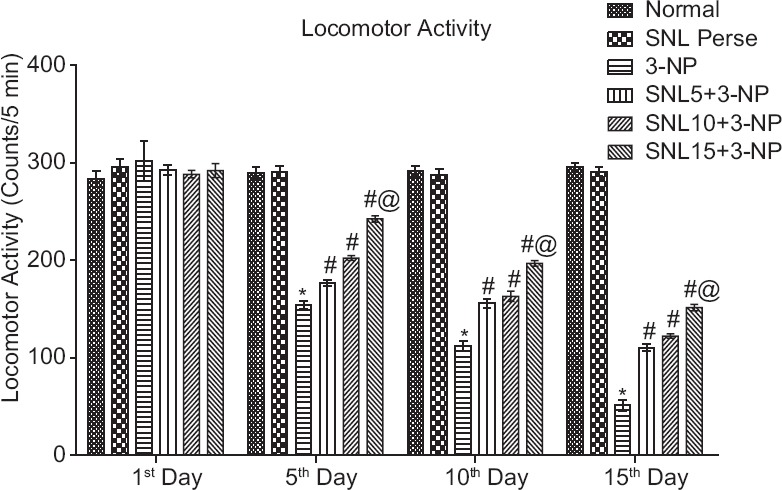
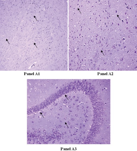
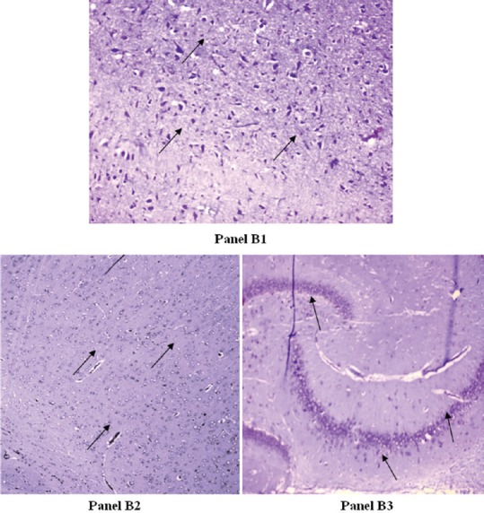
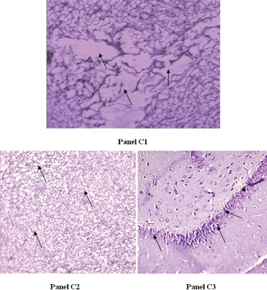
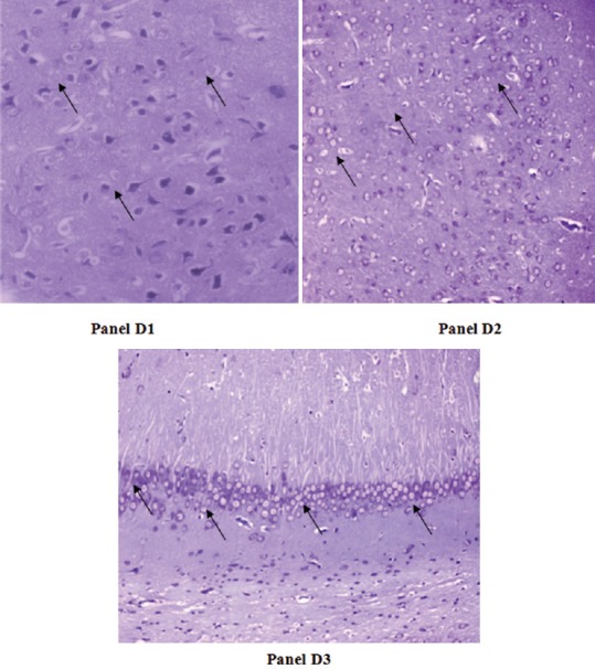
Results: Chronic 3-NP, 10 mg/kg i.p., caused physical and mental abnormalities in animals, including memory impairment, weak grip strength, abnormal posture, and cognitive deficit. Biochemical analysis of brain homogenate in 3-NP-treated rats showed altered mitochondrial complexes, oxidative stress, and elevated lipid biomarkers. Neurohistological alterations of hippocampus, basal ganglia, and cerebral cortex of 3-NP-treated rats exhibit severe neuronal space, irregular damaged cells, and dense pyknotic nuclei-associated marked focal diffused gliosis. SNL administered for 15 days significantly improved motor performance and cognitive behavior task and restored the histopathological changes. Further, SNL treatment significantly improved mitochondrial complexes such as coenzyme-Q10 enzyme activity and attenuated inflammatory and oxidative damage of rat brain.
Conclusion: In the present research work, SNL (5, 10, and 15 mg/kg p.o.) provided notable neuroprotective effect, which was confirmed by behavioral paradigms and biochemical test. It restored the behavioral and biochemical alteration caused by 3-NP and confirmed the strong neuroprotective mechanism of SNL in 3-NP-intoxicated memory and cognitive abnormalities.
Keywords: 3-nitropropionic acid; Coenzyme-Q10; Huntington's disease; mitochondrial dysfunction; solanesol.
Conflict of interest statement
There are no conflicts of interest.
Figures
Similar articles
-
Possible neuroprotective effect of Withania somnifera root extract against 3-nitropropionic acid-induced behavioral, biochemical, and mitochondrial dysfunction in an animal model of Huntington's disease.J Med Food. 2009 Jun;12(3):591-600. doi: 10.1089/jmf.2008.0028. J Med Food. 2009. PMID: 19627208
-
Neuroprotective activity of tetramethylpyrazine against 3-nitropropionic acid induced Huntington's disease-like symptoms in rats.Biomed Pharmacother. 2018 Sep;105:1254-1268. doi: 10.1016/j.biopha.2018.06.079. Epub 2018 Jun 22. Biomed Pharmacother. 2018. PMID: 30021362
-
Protective effect of rivastigmine against 3-nitropropionic acid-induced Huntington's disease like symptoms: possible behavioural, biochemical and cellular alterations.Eur J Pharmacol. 2009 Aug 1;615(1-3):91-101. doi: 10.1016/j.ejphar.2009.04.058. Epub 2009 May 13. Eur J Pharmacol. 2009. PMID: 19445928
-
Protective Effect of Natural Products against Huntington's Disease: An Overview of Scientific Evidence and Understanding Their Mechanism of Action.ACS Chem Neurosci. 2021 Feb 3;12(3):391-418. doi: 10.1021/acschemneuro.0c00824. Epub 2021 Jan 21. ACS Chem Neurosci. 2021. PMID: 33475334 Review.
-
Magnetic resonance imaging and spectroscopy in assessing 3-nitropropionic acid-induced brain lesions: an animal model of Huntington's disease.Prog Neurobiol. 2004 Feb;72(2):87-110. doi: 10.1016/j.pneurobio.2004.02.002. Prog Neurobiol. 2004. PMID: 15063527 Review.
Cited by
-
Nrf2/HO-1 Signaling Activator Acetyl-11-keto-beta Boswellic Acid (AKBA)-Mediated Neuroprotection in Methyl Mercury-Induced Experimental Model of ALS.Neurochem Res. 2021 Nov;46(11):2867-2884. doi: 10.1007/s11064-021-03366-2. Epub 2021 Jun 1. Neurochem Res. 2021. PMID: 34075522
-
Europinidin Mitigates 3-NPA-Induced Huntington's Disease Symptoms in Rats: A Comprehensive Analysis of Oxidative Stress, Mitochondrial Enzyme Complex Activity, Pro-Inflammatory Markers and Neurotransmitter Alterations.Biomedicines. 2024 Mar 12;12(3):625. doi: 10.3390/biomedicines12030625. Biomedicines. 2024. PMID: 38540238 Free PMC article.
-
Solanesol: a promising natural product.Front Pharmacol. 2025 Mar 24;16:1504245. doi: 10.3389/fphar.2025.1504245. eCollection 2025. Front Pharmacol. 2025. PMID: 40196358 Free PMC article. Review.
-
Oxytocin Anti-Apoptotic Potential Mediates Neuroprotection Against 3-Nitropropionic Acid-Induced Huntington's Disease-Like Pathophysiology in Rats: Involvement of Calpain-2/p25 Cdk5/MEF-2 Signaling Pathway.Neurochem Res. 2025 Apr 19;50(3):148. doi: 10.1007/s11064-025-04397-9. Neurochem Res. 2025. PMID: 40252127
-
Lysophosphatidylcholine promoting α-Synuclein aggregation in Parkinson's disease: disrupting GCase glycosylation and lysosomal α-Synuclein degradation.NPJ Parkinsons Dis. 2025 Mar 15;11(1):47. doi: 10.1038/s41531-025-00902-7. NPJ Parkinsons Dis. 2025. PMID: 40089519 Free PMC article.
References
-
- Zheng Z, Diamond MI. Huntington disease and the huntingtin protein. Prog Mol Biol Transl Sci. 2012;107:189–214. - PubMed
-
- Mehan S, Kaur G, Dudi R, Rajput M, Kalra S. Restoration of mitochondrial dysfunction in 6-hydroxydopamine induced Parkinson's disease: A complete review. Open J Park Dis Treat. 2017;1:1–26.
-
- Brouillet E, Guyot MC, Mittoux V, Altairac S, Condé F, Palfi S, et al. Partial inhibition of brain succinate dehydrogenase by 3-nitropropionic acid is sufficient to initiate striatal degeneration in rat. J Neurochem. 1998;70:794–805. - PubMed
MeSH terms
Substances
LinkOut - more resources
Full Text Sources
Medical
Miscellaneous
