. 2019 Mar;17(3):2373-2380.
doi: 10.3892/etm.2019.7167. Epub 2019 Jan 14.

Oestrogen inhibits PTPRO to prevent the apoptosis of renal podocytes

Affiliations

Oestrogen inhibits PTPRO to prevent the apoptosis of renal podocytes

Wei Ren et al. Exp Ther Med. 2019 Mar.

Abstract

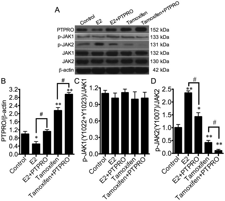
Podocytes are a major component of the glomerular filtration membrane, and their apoptosis is involved in a variety of nephrotic syndromes. In the current study, the effects and molecular mechanisms of oestrogen on the proliferation and apoptosis of podocytes were investigated to elucidate the role of oestrogen in the pathogenesis of childhood nephrotic syndrome. The cell proliferation of mouse renal podocytes (MPC-5) and human primary renal podocytes was promoted by 17β-oestradiol (E2) in what appear to be a time-dependent manner. Apoptosis was inhibited by E2 and promoted by the E2 antagonist, tamoxifen. The expression of protein tyrosine phosphatase receptor type O (PTPRO) decreased with the increasing dosage of E2, but increased with the increasing dosage tamoxifen in MPC-5 and human podocytes. The protein, oestrogen receptor (ER)α, was not expressed in MPC-5 and human podocytes. E2 binding to ERβ completely eliminated PTPRO expression in MPC-5. In podocytes, PTPRO was phosphorylated by E2 at the Y1007 and associated with tyrosine-protein kinase JAK2 (JAK2) activation, rather than JAK1 activation. PTPRO was involved in the binding of E2 to signal transducer and activator of transcription (STAT)3 at the Y705 and S727 sites, resulting in the phosphorylation of STAT3 in podocytes. Through PTPRO, E2 also regulated the proliferation and apoptosis of podocytes. In conclusion, oestrogen binding to ERβ, rather than ERα, promoted the proliferation of podocytes and inhibited the apoptosis of podocytes by inhibiting the expression of PTPRO. The mechanism may be associated with the activation of the JAK2/STAT3 signalling pathway. The current study may provide a novel direction for the treatment of childhood nephrotic syndrome.

Keywords: apoptosis; cell proliferation; oestrogen; protein tyrosine phosphatase receptor type O; renal podocytes.


Figures

Figure 1.
Figure 1.
Identification of the number of cycles for PCR analysis. The number of cycles used for the PCR was determined using semi-quantitative RT-PCR. *P<0.05 as indicated. RT-PCR, reverse transcription-polymerase chain reaction.
Figure 2.
Figure 2.
Oestrogen promotes cell viability and proliferation, and prevents apoptosis in podocytes. (A) The viability of MPC-5 cells treated with 5–100 nM E2 or 0.1–5 µM tamoxifen using a MTT assay. The (B) proliferation rate of and (C) percentage of apoptotic MPC-5 cells treated with 100 nM E2 or 5 µM tamoxifen using a MTT assay and flow cytometry, respectively. (D) The viability of human primary renal podocytes treated with 5–100 nM E2 or 0.1–5 µM tamoxifen using a MTT assay. The (E) proliferation rate of and (F) percentage of apoptotic human primary renal podocytes treated with 100 nM E2 or 5 µM tamoxifen using a MTT assay and flow cytometry, respectively. *P<0.05, **P<0.01 vs. Control. E2, 17β-oestradiol.
Figure 3.
Figure 3.
Oestrogen and its receptors inhibit PTPRO expression in podocytes. PTPRO mRNA expression in (A) MPC-5 cells and (B) human primary renal podocytes treated with 5–100 nM E2 or 0.1–5 µM tamoxifen using semi-quantitative reverse transcription-polymerase chain reaction. *P<0.05, **P<0.01 vs. Control. (C) The protein expression of ERα and ERβ were detected by western blotting in MPC-5 cells and human primary renal podocytes. The protein expression of ERβ and PTPRO were detected by western blotting in MPC-5 cells stimulated with 100 nM E2 and/or ERβ overexpression vectors. (D) The quantification of ERα and ERβ in MPC-5 cells and human primary renal podocytes. (E) The quantification of the ERβ protein in MPC-5 cells stimulated with 100 nM E2 and/or ERβ overexpression vectors. The quantification of (F) the PTPRO mRNA and (G) the PTPRO protein in MPC-5 cells stimulated with 100 nM E2 and/or an ERβ overexpression vector. **P<0.01 vs. Control-; ##P<0.01 vs. Control+. PTPRO, protein tyrosine phosphatase receptor type O; E2, 17β-oestradiol; ER, oestrogen receptor; 1, MPC-5 cells; 2, human primary renal podocytes.
Figure 4.
Figure 4.
Oestrogen activates JAK2 by inhibiting PTPRO in podocytes. MPC-5 cells were simulated with in E2 or tamoxifen and transfected with a PTPRO overexpression vector. (A) Protein expression in MPC5 cells. Quantification of (B) PTPRO, (C) p-JAK1 and (D) p-JAK2 proteins in MPC5 cells. *P<0.05, **P<0.01 vs. Control; #P<0.05 as indicated. PTPRO, protein tyrosine phosphatase receptor type O; E2, 17β-oestradiol; ER, oestrogen receptor; p-, phosphorylation; JAK, tyrosine-protein kinase JAK.
Figure 5.
Figure 5.
Oestrogen activates STAT3 by inhibiting PTPRO, which is involved in the regulation of the viability and apoptosis of podocytes through E2 binding. MPC-5 cells were simulated with in E2 or tamoxifen and transfected with a PTPRO overexpression vector. (A) Protein expression in MPC5 cells. Quantification of (B) p-STAT3(Y705) and (C) p-STAT3(S727) proteins. The (D) viability of and (E) percentage of apoptotic MPC-5 cells. *P<0.05, **P<0.01 vs. Control; #P<0.05, ##P<0.01. PTPRO, protein tyrosine phosphatase receptor type O; E2, 17β-oestradiol; p-, phosphorylation; OD, optical density; STAT, signal transducer and activator of transcription.

References

    1. Welsh GI, Saleem MA. Nephrin-signature molecule of the glomerular podocyte? J Pathol. 2010;220:328–237. - PubMed
    1. Niranjan T, Bielesz B, Gruenwald A, Ponda M P, Kopp JB, Thomas DB, Susztak K. The Notch pathway in podocytes plays a role in the development of glomerular disease. Nat Med. 2008;14:290–298. doi: 10.1038/nm1731. - DOI - PubMed
    1. Ding F, Wickman L, Wang SQ, Zhang Y, Wang F, Afshinnia F, Hodgin J, Ding J, Wiggins RC. Accelerated podocyte detachment and progressive podocyte loss from glomeruli with age in Alport Syndrome. Kidney Int. 2017;92:1515–1525. doi: 10.1016/j.kint.2017.05.017. - DOI - PMC - PubMed
    1. Mauvais-Jarvis F, Clegg DJ, Hevener AL. The role of estrogens in control of energy balance and glucose homeostasis. Endocr Rev. 2013;34:309–338. doi: 10.1210/er.2012-1055. - DOI - PMC - PubMed
    1. Mercantepe T, Unal D, Selli J, Mercantepe F, Unal B, Karabiyik TN. Protective effects of estrogen and bortezomib in kidney tissue of post-menopausal rats: An ultrastructural study. Ren Fail. 2016;38:1129–1135. doi: 10.1080/0886022X.2016.1184958. - DOI - PubMed