

Proximal Hamstring Repair: A Biomechanical Analysis of Variable Suture Anchor Constructs
- PMID: 30783606
- PMCID: PMC6365995
- DOI: 10.1177/2325967118824149
Proximal Hamstring Repair: A Biomechanical Analysis of Variable Suture Anchor Constructs
Abstract
Background: Despite an abundance of literature regarding construct strength for a myriad of anchors and anchor configurations in the shoulder, there remains a paucity of biomechanical studies detailing the efficacy of these implants for proximal hamstring repair.
Purpose: To biomechanically evaluate the ultimate failure load and failure mechanism of knotless and knotted anchor configurations for hamstring repair.
Study design: Controlled laboratory study.
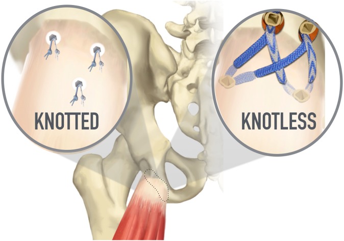
Methods: A total of 17 cadaveric specimens divided into 3 groups composed of intact hamstring tendons as well as 2 different anchor configurations (all-knotted and all-knotless) underwent first cyclic loading and subsequent maximal loading to failure. This protocol entailed a 10-N preload, followed by 100 cycles incrementally applied from 20 to 200 N at a frequency of 0.5 Hz, and ultimately followed by a load to failure with a loading rate of 33 mm/s. The ultimate failure load and mechanism of failure were recorded for each specimen, as was the maximal displacement of each bone-tendon interface subsequent to maximal loading. Analysis of variance was employed to calculate differences in the maximal load to failure as well as the maximal displacement between the 3 study groups. Holm-Sidak post hoc analysis was applied when necessary.
Results: The all-knotless suture anchor construct failed at the highest maximal load of the 3 groups (767.18 ± 93.50 N), including that for the intact tendon group (750.58 ± 172.22 N). There was no statistically significant difference between the all-knotless and intact tendon groups; however, there was a statistically significant difference in load to failure when the all-knotless construct was compared with the all-knotted technique (549.56 ± 20.74 N) (P = .024). The most common mode of failure in both repair groups was at the suture-tendon interface, whereas the intact tendon group most frequently failed via avulsion of the tendon from its insertion site.
Conclusion: Under biomechanical laboratory testing conditions, proximal hamstring repair using all-knotless suture anchors outperformed the all-knotted suture anchor configuration with regard to elongation during cyclic loading and maximal load to failure. Failure in the all-knotted repair group was at the suture-tendon interface in most cases, whereas the all-knotless construct failed most frequently at the musculotendinous junction.
Clinical relevance: No biomechanical studies have clearly identified the optimal anchor configuration to avert proximal hamstring repair failure. Delineating this ideal suture anchor construct and its strength compared with an intact hamstring tendon may alter the current standards for postoperative rehabilitation, which remain extremely conservative and onerous for these patients.
Keywords: anchor; biomechanics; hamstring; hamstring injury; tendon repair.
Conflict of interest statement
One or more of the authors has declared the following potential conflict of interest or source of funding: M.B.G. is a paid consultant for Arthrex, Medacta, and Ferring Pharmaceuticals; receives royalties from Arthrex; has received research grants from Arthrex; has received hospitality payments from Arthrex and Stryker; and is a shareholder in the Cedars-Sinai Kerlan-Jobe institute. B.S.A. has received research support from Smith & Nephew, Arthrex, DJO, Stryker, CDC Medical, and Zimmer Biomet. AOSSM checks author disclosures against the Open Payments Database (OPD). AOSSM has not conducted an independent investigation on the OPD and disclaims any liability or responsibility relating thereto.
Figures

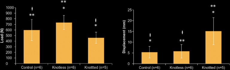
 P = .140. For the displacement bar graph: *P = .005, **P = .005, and
P = .866.
P = .140. For the displacement bar graph: *P = .005, **P = .005, and
P = .866.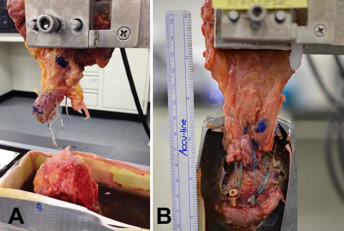
References
-
- Ahmad CS, Redler LH, Ciccotti MG, Maffulli N, Longo UG, Bradley J. Evaluation and management of hamstring injuries. Am J Sports Med. 2013;41(12):2933–2947. - PubMed
-
- Baums MH, Kostuj T, Klinger HM, Papalia R. [Rotator cuff repair: single- vs double-row. Clinical and biomechanical results]. Orthopade. 2016;45(2):118–124. - PubMed
-
- Campbell KA, Quirno M, Hamula M, et al. Suture anchor repair of complete proximal hamstring ruptures: a cadaveric biomechanical evaluation. Bull Hosp Jt Dis (2013). 2017;75(4):241–247. - PubMed
-
- Hamming MG, Philippon MJ, Rasmussen MT, et al. Structural properties of the intact proximal hamstring origin and evaluation of varying avulsion repair techniques: an in vitro biomechanical analysis. Am J Sports Med. 2015;43(3):721–728. - PubMed
LinkOut - more resources
Full Text Sources
