. 2019 May 7;47(8):4124-4135.
doi: 10.1093/nar/gkz110.

MRE11 UFMylation promotes ATM activation

Affiliations

MRE11 UFMylation promotes ATM activation

Zhifeng Wang et al. Nucleic Acids Res. .

Erratum in

Abstract

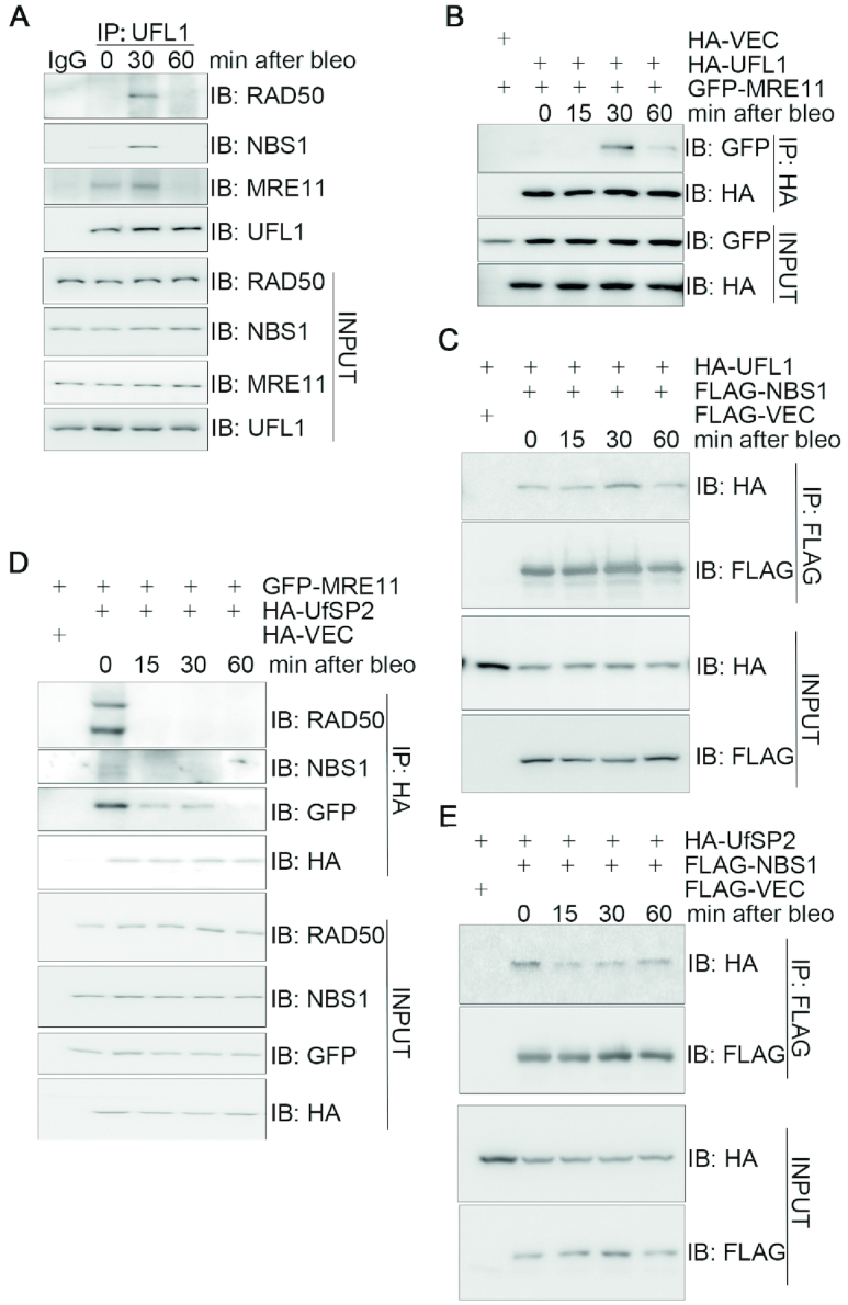
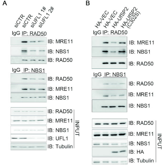
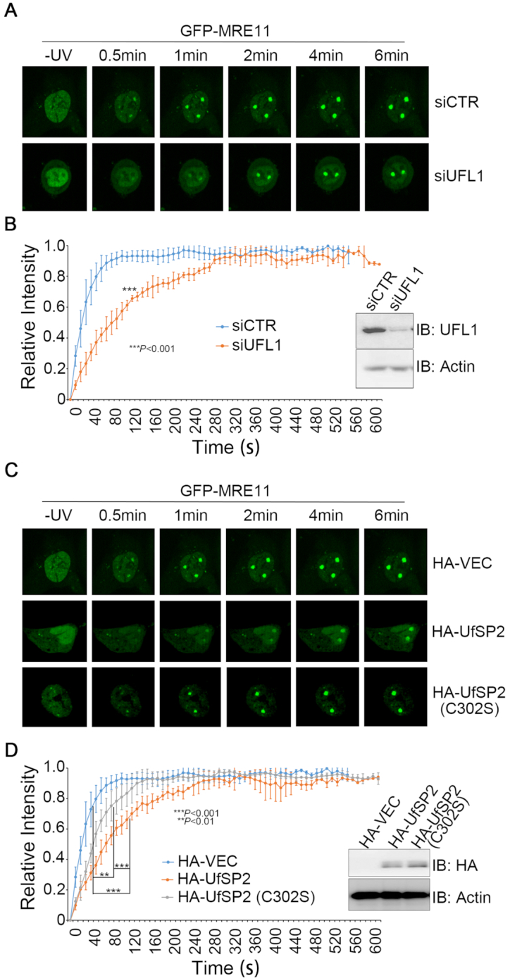
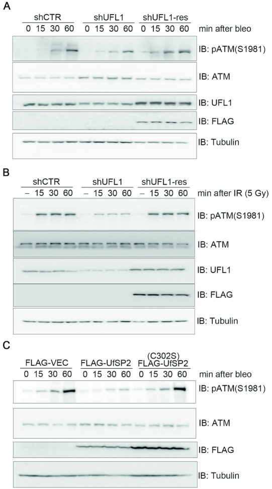
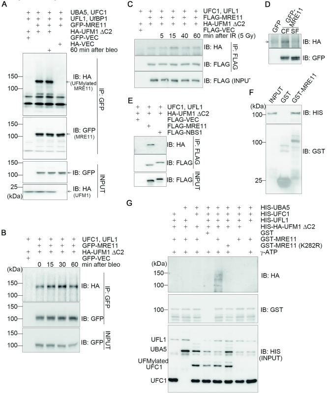
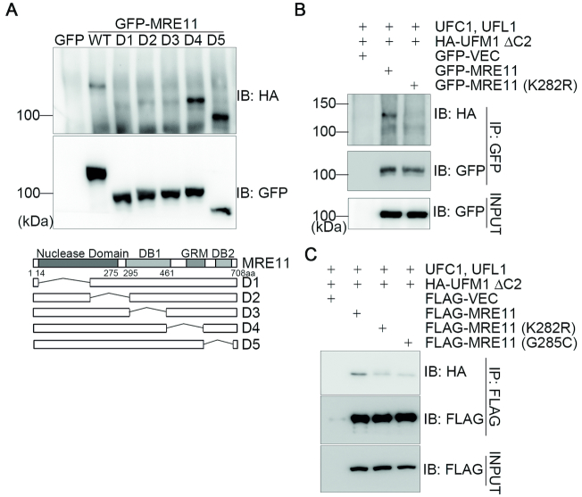
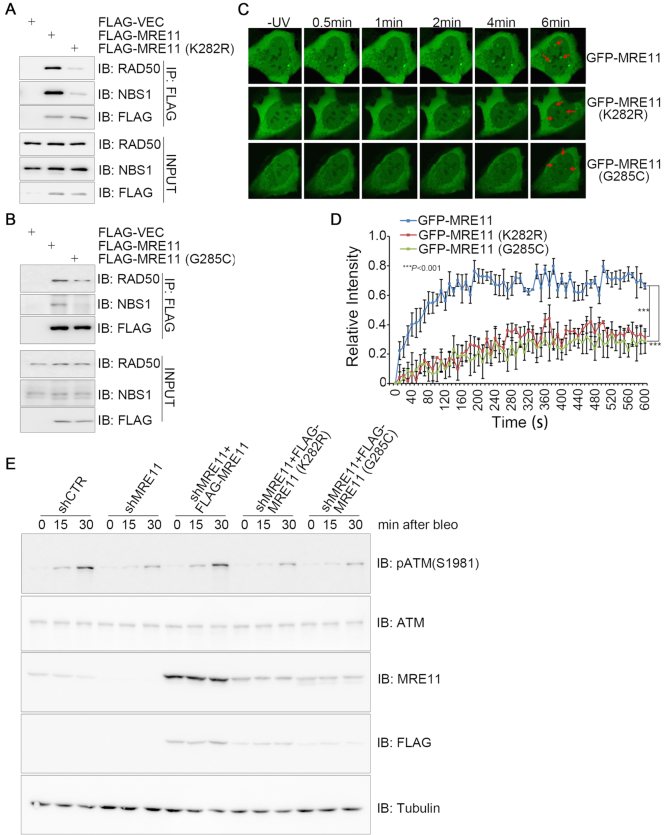
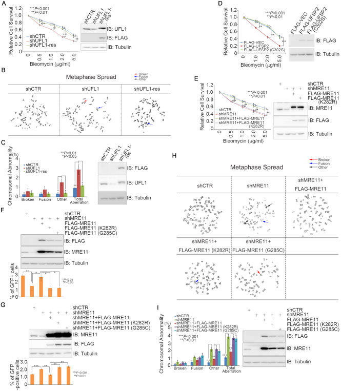
A proper DNA damage response (DDR) is essential to maintain genome integrity and prevent tumorigenesis. DNA double-strand breaks (DSBs) are the most toxic DNA lesion and their repair is orchestrated by the ATM kinase. ATM is activated via the MRE11-RAD50-NBS1 (MRN) complex along with its autophosphorylation at S1981 and acetylation at K3106. Activated ATM rapidly phosphorylates a vast number of substrates in local chromatin, providing a scaffold for the assembly of higher-order complexes that can repair damaged DNA. While reversible ubiquitination has an important role in the DSB response, modification of the newly identified ubiquitin-like protein ubiquitin-fold modifier 1 and the function of UFMylation in the DDR is largely unknown. Here, we found that MRE11 is UFMylated on K282 and this UFMylation is required for the MRN complex formation under unperturbed conditions and DSB-induced optimal ATM activation, homologous recombination-mediated repair and genome integrity. A pathogenic mutation MRE11(G285C) identified in uterine endometrioid carcinoma exhibited a similar cellular phenotype as the UFMylation-defective mutant MRE11(K282R). Taken together, MRE11 UFMylation promotes ATM activation, DSB repair and genome stability, and potentially serves as a therapeutic target.


Figures

Figure 1.
Figure 1.
UFL1/UfSP2 dynamically interacts with the MRN complex in response to DNA damage. (A) DSB-induced interaction between UFL1 and the MRN complex. HEK293T cells were treated with 10 μg/ml bleomycin and harvested at the indicated time points. Total cell lysates were subjected to IP followed by immunoblotting with the indicated antibodies. (B) The interaction between UFL1 and MRE11 peaked 30 min after bleomycin treatment. HEK293T cells co-expressing HA-UFL1 and GFP-MRE11 were treated with 10 μg/ml bleomycin at the indicated time points. Total cell lysates were harvested and subjected to IP and immunoblotting with the indicated antibodies. (C) The interaction between HA-UFL1 and FLAG-NBS1 peaked at 30 min after bleomycin treatment. HEK293T cells co-expressing HA-UFL1 and FLAG-NBS1 were treated with 10 μg/ml bleomycin. Total cell lysates were harvested at the indicated times and subjected to IP and immunoblotting with the indicated antibodies. (D and E) DNA damage disrupted the interaction between HA-UfSP2 and GFP-MRE11 or FLAG-NBS1. HEK293T cells co-expressing HA-UfSP2 and GFP-MRE11 or FLAG-NBS1 were treated with 10 μg/ml bleomycin. Total cell lysates were harvested at different times as indicated, and subjected to IP and immunoblotting with the indicated antibodies. Abbreviations: bleo, bleomycin; IB, immunoblot; IP, immunoprecipitation.
Figure 2.
Figure 2.
UFMylation promotes MRN complex formation under unperturbed conditions. (A) UFL1 depletion decreased MRN complex formation. Total lysates derived from HEK293T cells transfected with UFL1 (siUFL1 1# and 2#) or control (siCTR) siRNAs were immunoprecipitated with RAD50 or NBS1 antibodies. The precipitates were probed with the indicated antibodies. (B) Over-expression of UfSP2 compromised MRN complex formation. Total lysates derived from HEK293T cells overexpressing HA-VEC, HA-UfSP2 or HA-UfSP2(C302S) were subjected to IP and immunoblotting with the indicated antibodies. Abbreviations: bleo, bleomycin; IB, immunoblot; IP, immunoprecipitation; siCTR, small interfering RNA control.
Figure 3.
Figure 3.
UFMylation promotes MRE11 recruitment to the DNA damage stripes. (A and B) UFL1 depletion decreased the initial recruitment of MRE11 to DNA damage stripes. UFL1-depleted (siUFL1) or mock (siCTR)-depleted DU145 cells transiently expressed GFP-MRE11. (C and D) UfSP2 over-expression decreased MRE11 initial recruitment to DNA damage stripes. DU145 cells were co-transfected with an HA vector, HA-UfSP2 or HA-UfSP2(C302S) and GFP-MRE11 at a molar ratio of 10:1. GFP-positive cells were micro-irradiated with a UV laser (365 nm) and consecutive images were collected at 10-s interval for 10 min. Representative images of GFP-MRE11 recruitment are shown in A and C, and the statistical analysis of recruitment dynamics with the Spearman’s rank-order correlation test is shown in B and D.
Figure 4.
Figure 4.
Defective UFMylation compromises optimal ATM activation upon DNA damage. (A and B) Inhibition of UFL1 expression compromised optimal activation of ATM upon DNA damage. A549 cells that were mock depleted (shCTR), UFL1 depleted (shUFL1) or UFL1 depleted, and then rescued with an shUFL1-resistant form of UFL1 (shUFL1-res) were treated with 10 μg/ml bleomycin (A) or irradiated with 5-Gy IR (B). Total cell lysates were harvested at different times after treatment and subjected to immunoblotting with the indicated antibodies. (C) Over-expression of UfSP2 compromised optimal activation of ATM upon DNA damage. A549 cells expressing FLAG-VEC, FLAG-UfSP2 or FLAG-UfSP2(C302S) were treated with 10 μg/ml bleomycin. Total cell lysates were harvested and subjected to immunoblotting with the indicated antibodies.
Figure 5.
Figure 5.
MRE11 is UFMylated and this UFMylation transiently increases after DSB induction. (A) MRE11 is UFMylated in vivo. HEK293T cells co-expressing UFMylation factors (UBA5, UFC1, UFL1, UfBP1, HA-UFM1ΔC2) and GFP-MRE11 were lysed in 5% SDS-containing buffer. Total cell lysates were boiled for 5 min before IP and immunoblotting with the indicated antibodies. (B and C) MRE11 UFMylation is induced by DSBs. Experiments were performed as described in A except that HEK293T transfectants were treated with 10 μg/ml bleomycin (B) or 5-Gy IR (C) and total cell lysates were harvested at different time-points post-treatment. (D) UFMylated MRE11 is enriched on chromatin. Chromatin fractionation assays were performed with HEK293T cells co-expressing the UFMylation factors (UBA5, UFC1, UFL1, UfBP1, HA-UFM1ΔC2) and GFP-MRE11. Both the cytosolic fraction (SF) and chromatin-enriched fraction (CF) were subjected to IP with an anti-GFP antibody and immunoblotting with the indicated antibodies. (E) UFMylation of NBS1 was undetected. HEK293T cells co-expressing the UFMylation factors (UBA5, UFC1, UFL1, UfBP1, HA-UFM1ΔC2) and FLAG-MRE11 or FLAG-NBS1 were lysed in 5% SDS-containing buffer. Total cell lysates were boiled for 5 min before IP and immunoblotting with the indicated antibodies. (F) MRE11 directly interacted with UFL1 in vitro. Bacterially produced GST or GST-MRE11 was used to pull down bacterially produced HIS-UFL1. (G) MRE11 is UFMylated in vitro. Recombinant UFMylation factors (HIS-UBA5, HIS-UFC1, HIS-UFL1 and HIS-HA-UFM1ΔC2) with bacterially produced GST, GST-MRE11 or GST-MRE11(K282R) were incubated in UFMylation buffer at 30°C for 90 min. The reaction was terminated by adding SDS sample buffer and the samples were subjected to sodium dodecyl sulphate-polyacrylamide gel electrophoresis (SDS-PAGE) followed by immunoblotting with the indicated antibodies.
Figure 6.
Figure 6.
K282 is the essential UFMylation site in MRE11. (A) Mapping the essential regions for MRE11 UFMylation. A series of deletion mutants in the context of full-length MRE11 were generated as indicated. The mutants or the control plasmids were co-transfected with the UFMylation factors (UBA5, UFC1, UFL1, UfBP1, HA-UFM1ΔC2) into HEK293T cells. Experiments were performed as described in 5A but using these deletion mutants. (B) K282 is the major UFMylation site for MRE11. HEK293T cells co-expressing the UFMylation factors (UBA5, UFC1, UFL1, UfBP1, HA-UFM1ΔC2) and GFP-MRE11 or GFP-MRE11(K282R) were lysed in 5% SDS-containing buffer. Total cell lysates were boiled for 5 min before IP and immunoblotting with the indicated antibodies. (C) The pathogenic mutant MRE11(G285C) exhibited defective UFMylation. Experiments were performed as described in B except that GFP-MRE11(G285C) was also included.
Figure 7.
Figure 7.
Defective MRE11 UFMylation impairs MRN complex formation and DNA damage-induced ATM activation. (A and B) UFMylation-defective mutants compromised formation of the MRN complex. Total cell lysates derived from HEK293T cells expressing a FLAG vector, FLAG-MRE11, FLAG-MRE11(K282R) or FLAG-MRE11(G285C) were subjected to IP with an anti-FLAG antibody and immunoblotting with the indicated antibodies. (C and D) UFMylation-defective mutants compromised recruitment of UFMylation-defective mutants to DNA damage stripes. U2OS cells transiently expressed GFP-MRE11, GFP-MRE11(K282R) or GFP-MRE11(G285C). GFP-positive cells were micro-irradiated with a UV laser (365 nm) and consecutive images were captured at 10-s interval for 10 min. Representative images of GFP-MRE11 recruitment are shown in C, and the statistical analysis of recruitment dynamics with the Spearman’s rank-order correlation test is shown in (D). (E) UFMylation-defective mutants compromised DNA damage-induced ATM activation. A549 cells stably expressing shCTR or shMRE11 were re-introduced with FLAG-MRE11, FLAG-MRE11(K282R) or FLAG-MRE11(G285C). The cells were treated with 10 μg/ml bleomycin for 15 or 30 min. Total cell lysates were harvested and analyzed by SDS-PAGE and immunoblotting with the indicated antibodies.
Figure 8.
Figure 8.
MRE11 UFMylation promotes HR-medicated DSB repair and genome stability. (A) Depletion of UFL1-sensitized cells to bleomycin treatment. A549 cells stably expressing shCTR, shUFL1 or shUFL1 reconstituted with an shUFL1-resistant form of FLAG-UFL1 were treated with increasing concentrations of bleomycin, as indicated. The surviving colonies were analyzed after 10 days. (B and C) Depletion of UFL1 resulted in aberrant mitotic chromosomes. The cells generated in A were used for mitotic spread preparation, and >400 mitotic chromosomes per type of manipulated cells were analyzed. Representative images of mitotic spreads are shown in B, while the quantitative analysis is shown in (C). (D) Overexpression of UfSP2 sensitized cells to bleomycin treatment. A549 cells stably expressing a FLAG vector, FLAG-UfSP2 or FLAG-UfSP2(C302S) were treated with increasing concentrations of bleomycin, as indicated. The surviving colonies were analyzed after 10 days. (E) MRE11(K282R) failed to rescue MRE11-depletion-induced cellular sensitivity to bleomycin treatment. A549 cells stably expressing shCTR, shMRE11, shMRE11 reconstituted with FLAG-MRE11 or FLAG-MRE11(K282R) were treated with increasing concentrations of bleomycin, as indicated. The surviving colonies were analyzed after 10 days. (F) Defective MRE11 UFMylation compromised HR-mediated DSB repair. DRU2OS cells, in which a DR-GFP reporter cassette is integrated, stably expressing shCTR, shMRE11 or shMRE11 reconstituted with FLAG-MRE11, FLAG-MRE11(K282R) or FLAG-MRE11(G285C). HR assays were performed in triplicate. (G) Defective MRE11 UFMylation promoted NHEJ-mediated DSB repair. U2OS cells, in which an EJ5-GFP reporter cassette is integrated, stably expressing shCTR, shMRE11 or shMRE11 reconstituted with FLAG-MRE11, FLAG-MRE11(K282R) or FLAG-MRE11(G285C). NHEJ assays were performed in triplicate. (H and I) Defective MRE11 UFMylation contributed to aberrant mitotic chromosomes. A549 cells stably expressing shCTR, shMRE11 or shMRE11 reconstituted with FLAG-MRE11, FLAG-MRE11(K282R) or FLAG-MRE11(G285C) and then subjected to mitotic spread preparation; >400 mitotic chromosomes per type of manipulated cells were analyzed. All data were derived from three independent experiments. Representative images of mitotic spreads are shown in H, while the quantitative analysis is shown in I. A two-way ANOVA was performed to determine statistical significance. *: p<0.05; **: p<0.01; ***: p<0.001.

Similar articles

Cited by

References

    1. Lee J.H., Paull T.T.. Direct activation of the ATM protein kinase by the Mre11/Rad50/Nbs1 complex. Science. 2004; 304:93–96. - PubMed
    1. Lee J.H., Paull T.T.. ATM activation by DNA double-strand breaks through the Mre11-Rad50-Nbs1 complex. Science. 2005; 308:551–554. - PubMed
    1. Lee J.H., Paull T.T.. Activation and regulation of ATM kinase activity in response to DNA double-strand breaks. Oncogene. 2007; 26:7741–7748. - PubMed
    1. Bakkenist C.J., Kastan M.B.. DNA damage activates ATM through intermolecular autophosphorylation and dimer dissociation. Nature. 2003; 421:499–506. - PubMed
    1. Sun Y., Jiang X., Chen S., Fernandes N., Price B.D.. A role for the Tip60 histone acetyltransferase in the acetylation and activation of ATM. Proc. Natl. Acad. Sci. U.S.A. 2005; 102:13182–13187. - PMC - PubMed

Publication types

MeSH terms