

Evaluation of the yield of post-clipping angiography and nationwide current practice
- PMID: 30783804
- PMCID: PMC6431297
- DOI: 10.1007/s00701-019-03834-3
Evaluation of the yield of post-clipping angiography and nationwide current practice
Abstract
Background: Surgical treatment of intracranial saccular aneurysms aims to prevent (re)hemorrhage by complete occlusion of the aneurysmal lumen. It is unclear whether routine postoperative imaging, to assess aneurysmal occlusion, is necessary since intraoperative assessment by the neurosurgeon may be sufficient. We assessed routine clinical protocols for post-clipping imaging in the Netherlands and determined whether intraoperative assessment of aneurysm clippings sufficiently predicts aneurysm residuals.
Methods: A survey was conducted to assess postoperative imaging protocols in centers performing clipping of intracranial aneurysms in the Netherlands (n = 9). Furthermore, a retrospective single-center cohort study was performed to determine the predictive value of intraoperative assessment of aneurysm occlusion in relation to postoperative digital subtraction angiography (DSA) findings, between 2009 and 2017.
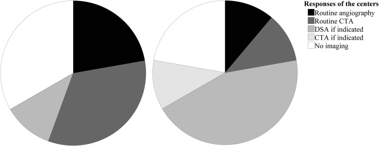
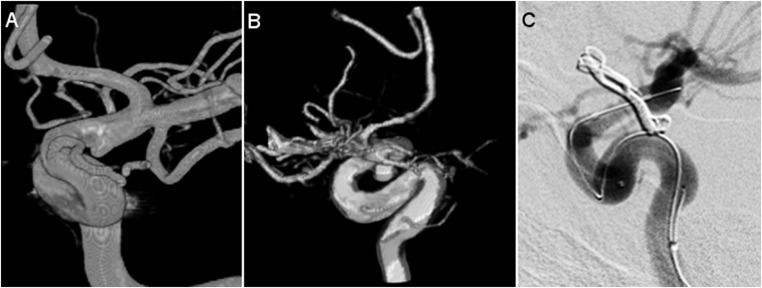
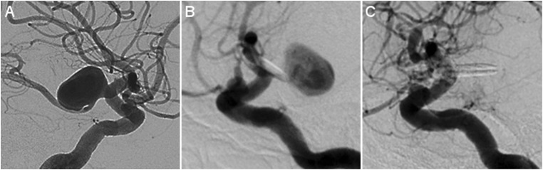
Results: No center performed intraoperative DSA in a hybrid OR, routinely. Respectively, four (44.4%), seven (77.8%), and three (33.3%) centers did not routinely perform early postoperative imaging, late follow-up imaging, or any routine imaging at all. Regarding our retrospective study, 106 patients with 132 clipped aneurysms were included. There were 23 residuals ≥ 1 mm (17.4%), of which 10 (43.5%) were unexpected. For the presence of these residuals, intraoperative assessment showed a sensitivity of 56.5%, a specificity of 86.2%, a positive predictive value of 46.4%, and a negative predictive value of 90.4%.
Conclusions: There is lack of consensus regarding the post-clipping imaging strategy in the Netherlands. Since intraoperative assessment is shown to be insufficient to predict postoperative aneurysm residuals, we advocate routine postoperative imaging after aneurysm clipping unless this is not warranted on the basis of patient age, clinical condition, and/or comorbidity.
Keywords: Residual; Retreatment; Ruptured; Saccular aneurysm; Surgery; Unruptured.
Conflict of interest statement
Conflict of interest
The authors declare that they have no conflict of interest.
Ethical approval
For this type of study, formal consent is not required.
Figures
Similar articles
-
Comparison of Intra- and Postoperative 3-Dimensional Digital Subtraction Angiography in Evaluation of the Surgical Result After Intracranial Aneurysm Treatment.Neurosurgery. 2020 Sep 15;87(4):689-696. doi: 10.1093/neuros/nyz487. Neurosurgery. 2020. PMID: 31748795
-
Impact of Intraoperative 3-Dimensional Volume-Rendering Rotational Angiography on Clip Repositioning Rates in Aneurysmal Surgery.World Neurosurg. 2018 Jun;114:e573-e580. doi: 10.1016/j.wneu.2018.03.035. Epub 2018 Mar 14. World Neurosurg. 2018. PMID: 29548950
-
Detection of remnants in clipped unruptured intracranial aneurysms by intraoperative CT-angiography and postoperative DSA: clinical relevance and follow-up.Acta Neurochir (Wien). 2025 Apr 17;167(1):109. doi: 10.1007/s00701-025-06518-3. Acta Neurochir (Wien). 2025. PMID: 40240681 Free PMC article.
-
Essentials in intraoperative indocyanine green videoangiography assessment for intracranial aneurysm surgery: conclusions from 295 consecutively clipped aneurysms and review of the literature.Neurosurg Focus. 2014 Feb;36(2):E7. doi: 10.3171/2013.11.FOCUS13475. Neurosurg Focus. 2014. PMID: 24484260 Review.
-
Indocyanine Green Videoangiography in Aneurysm Surgery: Systematic Review and Meta-Analysis.Neurosurgery. 2018 Aug 1;83(2):166-180. doi: 10.1093/neuros/nyx387. Neurosurgery. 2018. PMID: 28973404
Cited by
-
Value of 3-Dimensional Digital Subtraction Angiography for Detection and Classification of Intracranial Aneurysm Remnants After Clipping.Oper Neurosurg. 2021 Jul 15;21(2):63-72. doi: 10.1093/ons/opab087. Oper Neurosurg. 2021. PMID: 33861324 Free PMC article.
References
-
- Acevedo JC, Turjman F, Sindou M. Postoperative angiography in surgery for intracranial aneurysm. Prospective study in a consecutive series of 267 operated aneurysms. Neurochirurgie. 1997;43:275–284. - PubMed
-
- Bernat AL, Clarençon F, André A, Nouet A, Clémenceau S, Sourour NA, et al. Risk factors for angiographic recurrence after treatment of unruptured intracranial aneurysms: outcomes from a series of 178 unruptured aneurysms treated by regular coiling or surgery. J Neuroradiol. 2017;44:298–307. doi: 10.1016/j.neurad.2017.05.003. - DOI - PubMed
MeSH terms
LinkOut - more resources
Full Text Sources
Medical
