. 2019 Feb 19;26(8):2113-2125.e6.
doi: 10.1016/j.celrep.2019.01.086.

Dynamics of the Eukaryotic Replicative Helicase at Lagging-Strand Protein Barriers Support the Steric Exclusion Model

Affiliations

Dynamics of the Eukaryotic Replicative Helicase at Lagging-Strand Protein Barriers Support the Steric Exclusion Model

Hazal B Kose et al. Cell Rep. .

Abstract

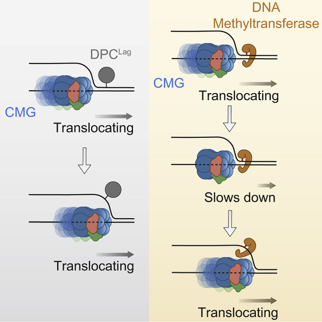
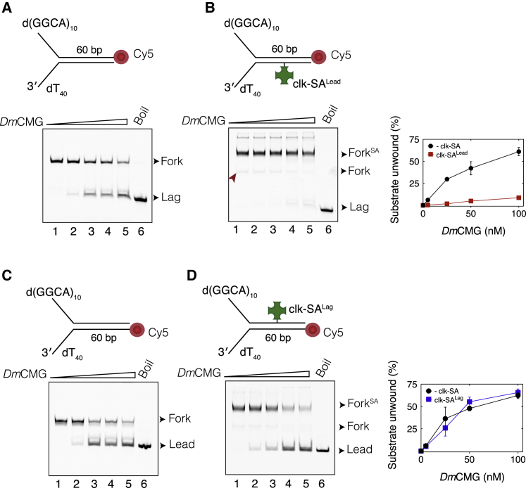
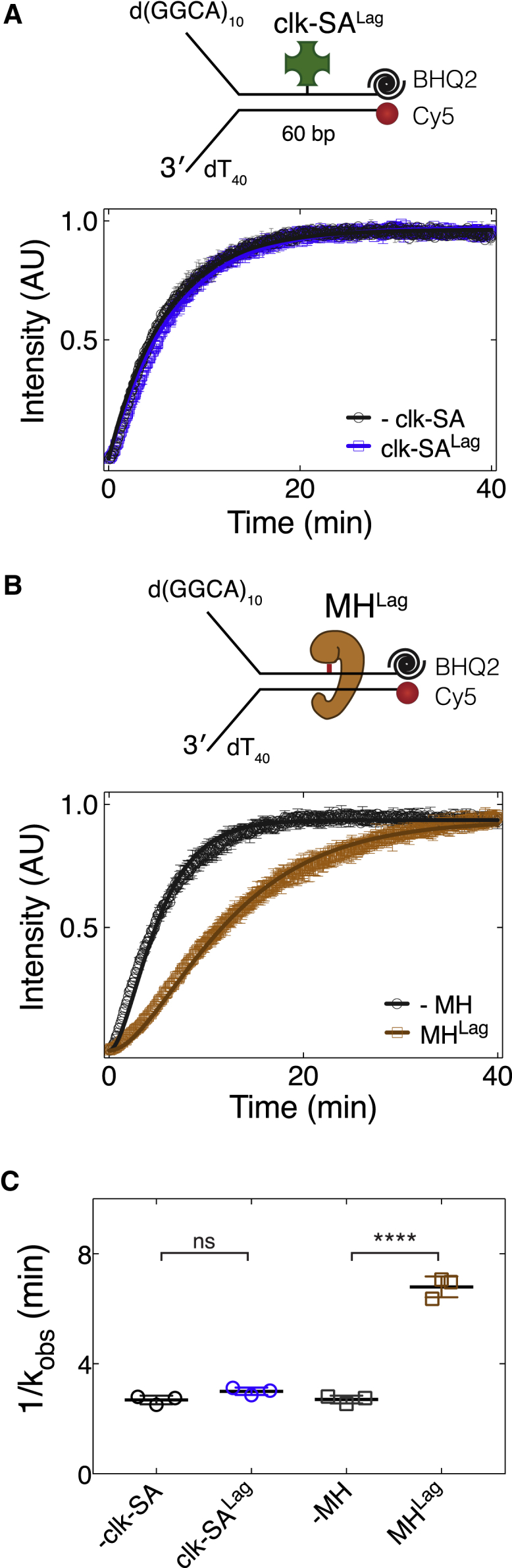
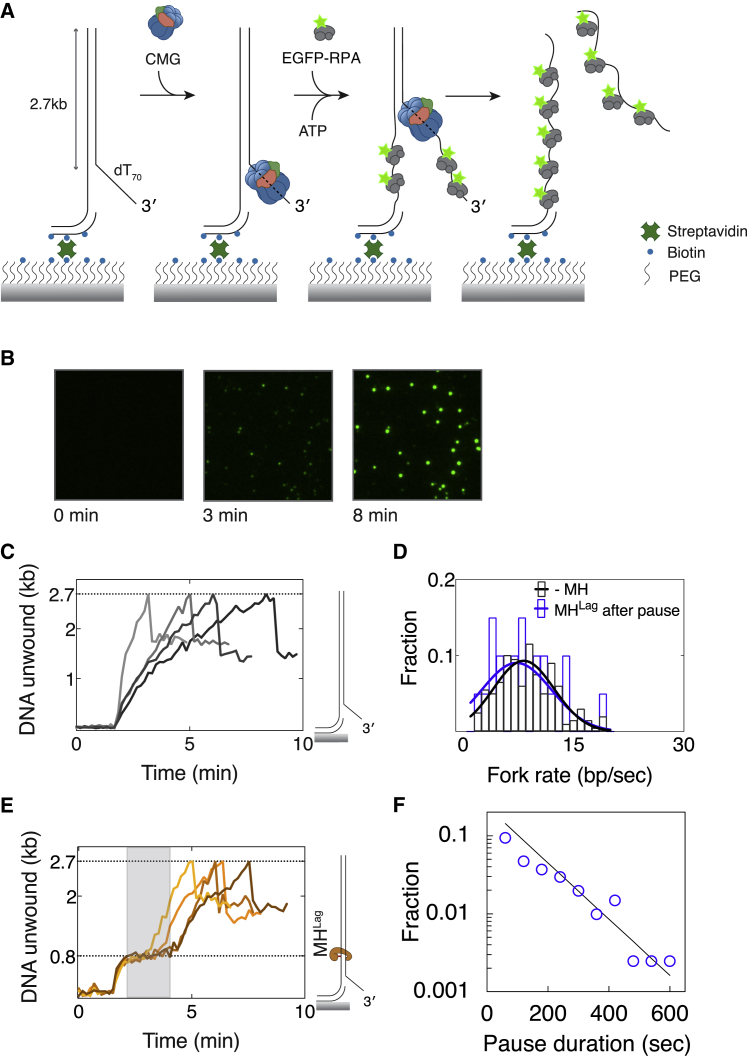
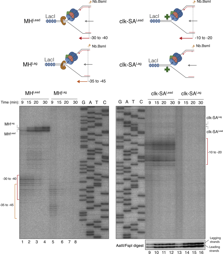
Progression of DNA replication depends on the ability of the replisome complex to overcome nucleoprotein barriers. During eukaryotic replication, the CMG helicase translocates along the leading-strand template and unwinds the DNA double helix. While proteins bound to the leading-strand template efficiently block the helicase, the impact of lagging-strand protein obstacles on helicase translocation and replisome progression remains controversial. Here, we show that CMG and replisome progressions are impaired when proteins crosslinked to the lagging-strand template enhance the stability of duplex DNA. In contrast, proteins that exclusively interact with the lagging-strand template influence neither the translocation of isolated CMG nor replisome progression in Xenopus egg extracts. Our data imply that CMG completely excludes the lagging-strand template from the helicase central channel while unwinding DNA at the replication fork, which clarifies how two CMG helicases could freely cross one another during replication initiation and termination.

Keywords: CMG; DNA-protein crosslink; eukaryotic DNA replication; replicative helicase; single-molecule imaging; steric exclusion.


Figures

None
Graphical abstract
Figure 1
Figure 1
Interaction of DmCMG with Strand-Specific Biotin-Streptavidin Complexes (A) Experimental approach used in unwinding assays. (B and C) DmCMG-mediated unwinding of fork DNA templates in the (B) absence or (C) presence of SALead. Right panel shows percentage of substrate unwound as a function of DmCMG concentration. The extended length on the 5′ tail (17 repeats of d(GGCA)) was necessary to discriminate SA-bound ssDNA (LeadSA) from naked dsDNA (Fork) when separated on polyacrylamide gel. (D and E) Unwinding of fork DNA templates in the (D) absence or (E) presence of SALag. Right panel shows percentage of substrate unwound as a function of DmCMG concentration. In all gel images, lanes 1–8 correspond to reactions containing 0, 1, 5, 15, 25, 50, 75, and 100 nM DmCMG. Lane 9 contains heat-denatured fork DNA that marks positions of the leading- (Lead) and lagging-strand (Lag) templates. Addition of SA to denatured DNA (lane 10) reveals positions of SA-bound leading- (LeadSA) and lagging-strand (LagSA) templates. Fork substrates were labeled at both 5′ ends with 32P. The radiolabel is shown as a red asterisk. Data on the right panels correspond to mean ± SD from three independent experiments. See also Figure S1.
Figure 2
Figure 2
ScCMG Bypasses a Biotin-Streptavidin Complex on the Excluded Strand (A and B) Unwinding of fork DNA by ScCMG in the (A) absence or (B) presence of SALead. Right panel shows percentage of substrate unwound against ScCMG concentration. (C and D) Unwinding of fork DNA by ScCMG in the (C) absence or (D) presence of SALag. Right panel shows percentage of substrate unwound against ScCMG concentration. In all gel images, lanes 1–4 correspond to reactions with 0, 5, 25, and 50 nM ScCMG. In panels (B) and (D), heat-denatured fork DNA was incubated with SA (lane 5) revealing the positions of SA-bound leading- (LeadSA) and lagging-strand (LagSA) templates. All fork DNA templates used in these assays were labeled at both 5′ ends with 32P. The radiolabel is shown as a red asterisk. Data represented here are mean ± SD from three independent experiments. See also Figure S2.
Figure 3
Figure 3
A Protein Attached to Fork DNA Can Hinder CMG Binding (A) Schematic representation of experimental approach used in unwinding assays. “CMG→TA” refers to the strategy where CMG was allowed to bind the fork substrate before addition of traptavidin (TA). “TA→CMG” corresponds to CMG binding to the fork DNA that was pre-bound to TA. (B and C) Unwinding of fork DNA bearing TALag by (B) DmCMG or (C) ScCMG. Right panels show percentage of substrate unwound in each reaction. In all gel images, lane 1 corresponds to reaction lacking CMG, lane 2 to reaction lacking TA, lanes 3 and 4 to reactions including CMG and TA in different orders as indicated. All reactions included 5 nM DNA substrate and 50 nM helicase. DNA templates are Cy5 labeled at the 5′ end of the leading-strand template. Data represented here are mean ± SD from three independent experiments. n.s., not significant; p < 0.05, ∗∗p < 0.01, ∗∗∗p < 0.001. See also Figure S3.
Figure 4
Figure 4
CMG Can Bypass a Covalent Lagging-Strand Protein Crosslink (A and B) DmCMG-mediated unwinding of fork DNA in the (A) absence or (B) presence of clk-SALead. DNA contained a Cy5 at the 3′ end of the lagging-strand template. Weak unwinding that was observed on the clk-SALead-modified substrate could be attributed to trace amounts of non-conjugated DNA substrate (red arrow) being unwound by CMG. Right panel shows the fraction of DNA unwound against DmCMG concentration from three independent experiments (mean ± SD). (C and D) DmCMG-mediated unwinding of fork DNA in the (C) absence or (D) presence of clk-SALag. DNA contained a Cy5 at the 5′ end of the leading-strand template. Right panel shows the percentage of DNA unwound against DmCMG concentration from three independent experiments (mean ± SD). In all gel images, lanes 1–5 correspond to reactions containing 0, 5, 25, 50, and 100 nM DmCMG. Lane 6 corresponds to heat-denatured fork DNA that marks the position of the Cy5-labeled strand. See also Figure S4.
Figure 5
Figure 5
Kinetics of CMG Translocation Is Dependent on the Nature of Lagging-Strand Protein Barrier (A) Single turn-over fluorescence time-course unwinding assays performed using fork DNA substrates with (blue) or without (black) clk-SALag. (B) Single turn-over fluorescence time-course unwinding of uncrosslinked (black) or MHLag-modified (brown) fork DNA. Fork substrates were labeled with Cy5 fluorophore at the 5′ end of the leading-strand template and contained a BHQ2 fluorescence quencher at the complementary 3′ end. (C) Observed rate constants measured by fitting the data in (A) and (B) to Equation 2 (see the STAR Methods). Data represented here are mean ± SD from three independent experiments. Solid lines in (A) and (B) represent fits to Equation 2. n.s., not significant; p < 0.05, ∗∗p < 0.01, ∗∗∗p < 0.001. See also Figure S5.
Figure 6
Figure 6
Single-Molecule Detection of CMG Pausing at a Lagging-Strand Methyltransferase Block (A) Schematic representation of experimental approach used in single-molecule DNA unwinding assays. (B) Images of a sample field of view showing accumulation of EGFP-RPA fluorescence signal at different time points from the addition of EGFP-RPA into the chamber. (C) Example unwinding traces of DNA substrates without a protein barrier. Traces exhibit a signal drop upon completion of unwinding due to dissociation of the leading-strand template (depicted in A). (D) Distribution of average fork rates measured in fully unwound substrates without MH (black) and after bypassing MHLag (blue). Number of molecules are n(-MH) = 199, n(MHLag after pause) = 20. (E) Sample unwinding traces of DNA substrates modified with MHLag. Pausing observed at 800 bp is highlighted with gray rectangle. (F) Distribution of pause durations observed in molecules exhibiting a pausing event (n = 109). The solid line is a fit to a single exponential. See also Figure S6.
Figure 7
Figure 7
Replication Fork Dynamics at Different DPCs in Xenopus Egg Extracts Plasmids modified with MHLead, MHLag, clk-SALead, and clk-SALag were replicated in egg extracts in the presence of LacI to ensure that a single fork encounters the DPC (Duxin et al., 2014). At the indicated time points, samples were digested with Nb.BsmI and analyzed on a denaturing polyacrylamide gel. The upper schematics depict the nascent leading-strand products liberated by Nb.BsmI digest. After replication, plasmids containing clk-SALead and clk-SALag were also digested with AatII and FspI (bottom radiograph) that cleave on either side of the DPC and allows to monitor the nascent leading- and lagging-strand extensions past the DPC. Note that ∼50% of the plasmid contained crosslinked SA (Figure S7).

References

    1. Abid Ali F., Renault L., Gannon J., Gahlon H.L., Kotecha A., Zhou J.C., Rueda D., Costa A. Cryo-EM structures of the eukaryotic replicative helicase bound to a translocation substrate. Nat. Commun. 2016;7:10708. - PMC - PubMed
    1. Abid Ali F., Douglas M.E., Locke J., Pye V.E., Nans A., Diffley J.F.X., Costa A. Cryo-EM structure of a licensed DNA replication origin. Nat. Commun. 2017;8:2241. - PMC - PubMed
    1. Baskin J.M., Prescher J.A., Laughlin S.T., Agard N.J., Chang P.V., Miller I.A., Lo A., Codelli J.A., Bertozzi C.R. Copper-free click chemistry for dynamic in vivo imaging. Proc. Natl. Acad. Sci. USA. 2007;104:16793–16797. - PMC - PubMed
    1. Bell S.P., Labib K. Chromosome duplication in Saccharomyces cerevisiae. Genetics. 2016;203:1027–1067. - PMC - PubMed
    1. Blow J.J., Laskey R.A. A role for the nuclear envelope in controlling DNA replication within the cell cycle. Nature. 1988;332:546–548. - PubMed

Publication types

LinkOut - more resources