Subcellular control of membrane excitability in the axon
- PMID: 30784979
- PMCID: PMC6919308
- DOI: 10.1016/j.conb.2019.01.020
Subcellular control of membrane excitability in the axon
Abstract
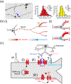
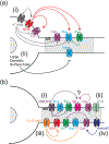
Ion channels are microscopic pore proteins in the membrane that open and close in response to chemical and electrical stimuli. This simple concept underlies rapid electrical signaling in the brain as well as several important aspects of neural plasticity. Although the soma accounts for less than 1% of many neurons by membrane area, it has been the major site of measuring ion channel function. However, the axon is one of the longest processes found in cellular biology and hosts a multitude of critical signaling functions in the brain. Not only does the axon initiate and rapidly propagate action potentials (APs) across the brain but it also forms the presynaptic terminals that convert these electrical inputs into chemical outputs. Here, we review recent advances in the physiological role of ion channels within the diverse landscape of the axon and presynaptic terminals.
Copyright © 2019 Elsevier Ltd. All rights reserved.
Conflict of interest statement
Conflict of interest statement
Nothing declared.
Figures


References
-
- Debanne D, et al., Axon physiology. Physiol Rev, 2011. 91(2): p. 555–602. - PubMed
-
- Cho IH, et al., Sodium Channel beta2 Subunits Prevent Action Potential Propagation Failures at Axonal Branch Points. J Neurosci, 2017. 37(39): p. 9519–9533. - PMC - PubMed
-
• **This paper uses genetically encoded voltage and calcium indicators to identify heterogeneous sodium channel activity in unmyelinated axonal arborizations as well as a critical role for Nav beta2 in AP propagation across branch points.
-
- Chereau R, et al., Superresolution imaging reveals activity-dependent plasticity of axon morphology linked to changes in action potential conduction velocity. Proc Natl Acad Sci U S A, 2017. 114(6): p. 1401–1406. - PMC - PubMed
-
• *This paper uses STED imaging to reveal dynamic changes in axon and synaptic terminal morphology to alter the timing of AP propagation.
-
- Debanne D, et al., Action-potential propagation gated by an axonal I(A)-like K+ conductance in hippocampus. Nature, 1997. 389(6648): p. 286–9. - PubMed
Publication types
MeSH terms
Substances
Grants and funding
LinkOut - more resources
Full Text Sources
Miscellaneous

