. 2019 Mar 1:14:609-616.
doi: 10.1016/j.omtn.2019.01.003. Epub 2019 Jan 15.

circRAPGEF5 Contributes to Papillary Thyroid Proliferation and Metastatis by Regulation miR-198/FGFR1

Affiliations

circRAPGEF5 Contributes to Papillary Thyroid Proliferation and Metastatis by Regulation miR-198/FGFR1

Weiwei Liu et al. Mol Ther Nucleic Acids. .

Retraction in

Abstract

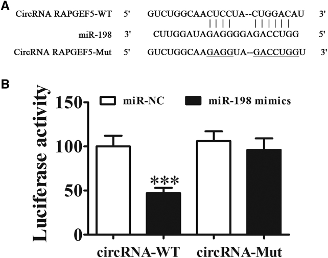
The circular RNA RAPGEF5 (circRAPGEF5) is generated from five exons of the RAPGEF5 gene and abnormal expression in papillary thyroid cancer (PTC). However, whether circRAPGEF5 plays a role in PTC tumorigenesis remains unclear. The aim of the present study was to investigate the role of circRAPGEF5 in PTC. The results showed that circRAPGEF5 was upregulated in PTC tissues and cell lines. circRAPGEF5 knockdown inhibited cell proliferation, migration, and invasion in vitro; and circRAPGEF5 silencing downregulated fibroblast growth factor receptor 1 (FGFR1) expression by "sponging" miR-198, suppressing the aggressive biological behaviors of PTC. Luciferase reporter assays confirmed that circRAPGEF5 interacted with miR-198 and that miR-198 interacted with the 3' UTR of FGFR1 to downregulate its expression. Xenograft experiments confirmed that circRAPGEF5 knockdown suppressed FGFR1-mediated tumor growth by promoting miR-198 expression. circRAPGEF5 acts as a tumor promoter via a novel circRAPGEF5/miR-198/FGFR1 axis, providing a potential biomarker and therapeutic target for the management of PTC.

Keywords: FGFR1; circRNA RAPGEF5; miR-198; papillary thyroid cancer; sponging.


Figures

Figure 1
Figure 1
The Expression of circRAPGEF5 Was Increased in Both PTC Tissues and PTC Cell Lines (A) The genomic loci of the RAPGEF5 gene and circRAPGEF5. Arrow indicates back-splicing. (B) Analysis of 30 paired tumor tissue samples and adjacent non-tumor tissue samples showed that the expression of circRAPGEF5 was increased in PTC tissues compared with adjacent normal tissues (n = 30). Data are indicated as the mean ± SD. ***p < 0.001 versus normal tissues. (C) The expression of circRAPGEF5 in PTC cells (BCPAP, KTC-1, and K1 cells) and normal PT cells (Nthy-ori 3-1) was detected by qRT-PCR. Data are indicated as the mean ± SD. ***p < 0.001 versus normal PTC cells. (D) Fluorescence in situ hybridization (FISH) assay was conducted to determine the subcellular localization of circRAPGEF5. Green indicates DAPI, and red indicates circRAPGEF5. Scale bars, 30 μm.
Figure 2
Figure 2
Knockdown of circRAPGEF5 Suppresses BCPAP Cell Proliferation, Invasion, and Migration (A) qRT-PCR detection of the expression of circRAPGEF5 after transfection with siRNA against circRAPGEF5 or negative control (NC) for 48 h. Data are indicated as the mean ± SD. n = 3. ***p < 0.001 versus NC. (B) Western blot detection of the expression of FGFR1. (C) Cell proliferation was measured by the CCK-8 assay. Data are indicated as the mean ± SD. n = 3. ***p < 0.001 versus NC. (D and E) Wound healing assays showed that downregulation of circRAPGEF5 prevents the closing of scratch wounds (D). Data are indicated as the mean ± SD (E). ***p < 0.001 versus NC. Scale bars, 100 μm. (F and G) Cell invasion was determined in BCPAP cells using the Transwell assay (F). Data are indicated as the mean ± SD (G). n = 3. ***p < 0.001 versus NC. Scale bars, 30 μm.
Figure 3
Figure 3
miR-198 Is a Target of circRAPGEF5 (A) Bioinformatics analysis predicted that the sequence of miR-198 matched the sequence of circRAPGEF5. The sequences of circRAPGEF5, including the wild-type (WT) and mutated (MUT) sequences (containing the miR-198 target sites), were cloned into the pmirGL0 vector. (B) The relative luciferase activity was determined at 48 h after transfection with circRAPGEF5 WT-Mut sequences and miR-198 mimics-NC in HEK293T cells. Data are indicated as the mean ± SD. n = 3. ***p < 0.001.
Figure 4
Figure 4
Downregulation of miR-198 Reversed the circRAPGEF5 Silencing-Induced Inhibition of Cell Proliferation, Invasion, and Migration in PTC Cells (A) qRT-PCR detection of the expression of miR-198 after transfection with siRNA against circRAPGEF5 and miR-198 inhibitor alone or in combination. (B and C) Western blot detection of the expression of FGFR1 (B). The relative protein levels were analyzed, and data are indicated as the mean ± SD (C). n = 3. ***p < 0.001 versus NC; ###p < 0.001 versus sicircRNA. (D) Cell proliferation was measured by the CCK-8 assay. Data are indicated as the mean ± SD. n = 3. ***p < 0.001 versus NC; ###p < 0.001 versus sicircRNA. (E and F) Wound healing assay showed that downregulation of circRAPGEF5 prevents the closing of scratch wounds (E). Data are indicated as the mean ± SD (F). ***p < 0.001 versus NC. Scale bars, 100 μm. (G and H) Cell invasion was determined in BCPAP cells using the Transwell assay (G). Data are indicated as the mean ± SD (H). n = 3. ***p < 0.001 versus NC; ###p < 0.001 versus sicircRNA. Scale bars, 30 μm.
Figure 5
Figure 5
FGFR1 Is a Target of miR-198 (A) Bioinformatics analysis predicted that the sequence of miR-198 matched the sequence of the 3′ UTR of FGFR1. The sequences of the FGFR1 3′ UTR, including the wild-type (WT) and mutated (MUT) sequences (containing the miR-198 target sites), were cloned into the pmirGL0 vector. (B) The relative luciferase activity was determined at 48 h after transfection with FGFR1 3′ UTR WT-Mut sequences and miR-198 mimics-NC in HEK293T cells. Data are indicated as the mean ± SD. n = 3. ***p < 0.001.
Figure 6
Figure 6
FGFR1 Overexpression Rescues the Effect of miR-198 on Suppressing Cell Proliferation, Invasion, and Migration in PTC Cells (A) qRT-PCR detection of the expression of miR-198 after transfection with miR-198 mimics and FGFR1 overexpression vector alone or in combination. n = 3. Data are indicated as the mean ± SD. ***p < 0.001 versus NC; ###p < 0.001 versus mimic. (B and C) Western blot detection of the expression of FGFR1 (B). The relative protein levels were analyzed, and data are indicated as the mean ± SD (C). n = 3. ***p < 0.001 versus NC; ###p < 0.001 versus mimic. (D) Cell proliferation was measured by the CCK-8 assay. Data are indicated as the mean ± SD. n = 3. ***p < 0.001 versus NC; ###p < 0.001 versus mimic. (E and F) Wound healing assays showed that downregulation of circRAPGEF5 prevents the closing of scratch wounds (E). Data are indicated as the mean ± SD (F). n = 3. ***p < 0.001 versus NC. Scale bars, 100 μm. (G and H) Cell migration and invasion were determined in BCPAP cells using the Transwell assay (G). Data are indicated as the mean ± SD (H). n = 3. ***p < 0.001 versus NC; ###p < 0.001 versus mimic. Scale bars, 30 μm.
Figure 7
Figure 7
Silencing of circRAPGEF5 Suppressed Tumor Formation in Xenograft Mice (A) Representative image of nude mice with tumors formed by injection with BCPAP cells (n = 6). (B) Tumor volume of mice was measured every week. Data are indicated as the mean ± SD. ***p < 0.001 versus NC. (C) qRT-PCR detection of the expression of miR-198. Data are indicated as the mean ± SD. n = 6. ***p < 0.001 versus NC. (D and E) Western blot detection of the expression of FGFR1 (D). Data are indicated as the mean ± SD (E). n = 6. ***p < 0.001 versus NC.

References

    1. Miller K.D., Siegel R.L., Lin C.C., Mariotto A.B., Kramer J.L., Rowland J.H., Stein K.D., Alteri R., Jemal A. Cancer treatment and survivorship statistics, 2016. CA Cancer J. Clin. 2016;66:271–289. - PubMed
    1. Cancer Genome Atlas Research Network Integrated genomic characterization of papillary thyroid carcinoma. Cell. 2014;159:676–690. - PMC - PubMed
    1. Zhu L., Li X.J., Kalimuthu S., Gangadaran P., Lee H.W., Oh J.M., Baek S.H., Jeong S.Y., Lee S.W., Lee J., Ahn B.C. Natural killer cell (NK-92MI)-based therapy for pulmonary metastasis of anaplastic thyroid cancer in a nude mouse model. Front. Immunol. 2017;8:816. - PMC - PubMed
    1. Cabanillas M.E., McFadden D.G., Durante C. Thyroid cancer. Lancet. 2016;388:2783–2795. - PubMed
    1. Dragomir M., Calin G.A. Circular RNAs in cancer - lessons learned from microRNAs. Front. Oncol. 2018;8:179. - PMC - PubMed

Publication types