. 2019 Mar 26;58(12):1616-1626.
doi: 10.1021/acs.biochem.9b00031. Epub 2019 Mar 5.

Crystal Structure and Conformational Dynamics of Pyrococcus furiosus Prolyl Oligopeptidase

Affiliations

Crystal Structure and Conformational Dynamics of Pyrococcus furiosus Prolyl Oligopeptidase

Ken Ellis-Guardiola et al. Biochemistry. .

Abstract

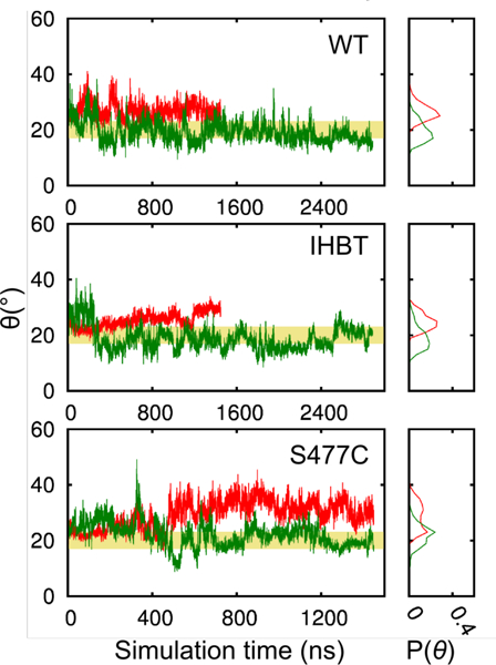
Enzymes in the prolyl oligopeptidase family possess unique structures and substrate specificities that are important for their biological activity and for potential biocatalytic applications. The crystal structures of Pyrococcus furiosus ( Pfu) prolyl oligopeptidase (POP) and the corresponding S477C mutant were determined to 1.9 and 2.2 Å resolution, respectively. The wild type enzyme crystallized in an open conformation, indicating that this state is readily accessible, and it contained bound chloride ions and a prolylproline ligand. These structures were used as starting points for molecular dynamics simulations of Pfu POP conformational dynamics. The simulations showed that large-scale domain opening and closing occurred spontaneously, providing facile substrate access to the active site. Movement of the loop containing the catalytically essential histidine into a conformation similar to those found in structures with fully formed catalytic triads also occurred. This movement was modulated by chloride binding, providing a rationale for experimentally observed activation of POP peptidase catalysis by chloride. Thus, the structures and simulations reported in this study, combined with existing biochemical data, provide a number of insights into POP catalysis.


Figures

Figure 1.
Figure 1.
A) General scheme for peptidase catalysis involving an enzyme (E) and a peptide substrate (S) to generate two product peptides (P1 and P2) via enzyme-substrate (ES) and enzyme-acyl (EA) intermediates. B) Potential domain opening and closing during Pfu POP peptidase catalysis.
Figure 2.
Figure 2.
A-C) Annotated ribbon diagram of Pfu POP showing inter-domain angle (θ), blade (b1–7) and hinge locations, and key active site residues and Cl ions. D) expanded view of POP active site showing residues in the catalytic triad, bound chloride ions, and the Pro-Pro ligand (omitted in A-C for clarity).
Figure 3.
Figure 3.
Electron density (contoured at 1.0 σ) for key features in the PfU POP crystal structure, including: A) the loop containing His592, B) the “latch loop”, C) a bound prolylproline ligand and bound chloride ions.
Figure 4.
Figure 4.
The open angle and its distribution in the MD simulations at 300 K (red) and 358.15 K (green) in replica 2 of the WT, IHBT, and S477C systems. The yellow banner indicates the intermediate open angle values between the open and the closed states.
Figure 5.
Figure 5.
The cartoon and surface representation showing the largest opening between propeller (light grey) blade b3 and the peptidase domain (dark grey). The snapshots are taken from the covalently-linked inhibitor simulation IHBT_3 to illustrate (A) the closed state and (B) the open, substrate-accessible state enzyme. The bottom panel shows a surface presentation of the same snapshot. The covalently bound inhibitor is colored green.
Figure 6.
Figure 6.
The time evolution of the S477 and H592 distance, DS477-H592. The hydroxyl oxygen (OG) in S477 and the imidazole amide hydrogen (HE2) are used to define the distance.
Figure 7.
Figure 7.
Sequential steps in the H-bond network H592 uses to get into the catalytic site (A-F). The contributing residues are shown in cyan sticks. The H592 look is in green and the residues H592 and S477 are colored in yellow.
Figure 8.
Figure 8.
The impact on S477-H592 distance from the binding of Cl to the R600 site. The site is occupied when the Cl is within 1.5 A to the crystal binding site of this Cl. The transparent, filled curves show the normalized population (Pnorm) distributions of DS477-H592 computed from the entire trajectory at 358.15 K. The thick, colored lines are the Pnorm distributions of DS477-H592 drawn from when Cl is bound to the R600 site. The three replicas are shown in red, green, and blue.
Figure 9.
Figure 9.
Overlay of closed crystal structures with a simulation snapshot with a closed H592 loop. Important residues are shown with sticks. The pKa values of the residues that correspond to H592 are 6.6, 6.6/6.9, 7.2, and 6.9 for 1QFS, 2BKL, 3IVM, and 4HVT. The pKa values are calculated using the program PROPKA31.
Figure 10.
Figure 10.
Conceptual summary of updated models for A) the inter-domain angle of the Pfu POP, B) the rate limiting step of Pfu POP peptidase catalysis, C) substrate entry into Pfu POP, and D) effects of chloride binding within Pfu POP (S = substrate).

Similar articles

Cited by

References

    1. Polgár L The Prolyl Oligopeptidase Family. Cell Mol Life Sci 2002, 59 (2), 349–362. - PMC - PubMed
    1. Szeltner Z; Polgar L Structure, Function and Biological Relevance of Prolyl Oligopeptidase. Curr. Protein Pept. Sci. 2008, 9 (1), 96–107. - PubMed
    1. Rea D; Fülöp V Structure-Function Properties of Prolyl Oligopeptidase Family Enzymes. Cell Biochem. Biophys. 2006, 44 (3), 349–365. - PubMed
    1. Ehren J; Govindarajan S; Moron B; Minshull J; Khosla C Protein Engineering of Improved Prolyl Endopeptidases for Celiac Sprue Therapy. Prot. Eng. Des. Sel. 2008, 21 (12), 699–707. - PMC - PubMed
    1. Walter R; Shlank H; Glass JD; Schwartz IL; Kerenyi TD Leucylglycinamide Released From Oxytocin by Human Uterine Enzyme. Science 1971, 173 (3999), 827–829. - PubMed

Publication types