. 2019 Mar 28;123(12):2609-2622.
doi: 10.1021/acs.jpcb.8b10220. Epub 2019 Mar 18.

Determinants of Orexin Receptor Binding and Activation-A Molecular Dynamics Study

Affiliations

Determinants of Orexin Receptor Binding and Activation-A Molecular Dynamics Study

Lasse Karhu et al. J Phys Chem B. .

Abstract

We assess the stability of two previously suggested binding modes for the neuropeptide orexin-A in the OX2 receptor through extensive molecular dynamics simulations. As the activation determinants of the receptor remain unknown, we simulated an unliganded receptor and two small-molecular ligands, the antagonist suvorexant and the agonist Nag26 for comparison. Each system was simulated in pure POPC membrane as well as in the 25% cholesterol-POPC membrane. In total, we carried out 36 μs of simulations. Through this set of simulations, we report a stable binding mode for the C-terminus of orexin-A. In addition, we suggest interactions that would promote orexin receptor activation, as well as others that would stabilize the inactive state.


Conflict of interest statement

The authors declare no competing financial interest.

Figures

Figure 1
Figure 1
Orexin peptides. Sequences are colored consistently with the figures. X: Pyroglutamoyl; lines denote disulfide bonds. For orexin-B (top right), one conformation has been published, while for orexin-A, three conformations have been observed.,
Figure 2
Figure 2
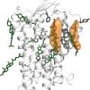
Cholesterol around the receptor. In green, the initial locations; in orange, the stable locations.
Figure 3
Figure 3
End point of orexin-A simulations. (A–D) TM5-mode, simulations 14, respectively; (E–H) TM7-mode, simulations 58, respectively.
Figure 4
Figure 4
RMSD of the peptide C-terminus. In green and orange, the “external” RMSD, and in blue and red, the “internal” RMSD. (A) Simulations 12, (B) 3-4, (C) 56, and (D) 78. See Methods, Analysis, for definitions for the external and internal RMSD.
Figure 5
Figure 5
Heatmap of binding interactions of the peptide C-terminus, averaged across all simulations sharing the same binding mode (14 for TM5-mode, 58 for TM7-mode). The heatmaps display the fraction of simulation time that any interatomic distance was below 4 Å. H:xx denotes the presence of a hydrogen bond and W:xx the presence of a water-mediated hydrogen bond. These are shown only when the frequency was over 20%.
Figure 6
Figure 6
Orexin-A binding interactions in the TM5-mode. (A) Binding site with close receptor residues shown as sticks. (B) Hydrogen bonds between the peptide C-terminus and the receptor. The atomic coordinates for the TM5-mode receptor complex presented here are available as Supporting Information.
Figure 7
Figure 7
Simulation-derived bent conformation of orexin-A overlaid with the recently published OX1 structure, which displays the N-terminal receptor helix.
Figure 8
Figure 8
RMSD of the small molecular ligands. In green and orange, the “external” RMSD, and in blue and red, the “internal” RMSD.
Figure 9
Figure 9
Heatmap of binding interactions of the small molecular ligands. For the mapping, the small molecules were divided into groups as displayed below the heatmaps. The heatmaps display the fraction of simulation time that any interatomic distance was below 4 Å. H:xx denotes the presence of a hydrogen bond and W:xx the presence of a water-mediated hydrogen bond. These are shown only when the frequency was over 20%.
Figure 10
Figure 10
Small molecular agonist Nag26 was mobile during the simulations, rendering the binding mode identification difficult. Representatives of the three most visited conformations are shown.
Figure 11
Figure 11
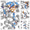
Several water molecules remain in the binding site throughout the simulations. (A) Overview of the binding-site water. The circles with lowercase letters refer to the panels on the right. (B) TM5–6 interface. (C) TM4–5 interface. (D) Asp1002x50.

References

    1. Kukkonen J. P.; Leonard C. S. Orexin/Hypocretin Receptor Signalling Cascades. Br. J. Pharmacol. 2014, 171, 314–331. 10.1111/bph.12324. - DOI - PMC - PubMed
    1. Cox C. D.; Breslin M. J.; Whitman D. B.; Schreier J. D.; McGaughey G. B.; Bogusky M. J.; Roecker A. J.; Mercer S. P.; Bednar R. a; Lemaire W.; et al. Discovery of the Dual Orexin Receptor Antagonist [(7R)-4-(5-Chloro-1,3-Benzoxazol-2-Yl)-7-Methyl-1,4-Diazepan-1-Yl][5-Methyl-2-(2H-1,2,3-Triazol-2-Yl)Phenyl]Methanone (MK-4305) for the Treatment of Insomnia. J. Med. Chem. 2010, 53, 5320–5332. 10.1021/jm100541c. - DOI - PubMed
    1. Roecker A. J.; Cox C. D.; Coleman P. J. Orexin Receptor Antagonists: New Therapeutic Agents for the Treatment of Insomnia. J. Med. Chem. 2016, 59, 504–530. 10.1021/acs.jmedchem.5b00832. - DOI - PubMed
    1. Nishino S.; Ripley B.; Overeem S.; Lammers G. J.; Mignot E. Hypocretin (Orexin) Deficiency in Human Narcolepsy. Lancet 2000, 355, 39–40. 10.1016/S0140-6736(99)05582-8. - DOI - PubMed
    1. Scammell T. E.; Winrow C. J. Orexin Receptors: Pharmacology and Therapeutic Opportunities. Annu. Rev. Pharmacol. Toxicol. 2011, 51, 243–266. 10.1146/annurev-pharmtox-010510-100528. - DOI - PMC - PubMed

Publication types