

Morphology of the Musculus Uvulae In Vivo Using MRI and 3D Modeling Among Adults With Normal Anatomy and Preliminary Comparisons to Cleft Palate Anatomy
- PMID: 30786757
- PMCID: PMC6693950
- DOI: 10.1177/1055665619828226
Morphology of the Musculus Uvulae In Vivo Using MRI and 3D Modeling Among Adults With Normal Anatomy and Preliminary Comparisons to Cleft Palate Anatomy
Abstract
Purpose: To investigate the musculus uvulae morphology in vivo in adults with normal velopharyngeal anatomy and to examine sex and race effects on the muscle morphology. We also sought to provide a preliminary comparison of musculus uvulae morphology in adults with normal velopharyngeal anatomy to adults with repaired cleft palate.
Methods: Three-dimensional magnetic resonance imaging data and Amira 5.5 Visualization Modeling software were used to evaluate the musculus uvulae in 70 participants without cleft palate and 6 participants with cleft palate. Muscle length, thickness, width, and volume were compared among participant groups.
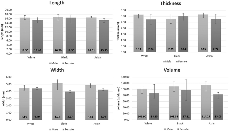
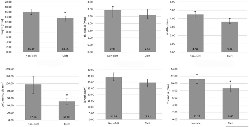
Results: Analysis of covariance analysis did not yield statistically significant differences in musculus uvulae length, thickness, width, or volume by race or sex among participants without cleft palate when the effect of body size was accounted for. Two-sample t test revealed that the musculus uvulae in participants with repaired cleft palate is significantly shorter (P = .008, 13.65 mm vs 16.07 mm) and has less volume (P = .002, 51.08 mm3 vs 97.62 mm3) than participants without cleft palate.
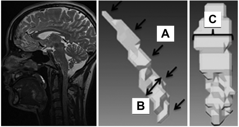
Conclusion: In adults with normal velopharyngeal anatomy, the musculus uvulae is a cylindrical oblong-shaped muscle lying on the nasal surface of the soft palate, with its greatest bulk located just nasal to the levator veli palatini muscle sling. In participants with repaired cleft palate, the musculus uvulae is substantially reduced in volume. This diminished muscle bulk located just at the point where the palate contacts the posterior pharyngeal wall may contribute to velopharyngeal insufficiency in children with repaired cleft palate.
Keywords: 3-dimensional reconstruction; cleft palate; magnetic resonance imaging; morphology; musculus uvulae; race; sex.
Figures
Similar articles
-
Velopharyngeal Muscle Morphology in Children With Unrepaired Submucous Cleft Palate: An Imaging Study.Cleft Palate Craniofac J. 2021 Mar;58(3):313-323. doi: 10.1177/1055665620954749. Epub 2020 Sep 10. Cleft Palate Craniofac J. 2021. PMID: 32909827
-
Asymmetry and Positioning of the Levator Veli Palatini Muscle in Children With Repaired Cleft Palate.J Speech Lang Hear Res. 2020 May 22;63(5):1317-1325. doi: 10.1044/2020_JSLHR-19-00240. Epub 2020 May 13. J Speech Lang Hear Res. 2020. PMID: 32402223 Free PMC article.
-
Morphology of the Levator Veli Palatini Muscle in Adults With Repaired Cleft Palate.J Craniofac Surg. 2017 May;28(3):833-837. doi: 10.1097/SCS.0000000000003373. J Craniofac Surg. 2017. PMID: 28060090 Free PMC article.
-
Anatomy and physiology of the velopharyngeal mechanism.Semin Speech Lang. 2011 May;32(2):83-92. doi: 10.1055/s-0031-1277712. Epub 2011 Sep 26. Semin Speech Lang. 2011. PMID: 21948636 Review.
-
Functional anatomy of the soft palate applied to wind playing.Med Probl Perform Art. 2010 Dec;25(4):183-9. Med Probl Perform Art. 2010. PMID: 21170481 Review.
Cited by
-
Obstructive Sleep Apnea and Role of the Diaphragm.Cureus. 2022 Sep 10;14(9):e29004. doi: 10.7759/cureus.29004. eCollection 2022 Sep. Cureus. 2022. PMID: 36159353 Free PMC article. Review.
-
An Exploratory Investigation of the Palatoglossus Muscle in Children Using Magnetic Resonance Imaging.J Speech Lang Hear Res. 2022 Nov 17;65(11):4151-4158. doi: 10.1044/2022_JSLHR-22-00303. Epub 2022 Oct 25. J Speech Lang Hear Res. 2022. PMID: 36283682 Free PMC article.
-
Simulation of Velopharyngeal Biomechanics Identifies Differences in Sphincter Pharyngoplasty Outcomes: A Matched Case-Control Study.Cleft Palate Craniofac J. 2024 Feb;61(2):339-349. doi: 10.1177/10556656221122634. Epub 2022 Aug 22. Cleft Palate Craniofac J. 2024. PMID: 35996316 Free PMC article.
-
Establishing a Clinical Protocol for Velopharyngeal MRI and Interpreting Imaging Findings.Cleft Palate Craniofac J. 2024 May;61(5):748-758. doi: 10.1177/10556656221141188. Epub 2022 Nov 30. Cleft Palate Craniofac J. 2024. PMID: 36448363 Free PMC article.
-
Differential Diagnosis of a Pharyngeal Fricative and Therapeutic Monitoring of Velopharyngeal Function Using Magnetic Resonance Imaging.Am J Speech Lang Pathol. 2025 Jan 7;34(1):1-11. doi: 10.1044/2024_AJSLP-24-00292. Epub 2024 Dec 5. Am J Speech Lang Pathol. 2025. PMID: 39637256 Free PMC article.
References
-
- Azzam N, Kuehn DP. The morphology of musculus uvulae. Cleft Palate J. 1977;14(1):78–87. - PubMed
-
- Boorman JG, Sommerlad BC. Musculus uvulae and levator palati: Their anatomical and functional relationship in velopharyngeal closure. Br J Plast Surg. 1985;38(3):333–338. - PubMed
-
- Ha S, Kuehn DP, Cohen M, Alperin N. Magnetic resonance imaging of the levator veli palatini muscle in speakers with repaired cleft palate. Cleft Palate Craniofac J. 2007;44(5):494–505. - PubMed
-
- Huang MH, Lee ST, Rajendran K. A fresh cadaveric study of the paratubal muscles: Implications for eustachian tube function in cleft palate. Plast Reconstr Surg. 1997;100(4):833–842. - PubMed
Publication types
MeSH terms
Grants and funding
LinkOut - more resources
Full Text Sources
Medical
