. 2019 Feb 20;20(1):6.
doi: 10.1186/s12868-019-0489-5.

Astrocytes infected with Chlamydia pneumoniae demonstrate altered expression and activity of secretases involved in the generation of β-amyloid found in Alzheimer disease

Affiliations

Astrocytes infected with Chlamydia pneumoniae demonstrate altered expression and activity of secretases involved in the generation of β-amyloid found in Alzheimer disease

Zein Al-Atrache et al. BMC Neurosci. .

Abstract

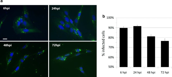
Background: Epidemiologic studies strongly suggest that the pathophysiology of late-onset Alzheimer disease (AD) versus early-onset AD has environmental rather than genetic causes, thus revealing potentially novel therapeutic targets to limit disease progression. Several studies supporting the "pathogen hypothesis" of AD demonstrate a strong association between pathogens and the production of β-amyloid, the pathologic hallmark of AD. Although the mechanism of pathogen-induced neurodegeneration of AD remains unclear, astrocytes, a key player of the CNS innate immune response and producer/metabolizer of β-amyloid, have been implicated. We hypothesized that Chlamydia pneumoniae infection of human astrocytes alters the expression of the amyloid precursor protein (APP)-processing secretases, ADAM10, BACE1, and PSEN1, to promote β-amyloid formation. Utilizing immunofluorescent microscopy, molecular, and biochemical approaches, these studies explore the role of an intracellular respiratory pathogen, Chlamydia pneumoniae, as an environmental trigger for AD pathology. Human astrocytoma cells in vitro were infected with Chlamydia pneumoniae over the course of 6-72 h. The gene and protein expression, as well as the enzymatic activity of non-amyloidogenic (ADAM10), and pro-amyloidogenic (BACE1 and PSEN1) secretases were qualitatively and quantitatively assessed. In addition, the formation of toxic amyloid products as an outcome of pro-amyloidogenic APP processing was evaluated through various modalities.

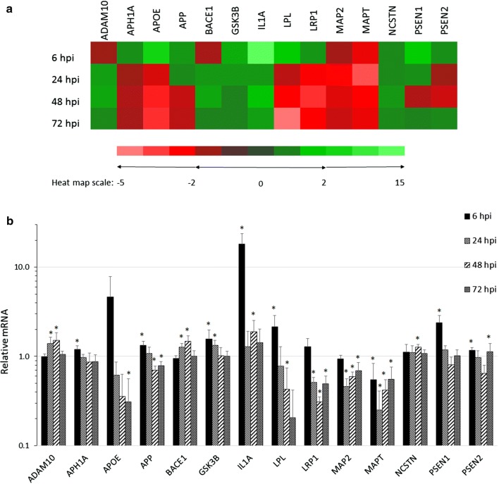
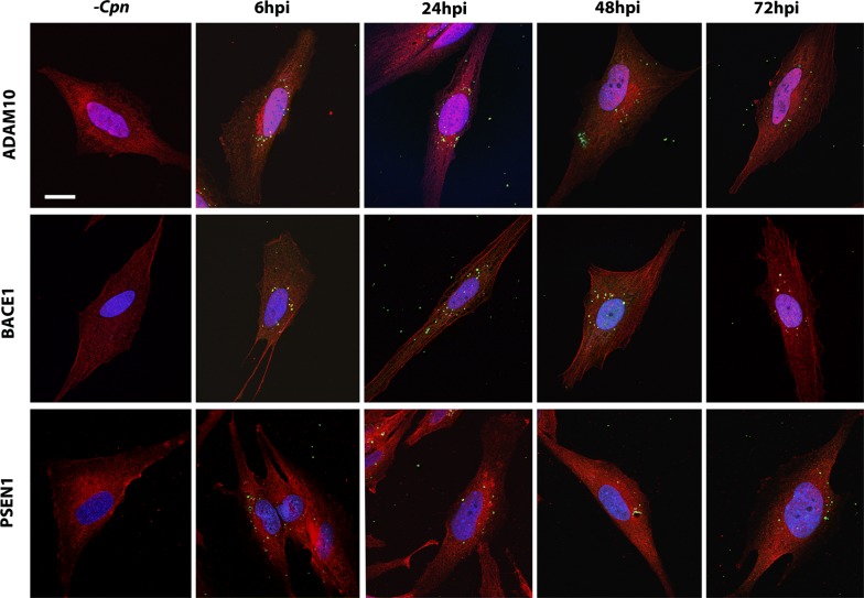
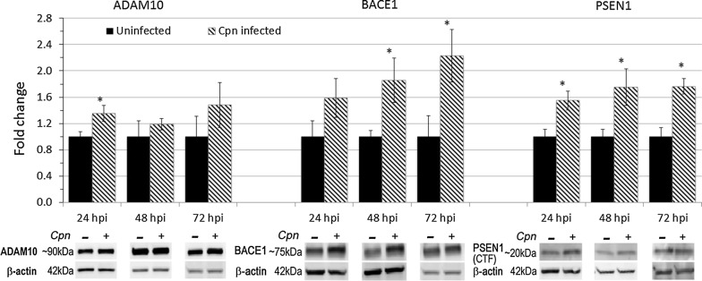
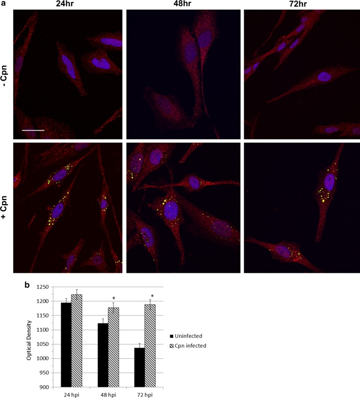
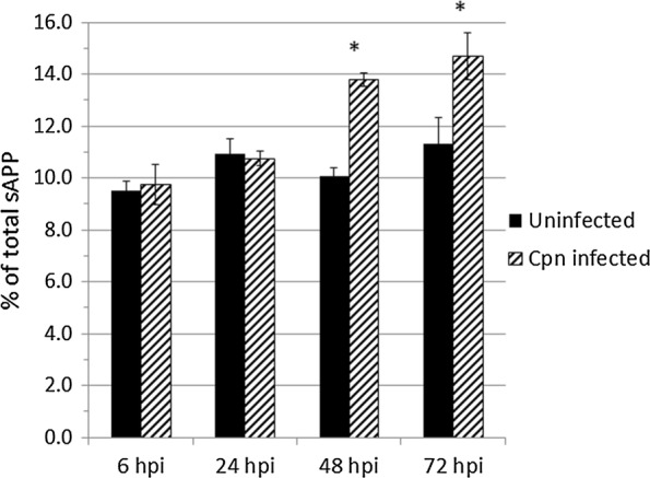
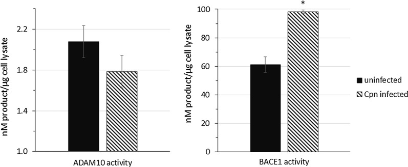
Results: Chlamydia pneumoniae infection of human astrocytoma cells promoted the transcriptional upregulation of numerous genes implicated in host neuroinflammation, lipid homeostasis, microtubule function, and APP processing. Relative to that of uninfected astrocytes, BACE1 and PSEN1 protein levels were enhanced by nearly twofold at 48-72 h post-Chlamydia pneumoniae infection. The processing of APP in Chlamydia pneumoniae-infected astrocytes favors the pro-amyloidogenic pathway, as demonstrated by an increase in enzymatic activity of BACE1, while that of ADAM10 was decreased. Fluorescence intensity of β-amyloid and ELISA-quantified levels of soluble-APP by products revealed temporally similar increases, confirming a BACE1/PSEN1-mediated processing of APP.

Conclusions: Our findings suggest that Chlamydia pneumoniae infection of human astrocytes promotes the pro-amyloidogenic pathway of APP processing through the upregulation of expression and activity of β-secretase, upregulated expression of γ-secretase, and decreased activity of α-secretase. These effects of astrocyte infection provide evidence for a direct link between Chlamydia pneumoniae and AD pathology.

Keywords: Alzheimer disease; Amyloid; Astrocytes; BACE1; Chlamydia pneumoniae; Neurodegeneration; Neuroinflammation; Pathogens; Secretase.


Figures

Fig. 1
Fig. 1
Chlamydia pneumoniae infects human astrocytes in vitro. STTG1 human astrocytes infected with Cpn strain AR39 at an MOI of 1 demonstrated a diffuse punctate labeling of Cpn (green) from 6 to 72 hpi. Nuclei are labeled with DAPI (blue). Scale bar represents 20 μm (a). Infected versus uninfected cell counts were averaged across approximately N = 2000–2500 cells per timepoint and in biological triplicate across two independent infections. Numerical data is expressed as percent infected cells (b). Percentages of infected cells were significantly different between 6 hpi versus 48 hpi and 72 hpi, 24 hpi versus 48 hpi and 72 hpi. Comparisons between populations were determined through one-way ANOVA, where significance was defined as p < 0.05, and confirmed using Tukey HSD post hoc analysis. Error bars represent standard deviation of the mean
Fig. 2
Fig. 2
Chlamydia pneumoniae infection of human astrocytes alters the transcript expression of AD-related genes. Gene transcripts from Cpn-infected and uninfected cells analyzed at all four timepoints post-infection revealed significant fold changes in genes closely related to AD pathology. The fold changes of fourteen genes implicated in known pathways of AD pathology are presented in a. Histograms of fold changes of these AD-associated genes are presented in b. All expression data was normalized to β-actin and Cpn-infected and uninfected cDNA samples were repeated in biological (N = 3) and technical triplicate for each timepoint. Asterisk indicates p < 0.05. ADAM10, A disintegrin and metalloproteinase 10; APH1A, anterior pharynx defective protein 1A; APOE, apolipoprotein E; APP, amyloid precursor protein; BACE1, βAPP-cleaving enzyme 1; GSK3B, glucogen synthase kinase 3-β; IL1A, interleukin 1α; LPL, lipoprotein lipase; lipoprotein receptor-related protein 1, LRP1; MAP2, microtubule associated protein 2; MAPT, microtubule associated protein tau; NCSTN, nicastrin; PSEN1, presenilin-1, PSEN2, presenilin-2
Fig. 3
Fig. 3
Chlamydia pneumoniae infection of astrocytes alters labeling of secretases. Astrocytes infected with Cpn from 6 to 72 hpi were double labeled for Cpn (green) and secretases ADAM10, BACE1 or PSEN1 C-terminal fragment (red). 10–15 cells per biological replicate were imaged against an equal number of uninfected control cells. Cells were visualized using laser scanning confocal microscopy, maintaining the voltage settings of each color channel identical across biological replicates. DAPI was used to visualize the nucleus. Representative images are included this figure. Scale bar represents 20 μm
Fig. 4
Fig. 4
Protein expression of ADAM10, BACE1 and PSEN1 in Chlamydia pneumoniae-infected and uninfected astrocytes. Whole cell lysate was harvested from Cpn-infected and uninfected astrocytoma cells, resolved via SDS-PAGE gel electrophoresis, and labeled for secretase proteins. Fold change represents densitometry analysis for protein levels of full-length ADAM10, BACE1 and PSEN1 C-terminal fragment in Cpn-infected cells compared to that of uninfected cells at the same timepoint post infection. All densitometry values were normalized to that of β-actin for each biological replicate (N = 5–7). Statistical analysis was conducted using student’s T-test of fold change within each timepoint (asterisk indicates p < 0.05). Error bars represent standard error of the mean
Fig. 5
Fig. 5
Fluorescence intensity of Aβ1-42 is increased in Chlamydia pneumoniae-infected astrocytes. Aβ1-42 (red) and Cpn (green) were visualized by laser scanning, confocal microscopy (a). To analyze the Z-images using FIJI software, a defined threshold subtraction was applied equally to each image to determine Aβ1-42 fluorescence intensity (b); mean fluorescence intensity was calculated for infected and uninfected astrocytes at 24, 48 and 72 hpi. Cells (N = 25–30) were analyzed across three biological replicates to reliably conduct a student’s t-test on the fluorescence intensities of Aβ1-2 in uninfected and Cpn-infected cells. Error bars represent standard error of the mean. Asterisk represents p < 0.05
Fig. 6
Fig. 6
Quantification of sAPPβ/total sAPP in media of uninfected and Chlamydia pneumoniae infected astrocytes using MSD ELISA. Conditioned media of uninfected and Cpn-infected cells at each timepoint post-infection was collected, concentrated, and assayed in equal volumes for sAPPβ and sAPPα levels. Standard curves of known concentrations of sAPPβ and sAPPα was used to determine the concentration of these individual Aβ species. Conditioned media was obtained from three biological replicates and the assay was conducted in technical triplicate. Student’s t-test was calculated using the average sAPPβ/total sAPP ratio of Cpn-infected conditioned media compared to that of uninfected conditioned media. Asterisk represents p < 0.05
Fig. 7
Fig. 7
BACE1 activity is increased and ADAM10 activity is decreased in Chlamydia pneumoniae infected astrocytes. FRET-based assays were used to quantify the activity of ADAM10 and BACE1 enzyme activity generated by Cpn-infected and uninfected cell lysates at 48 hpi. Fluorescence of the 5-FAM or HiLyte Fluor 488 dyes conjugated to ADAM10 or BACE1-specific substrates was detected as a result of cleavage by the respective enzymes and compared back to fluorescence intensity of known dye concentrations. The quantified dye concentration from ADAM10 and BACE1 fluorescent substrate cleavage from (N = 4) biological replicates of Cpn-infected and uninfected cell lysates is presented as an average in the histograms. Error bars represent standard error of the mean. Asterisk represents p < 0.05

Similar articles

Cited by

References

    1. Hebert LE, Weuve J, Scherr PA, Evans DA. Alzheimer disease in the United States (2010-2050) estimated using the 2010 census. Neurology. 2013;80:1778–1783. doi: 10.1212/WNL.0b013e31828726f5. - DOI - PMC - PubMed
    1. Querfurth HW, LaFerla FM. Alzheimer’s disease. N Engl J Med. 2010;362:329–344. doi: 10.1056/NEJMra0909142. - DOI - PubMed
    1. Sanabria-Castro A, Alvarado-Echeverría I, Monge-Bonilla C. Molecular pathogenesis of Alzheimer’s disease: an update. Ann Neurosci. 2017;24:46–54. doi: 10.1159/000464422. - DOI - PMC - PubMed
    1. Bertram L, Lill CM, Tanzi RE. The genetics of Alzheimer disease: back to the future. Neuron. 2010;68:270–281. doi: 10.1016/j.neuron.2010.10.013. - DOI - PubMed
    1. Goate A, Hardy J. Twenty years of Alzheimer’s disease-causing mutations. J Neurochem. 2012;120:3–8. doi: 10.1111/j.1471-4159.2011.07575.x. - DOI - PubMed

Publication types

MeSH terms