. 2019 Feb 20;7(1):24.
doi: 10.1186/s40478-019-0668-8.

Methylation array profiling of adult brain tumours: diagnostic outcomes in a large, single centre

Affiliations

Methylation array profiling of adult brain tumours: diagnostic outcomes in a large, single centre

Zane Jaunmuktane et al. Acta Neuropathol Commun. .

Abstract

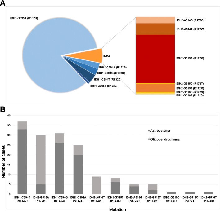
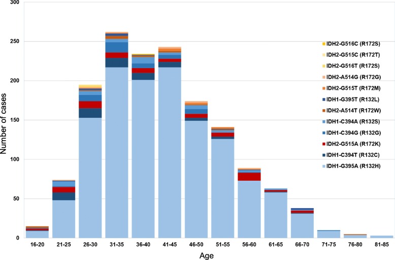
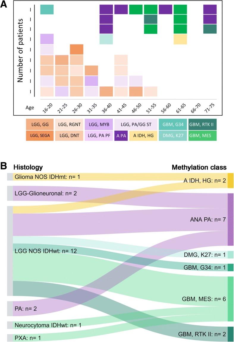
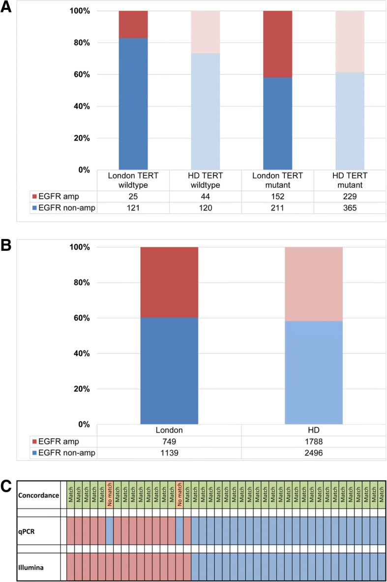
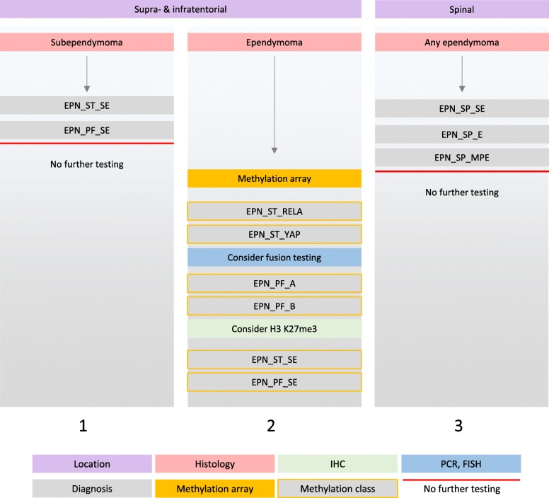
The introduction of the classification of brain tumours based on their DNA methylation profile has significantly changed the diagnostic approach for cases with ambiguous histology, non-informative or contradictory molecular profiles or for entities where methylation profiling provides useful information for patient risk stratification, for example in medulloblastoma and ependymoma. We present our experience that combines a conventional molecular diagnostic approach with the complementary use of a DNA methylation-based classification tool, for adult brain tumours originating from local as well as national referrals. We report the frequency of IDH mutations in a large cohort of nearly 1550 patients, EGFR amplifications in almost 1900 IDH-wildtype glioblastomas, and histone mutations in 70 adult gliomas. We demonstrate how additional methylation-based classification has changed and improved our diagnostic approach. Of the 325 cases referred for methylome testing, 179 (56%) had a calibrated score of 0.84 and higher and were included in the evaluation. In these 179 samples, the diagnosis was changed in 45 (25%), refined in 86 (48%) and confirmed in 44 cases (25%). In addition, the methylation arrays contain copy number information that usefully complements the methylation profile. For example, EGFR amplification which is 95% concordant with our Real-Time PCR-based copy number assays. We propose here a diagnostic algorithm that integrates histology, conventional molecular tests and methylation arrays.

Keywords: Adult brain tumours; BRAF; Brain tumour classification; DKFZ classifier; Ependymoma; Glioma; H3 K27M; Histone mutation; IDH1; IDH2; Illumina array; Methylation array; Methylation classifier; Molecular diagnostics.


Conflict of interest statement

Competing interests

The authors declare that they have no competing interests.

Publisher’s Note

Springer Nature remains neutral with regard to jurisdictional claims in published maps and institutional affiliations.

Figures

Fig. 1
Fig. 1
a, the association of methylation array testing rationale (left) with the outcome of the methylome-based classification (right). 179 cases with a calibrated score of 0.84 and higher were included in this graph. b, distribution of calibrated scores in 325 diagnostic samples examined (excluding research samples) demonstrating that for > 55% (179/325) of the predictions the classifier has had high confidence with estimated class probabilities of 94 > 99%. c, Scattergram of turnaround times (TAT) of tests between 2015 and 2018. Whilst the TAT in the first 2 years of the setup phase often comprised 50% of latencies over 30 days, these long TAT have been progressively reduced in 2018. In quarters 2–4 of 2018, the majority of the cases was completed within 4 weeks. For the first four months in 2015 we did not record the dates of requesting tests, therefore no TAT are shown between February and June 2015
Fig. 2
Fig. 2
a, the frequency of IDH mutations in our cohort (n = 1546). Blue, IDH1 mutations; orange and red, IDH2 mutations. The IDH1/IDH2 frequency in our cohort is slightly skewed toward rarer mutations, due to a proportion of referrals received specifically for sequencing studies. b, the frequency of IDH1 and IDH2 mutations and associated tumour types (n = 441 astrocytomas (of which 339 have IDH1-G395A); n = 363 oligodendrogliomas (of which 303 have IDH1-G395A)). Dark grey, astrocytomas; light grey, oligodendrogliomas. The mutations are sorted in descending order by overall frequency, excluding the most common IDH1-G395A mutation. The graph confirms the established association of certain mutations, in particular in the IDH2 gene, with oligodendroglial or astrocytic tumours
Fig. 3
Fig. 3
Age distribution of IDH mutations in our cohort (n = 1546) demonstrates that 15.7% of all IDH1/IDH2 mutations occur in patients 55 years and older, justifying routine testing for these mutations in this age cohort
Fig. 4
Fig. 4
Occurrence of histone H3.3 K27M- (upper panel) and H3.3 G34-mutant gliomas in our cohort and the association with ATRX protein loss. Rarely, a biphasic pattern of ATRX expression is observed both in H3 K27M- and H3 G34R-mutant gliomas (light purple boxes). On one occasion we have identified H3F3A G34V mutation (asterisk in the age group 16–20 years). In another recurrent high-grade glioma, both primary and recurrent tumours were classified as H3 G34-mutant glioblastoma, although no mutations could be found in H3F3A, HIST1H3B and HIST1H3C genes (asterisk in the age group 36–40)
Fig. 5
Fig. 5
CNS tumours of varied histology resolving into new entities defined by their methylation profile. IDHwt: IDH-wildtype; GBM IDHwt: Glioblastoma, IDH-wildtype; LGG NOS IDHwt: Low-grade glioma not otherwise specified, IDH-wildtype; LG-Glioneuronal: Low-grade glioneuronal tumour; PA: Pilocytic astrocytoma; HGG NOS IDHwt: High-grade glioma not otherwise specified, IDH-wildtype; HGNET, MN1: CNS high-grade neuroepithelial tumour with MN1 alteration; ANA PA: Anaplastic pilocytic astrocytoma (anaplastic astrocytoma with piloid features); LGG, MYB: Low-grade glioma with MYB alteration; DLGNT: diffuse leptomeningeal glioneuronal tumour
Fig. 6
Fig. 6
a, the outcome of methylation profiling of 44 IDH-wildtype CNS tumours with low-grade histology. Of these, 26 resolved into methylation classes associated with low-grade behaviour, and 18 resolved into entities associated with high-grade behaviour. b, shows how various tumours manifesting with low-grade histology resolve into distinct high-grade methylation classes. Abbreviations in a: LGG, GG: Low-grade glioma, ganglioglioma; LGG, SEGA: Low-grade glioma, subependymal giant cell astrocytoma; LGG, RGNT: Low-grade glioma, rosette forming glioneuronal tumour; LGG, DNT: Low-grade glioma, dysembryoplastic neuroepithelial tumour; LGG, MYB: Low-grade glioma with MYB alteration; LGG, PA PF: Low-grade glioma, pilocytic astrocytoma in posterior fossa; LGG, PA/GG ST: Low-grade glioma, pilocytic astrocytoma ganglioglioma spectrum in supratentorial compartment; A PA: Anaplastic pilocytic astrocytoma (anaplastic astrocytoma with piloid features); A IDH, HG: IDH-mutant high-grade astrocytoma; GBM, G34: H3 G34-mutant glioblastoma; DMG, K27: H3 K27-mutant diffuse midline glioma; GBM, RTK II: IDH-wildtype glioblastoma, RTK II subclass; GBM, MES: IDH-wildtype glioblastoma, mesenchymal subclass. Abbreviations in b: Glioma NOS IDHmt: IDH-mutant glioma, not otherwise specified; LGG-Glioneuronal: Low-grade glioma or glioneuronal tumour; LGG NOS IDHwt: IDH-wildtype low-grade glioma not otherwise specified; PA: Pilocytic astrocytoma; Neurocytoma, IDHwt: IDH-wildtype neurocytoma; PXA: pleomorphic xanthoastrocytoma; A IDH, HG: IDH-mutant high-grade astrocytoma; ANA PA: Anaplastic pilocytic astrocytoma (anaplastic astrocytoma with piloid features); DMG, K27: H3 K27-mutant diffuse midline glioma; GBM, G34: H3 G34-mutant glioblastoma; GBM, MES: IDH-wildtype glioblastoma, mesenchymal subclass; GBM, RTK II: IDH-wildtype glioblastoma, RTK II subclass
Fig. 7
Fig. 7
EGFR amplification in IDH-wildtype glioblastoma: a, comparison of our dataset with a previously published dataset [39] shows that the ratio of EGFR amplified and non-amplified, TERT-mutant GBM is similar to the published cohort (p = 0.3). Instead, the ratio of EGFR amplified and non-amplified, TERT-wildtype GBM is different between both cohorts (p = 0.04). b, comparison of the prevalence of EGFR status in GBM in our cohort (London, RT-PCR quantification) with those from the published dataset (“HD”, determined with the copy number readout from the methylation arrays) [39], shows no statistically significant difference (χ2 2.3, p = 0.13). c, comparison of EGFR status in our cohort determined with Illumina arrays and with RT-PCR. There is a 95% concordance between both methods (χ2 0.21, p = 0.64). EGFR was determined as amplified by RT-PCR where 6 and more copies were calculated with the CopyCaller™ software. EGFR data extracted from the copy number variation plot (downloadable from www.molecularneuropathology.org) were called amplified if the intensity was higher than 0.6 on a log2-scale [39]
Fig. 8
Fig. 8
Diagnostic testing algorithm for gliomas in adults. The first layer is the histological assessment. The histological identification of a glial tumour is followed by the standard application of the antibodies IDH1 (R132H) and ATRX. This identifies a majority of IDH-mutant gliomas (column 1, 2). IDH-mutant astrocytomas with ATRX loss are further tested for CDKN2A/B homozygous deletion to stratify high risk from lower risk astrocytomas (column 1). Lower risk IDH-mutant astrocytomas are also assessed for copy number variation, a suggested prognostic factor. This is achieved by the readout of the copy number variation (CNV) component of the methylation arrays. IDH-mutant gliomas with retained ATRX expression (column 2) are further tested for 1p/19q co-deletion with a conventional copy number assay (in our practice combined with TERT promoter mutation analysis). Those IDH-mutant tumours which have retained ATRX expression and either no co-deletion or an ambiguous copy number result, are further tested with methylation array. This helps to differentiate IDH-mutant oligodendrogliomas from IDH-mutant astrocytomas or glioblastomas with retained ATRX protein expression. Gliomas which are negative for IDH1 R132H are further tested for a panel of biomarkers: IDH1, IDH2, H3 K27 and G34, BRAF, TERT promoter, EGFR and CDKN2A/B. IDH-mutant gliomas are shown in columns 3–5. The subsequent testing algorithm in column 3 is the same as in column 1. The outcomes from histone mutation testing are in columns 6, 7. A significant proportion of IDH-wildtype, EGFR-amplified and TERT promoter mutant glioblastomas are represented in column 8. These molecular entities do not require further testing at present. Also, the detection of a BRAF V600E mutation usually does not require further methylation array analysis (column 9). Those glial tumours with unequivocal histology (e.g. DNET, RGNT, ganglioglioma, IDH-wildtype GBM) are usually not further tested. Instead, those with non-characteristic and non-specific low-grade or high-grade histology and inconclusive molecular profile undergo methylation array analysis to inform of the methylation class which may also suggest candidate mutations that can be further tested for subsequent validation, such as rare mutations in histone variant encoding genes other than H3F3A (column 10). Often the methylation analysis also serves as a risk stratifier
Fig. 9
Fig. 9
Diagnostic algorithm for ependymomas. In our diagnostic practice ependymomas in adults are infrequent. We first stratify the tumours by location and histological appearance. Identification of subependymomas is histologically straightforward and these tumours undergo no further testing (column 1). Supra- and infra-tentorial ependymomas are directly tested with methylation array (column 2). RELA and YAP fusions (and p65 and L1CAM IHC) may be further tested depending on the Classifier result. In our practice, EPN_PF_A are practically non-existent in the adult population, but H3 K27me3 expression status is technically straightforward and affordable and can be tested with IHC for completeness. A small proportion of supratentorial ependymomas with “classical” histology may be reclassified as subependymoma. Spinal tumours (column 3) are clinically low risk and their outcome is mainly determined by the extent of the surgical removal. Unless there is a specific clinical need or unusual histology, spinal tumours are not further tested with methylation arrays. Abbreviations: EPN_ST_SE: supratentorial subependymoma; EPN_PF_SE: posterior fossa subependymoma; EPN_ST_RELA: supratentorial ependymoma with RELA fusion; EPN_ST_YAP: supratentorial ependymoma with YAP fusion; EPN_PF_A: posterior fossa ependymoma group A; EPN_PF_B: posterior fossa ependymoma group B; EPN_SP_SE: spinal subependymoma; EPN_SP_E: spinal ependymoma; EPN_SP_MPE: spinal myxopapillary ependymoma
Fig. 10
Fig. 10
Refinement of diagnosis or identification of ependymomas through methylation profiling. Tumours with the histological diagnosis (left) of ependymoma or low-grade glioma, NOS were analysed for risk stratification or to establish a diagnosis. Only the tumours confirmed by the Classifier as ependymal are included in this diagram. Abbreviations: ST: supratentorial; PF: posterior fossa; SP: spinal; HGG NOS IDHwt ST: supratentorial high-grade glioma, not otherwise specified, IDH-wildtype; LGG NOS IDHwt PF: posterior fossa low-grade glioma, not otherwise specified, IDH-wildtype; EPN_ST, RELA: supratentorial ependymoma with RELA fusion (EPN_ST_RELA); SUBEPN, ST: supratentorial subependymoma (EPN_ST_SE); EPN, PF B: posterior fossa ependymoma group B (EPN_PF_B); SUBEPN, PF: posterior fossa subependymoma (EPN_PF_SE); EPN, MPE: spinal myxopapillary ependymoma (EPN_SP_MPE); EPN, SPINE: spinal ependymoma (EPN_SP_E)

Similar articles

Cited by

References

    1. Balss J, Meyer J, Mueller W, Korshunov A, Hartmann C, von Deimling A. Analysis of the IDH1 codon 132 mutation in brain tumors. Acta Neuropathol. 2008;116:597–602. doi: 10.1007/s00401-008-0455-2. - DOI - PubMed
    1. Capper D, Jones DTW, Sill M, Hovestadt V, Schrimpf D, Sturm D, Koelsche C, Sahm F, Chavez L, Reuss DE, Kratz A, Wefers AK, Huang K, Pajtler KW, Schweizer L, Stichel D, Olar A, Engel NW, Lindenberg K, Harter PN, Braczynski AK, Plate KH, Dohmen H, Garvalov BK, Coras R, Holsken A, Hewer E, Bewerunge-Hudler M, Schick M, Fischer R, Beschorner R, Schittenhelm J, Staszewski O, Wani K, Varlet P, Pages M, Temming P, Lohmann D, Selt F, Witt H, Milde T, Witt O, Aronica E, Giangaspero F, Rushing E, Scheurlen W, Geisenberger C, Rodriguez FJ, Becker A, Preusser M, Haberler C, Bjerkvig R, Cryan J, Farrell M, Deckert M, Hench J, Frank S, Serrano J, Kannan K, Tsirigos A, Bruck W, Hofer S, Brehmer S, Seiz-Rosenhagen M, Hanggi D, Hans V, Rozsnoki S, Hansford JR, Kohlhof P, Kristensen BW, Lechner M, Lopes B, Mawrin C, Ketter R, Kulozik A, Khatib Z, Heppner F, Koch A, Jouvet A, Keohane C, Muhleisen H, Mueller W, Pohl U, Prinz M, Benner A, Zapatka M, Gottardo NG, Driever PH, Kramm CM, Muller HL, Rutkowski S, von Hoff K, Fruhwald MC, Gnekow A, Fleischhack G, Tippelt S, Calaminus G, Monoranu CM, Perry A, Jones C, Jacques TS, Radlwimmer B, Gessi M, Pietsch T, Schramm J, Schackert G, Westphal M, Reifenberger G, Wesseling P, Weller M, Collins VP, Blumcke I, Bendszus M, Debus J, Huang A, Jabado N, Northcott PA, Paulus W, Gajjar A, Robinson GW, Taylor MD, Jaunmuktane Z, Ryzhova M, Platten M, Unterberg A, Wick W, Karajannis MA, Mittelbronn M, Acker T, Hartmann C, Aldape K, Schuller U, Buslei R, Lichter P, Kool M, Herold-Mende C, Ellison DW, Hasselblatt M, Snuderl M, Brandner S, Korshunov A, von Deimling A, Pfister SM. DNA methylation-based classification of central nervous system tumours. Nature. 2018;555:469–474. doi: 10.1038/nature26000. - DOI - PMC - PubMed
    1. Capper D, Preusser M, Habel A, Sahm F, Ackermann U, Schindler G, Pusch S, Mechtersheimer G, Zentgraf H, von Deimling A. Assessment of BRAF V600E mutation status by immunohistochemistry with a mutation-specific monoclonal antibody. Acta Neuropathol. 2011;122:11–19. doi: 10.1007/s00401-011-0841-z. - DOI - PubMed
    1. Capper D, Stichel D, Sahm F, Jones DTW, Schrimpf D, Sill M, Schmid S, Hovestadt V, Reuss DE, Koelsche C, Reinhardt A, Wefers AK, Huang K, Sievers P, Ebrahimi A, Scholer A, Teichmann D, Koch A, Hanggi D, Unterberg A, Platten M, Wick W, Witt O, Milde T, Korshunov A, Pfister SM, von Deimling A. Practical implementation of DNA methylation and copy-number-based CNS tumor diagnostics: the Heidelberg experience. Acta Neuropathol. 2018;136:181–210. doi: 10.1007/s00401-018-1879-y. - DOI - PMC - PubMed
    1. Capper D, Zentgraf H, Balss J, Hartmann C, von Deimling A. Monoclonal antibody specific for IDH1 R132H mutation. Acta Neuropathol. 2009;118:599–601. doi: 10.1007/s00401-009-0595-z. - DOI - PubMed

Publication types

Substances