. 2019 Feb 20;4(1):e00632-18.
doi: 10.1128/mSphere.00632-18.

The Antibiotic Neomycin Enhances Coxsackievirus Plaque Formation

Affiliations

The Antibiotic Neomycin Enhances Coxsackievirus Plaque Formation

Mikal A Woods Acevedo et al. mSphere. .

Abstract

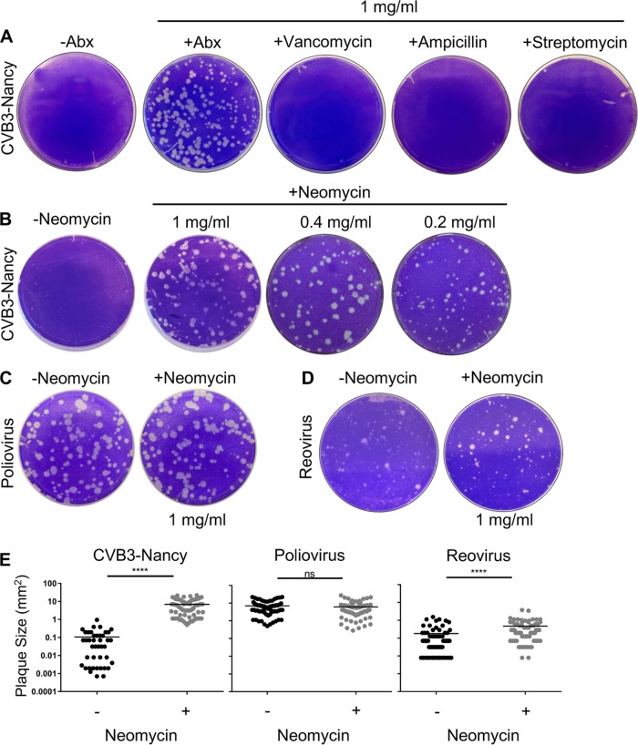
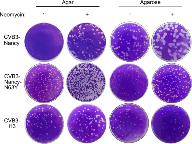
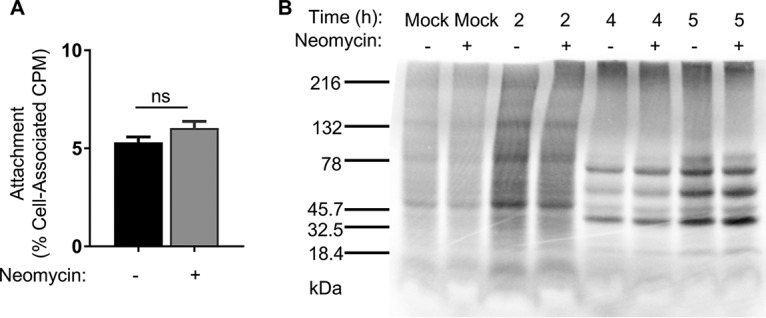
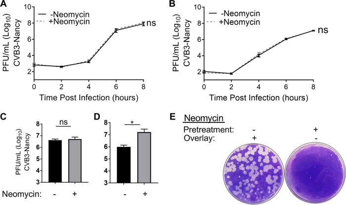
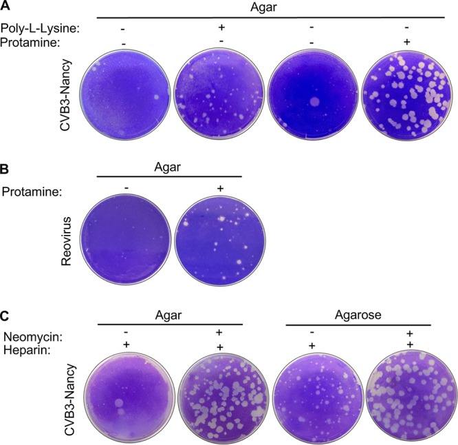
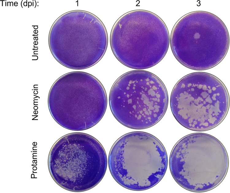
Coxsackievirus typically infects humans via the gastrointestinal tract, which has a large number of microorganisms collectively referred to as the microbiota. To study how the intestinal microbiota influences enteric virus infection, several groups have used an antibiotic regimen in mice to deplete bacteria. These studies have shown that bacteria promote infection with several enteric viruses. However, very little is known about whether antibiotics influence viruses in a microbiota-independent manner. In this study, we sought to determine the effects of antibiotics on coxsackievirus B3 (CVB3) using an in vitro cell culture model in the absence of bacteria. We determined that an aminoglycoside antibiotic, neomycin, enhanced the plaque size of CVB3 strain Nancy. Neomycin treatment did not alter viral attachment, translation, or replication. However, we found that the positive charge of neomycin and other positively charged compounds enhanced viral diffusion by overcoming the negative inhibitory effect of sulfated polysaccharides present in agar overlays. Neomycin and the positively charged compound protamine also enhanced plaque formation of reovirus. Overall, these data provide further evidence that antibiotics can play noncanonical roles in viral infections and that this should be considered when studying enteric virus-microbiota interactions.IMPORTANCE Coxsackieviruses primarily infect the gastrointestinal tract of humans, but they can disseminate systemically and cause severe disease. Using antibiotic treatment regimens to deplete intestinal microbes in mice, several groups have shown the bacteria promote infection with a variety of enteric viruses. However, it is possible that antibiotics have microbiota-independent effects on viruses. Here we show that an aminoglycoside antibiotic, neomycin, can influence quantification of coxsackievirus in cultured cells in the absence of bacteria.

Keywords: antibiotics; coxsackievirus; microbiota; neomycin; reovirus.


Figures

FIG 1
FIG 1
Effect of neomycin on plaque formation of CVB3-Nancy, poliovirus, and reovirus. (A) Effects of antibiotics on CVB3-Nancy plaque formation. HeLa cells were pretreated with or without 1 mg/ml of the indicated antibiotics prior to plating of 100 PFU of CVB3-Nancy on cells with agar overlays with or without 1 mg/ml of each antibiotic. Plates were stained with crystal violet. Abx, ampicillin, neomycin, streptomycin, and vancomycin mixture. (B) Effect of neomycin on CVB3-Nancy plaque formation. The experiment was performed as for panel A, in the presence or absence of neomycin at various concentrations. (C and D) Effects of neomycin on poliovirus (C) or type 3 Dearing reovirus (D). (E) Plaque size quantification of CVB3-Nancy, poliovirus, and reovirus in the presence or absence of 1 mg/ml of neomycin. Each symbol represents a plaque. ****, P < 0.0001 (unpaired two-tailed Student t tests). ns, not significant. All images are representative of results from at least 2 or 3 independent experiments.
FIG 2
FIG 2
Effect of neomycin on plaque formation of different CVB3 strains. HeLa cells were pretreated or not with neomycin and were infected with approximately 100 PFU of CVB3-Nancy, CVB3-Nancy-N63Y, or CVB3-H3, and then agar or agarose overlays, containing or lacking 1 mg/ml of neomycin, were added. All images are representative of results from at least two independent experiments.
FIG 3
FIG 3
Effects of neomycin on early stages of the CVB3-Nancy replication cycle. (A) Cell attachment assay. A total of 1 × 106 HeLa cells were pretreated or not with neomycin prior to incubation with 6,000 cpm (6 × 106 PFU) of 35S-labeled CVB3-Nancy or no virus (mock) at 4°C for 20 min to promote viral binding. Cells were washed and 35S was quantified in a scintillation counter. Data are means ± SEMs (unpaired two-tailed Student t tests; n = 5 total data points from two independent experiments). (B) Viral protein synthesis assay. A total of 2.5 × 106 HeLa cells that had been pretreated or not with neomycin were inoculated with CVB3-Nancy at an MOI of 20 for 30 min at 37°C. At the indicated time points, cells were washed and incubated in 1 ml of DMEM lacking methionine and cysteine supplemented with [35S]l-methionine and [35S]l-cysteine for 15 min at 37°C. Cell lysates from equal cell numbers were analyzed on an SDS-PAGE gel. Radiolabeled proteins were visualized using a phosphorlmager. The image is representative of results from three independent experiments.
FIG 4
FIG 4
Effects of neomycin on CVB3-Nancy replication kinetics. Shown is the growth curve in the presence of liquid media (A) or agar overlay (B). Briefly, 1 × 106 HeLa cells that had been pretreated or not with neomycin were inoculated with CVB3-Nancy at an MOI of 0.01. Virus was incubated for 30 min at 37°C, and either DMEM liquid (A) or 1% agar–1% DMEM mixture (B) with or without neomycin was added. At the indicated time points, intracellular virus was harvested and quantified by plaque assay. For panels A and B, n = 4 or 5 total data points from two independent experiments. To examine multicycle replication and spread, cells were infected with 100 PFU of CVB3-Nancy and either DMEM (C) or 1% agar–1% DMEM mixture (D) with or without neomycin, followed by plaque assay of cell-associated virus. For panels C and D, n = 6 total data points from three independent experiments. (E) HeLa cells were pretreated or not with 1 mg/ml of neomycin prior to plating of 100 PFU of CVB3-Nancy on cells with agar overlays with or without 1 mg/ml of neomycin. Plates were stained with crystal violet 48 hpi. *, P < 0.05 (unpaired two-tailed Student t tests). Images are representative of results from two independent experiments.
FIG 5
FIG 5
Effect of positively or negatively charged compounds on CVB3-Nancy and reovirus plaque formation. (A) A total of 8.8 × 106 HeLa cells were infected with 100 PFU of CVB3-Nancy and agar overlays were added with or without 0.1 µM poly-l-lysine or 0.8 mg/ml of protamine (positively charged compounds). (B) A total of 1 × 106 L929 cells were infected with 100 PFU of type 3 Dearing reovirus and agar overlays were added with or without 0.8 mg/ml of protamine. (C) A total of 8.8 × 106 HeLa cells were infected with 100 PFU of CVB3-Nancy and neomycin-containing agar or agarose overlays were added with or without 1 mg/ml of heparin (negatively charged compound). Images are representative of results from two independent experiments.
FIG 6
FIG 6
Effects of positively charged compounds on CVB3-Nancy diffusion. A total of 1 × 106 HeLa cells in 60-mm tissue culture plates were overlaid with 4 ml of 1% agar–1% DMEM mixture that contained or lacked 1 mg/ml of neomycin or 0.8 mg/ml of protamine. Once the overlay solidified, 5 × 104 PFU of CVB3-Nancy in 200 µl was added dropwise to the top of the overlay. Cells were placed at 37°C to allow diffusion of the virus through the overlay to the cell monolayer, and plates were stained with crystal violet at 1, 2, or 3 days postinfection (dpi) to reveal the extent of cell death from viral replication. Images are representative of results from two independent experiments.

References

    1. Tam PE. 2006. Coxsackievirus myocarditis: interplay between virus and host in the pathogenesis of heart disease. Viral Immunol 19:133–146. doi:10.1089/vim.2006.19.133. - DOI - PubMed
    1. Tracy S, Höfling K, Pirruccello S, Lane PH, Reyna SM, Gauntt CJ. 2000. Group B coxsackievirus myocarditis and pancreatitis: connection between viral virulence phenotypes in mice. J Med Virol 62:70–81. doi:10.1002/1096-9071(200009)62:1<70::AID-JMV11>3.0.CO;2-R. - DOI - PubMed
    1. Tracy S, Drescher KM. 2007. Coxsackievirus infections and NOD mice: relevant models of protection from, and induction of, type 1 diabetes. Ann N Y Acad Sci 1103:143–151. doi:10.1196/annals.1394.009. - DOI - PubMed
    1. Tracy S, Drescher KM, Chapman NM, Kim KS, Carson SD, Pirruccello S, Lane PH, Romero JR, Leser JS. 2002. Toward testing the hypothesis that group B coxsackieviruses (CVB) trigger insulin-dependent diabetes: inoculating nonobese diabetic mice with CVB markedly lowers diabetes incidence. J Virol 76:12097–12111. doi:10.1128/JVI.76.23.12097-12111.2002. - DOI - PMC - PubMed
    1. Chen P, Tao Z, Song Y, Liu G, Wang H, Liu Y, Song L, Li Y, Lin X, Cui N, Xu A. 2013. A coxsackievirus B5-associated aseptic meningitis outbreak in Shandong Province, China in 2009. J Med Virol 85:483–489. doi:10.1002/jmv.23478. - DOI - PubMed

Publication types