. 2019 Feb 20;9(1):2389.
doi: 10.1038/s41598-019-38718-0.

Targeted editing of the PSIP1 gene encoding LEDGF/p75 protects cells against HIV infection

Affiliations

Targeted editing of the PSIP1 gene encoding LEDGF/p75 protects cells against HIV infection

Yulia Lampi et al. Sci Rep. .

Abstract

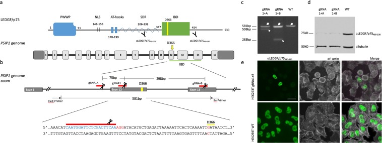
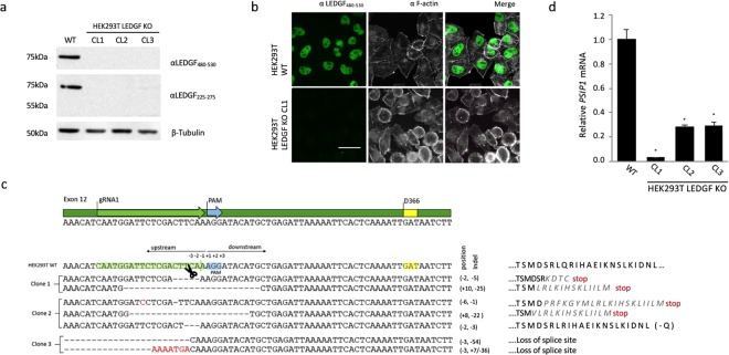
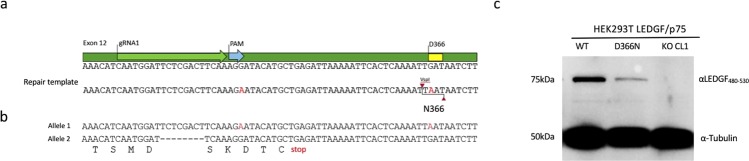
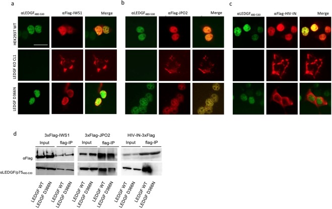
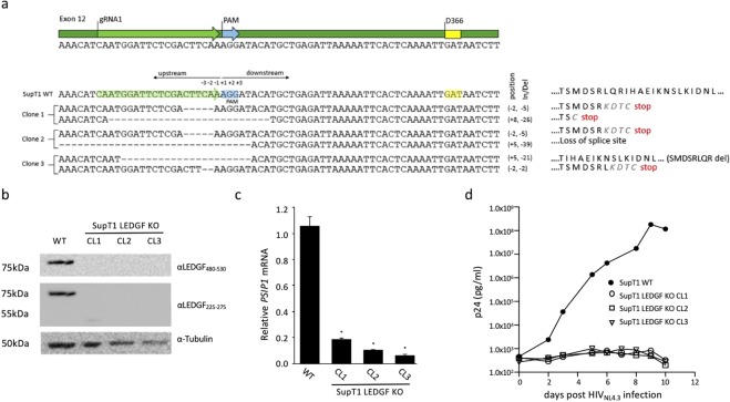
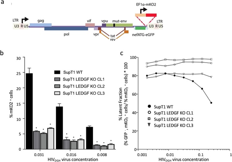
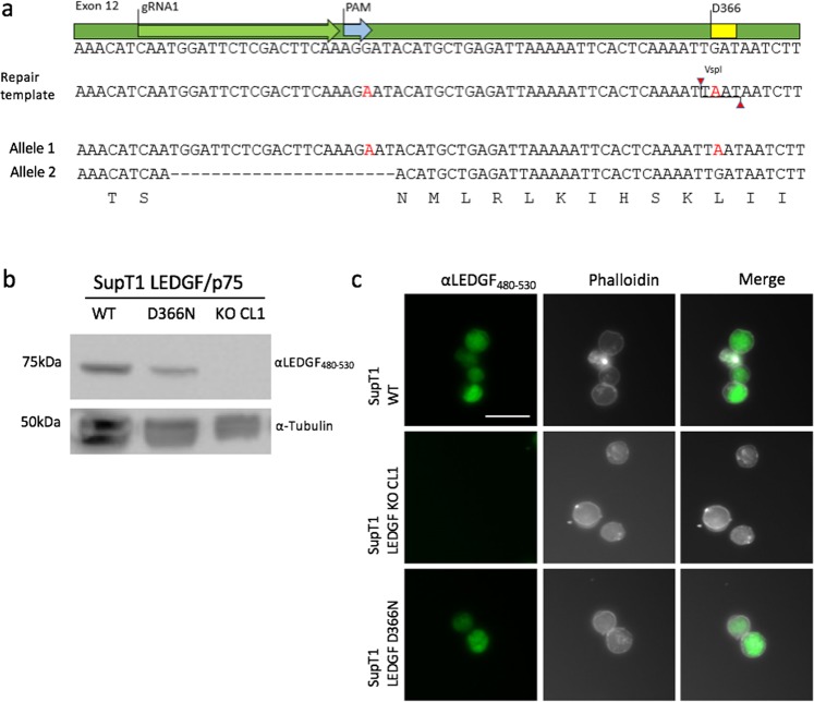
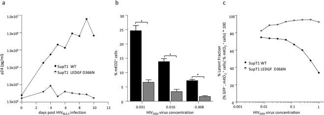
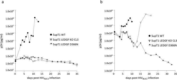
To fulfill a productive infection cycle the human immunodeficiency virus (HIV) relies on host-cell factors. Interference with these co-factors holds great promise in protecting cells against HIV infection. LEDGF/p75, encoded by the PSIP1 gene, is used by the integrase (IN) protein in the pre-integration complex of HIV to bind host-cell chromatin facilitating proviral integration. LEDGF/p75 depletion results in defective HIV replication. However, as part of its cellular function LEDGF/p75 tethers cellular proteins to the host-cell genome. We used site-specific editing of the PSIP1 locus using CRISPR/Cas to target the aspartic acid residue in position 366 and mutated it to asparagine (D366N) to disrupt the interaction with HIV IN but retain LEDGF/p75 cellular function. The resulting cell lines demonstrated successful disruption of the LEDGF/p75 HIV-IN interface without affecting interaction with cellular binding partners. In line with LEDGF/p75 depleted cells, D366N cells did not support HIV replication, in part due to decreased integration efficiency. In addition, we confirm the remaining integrated provirus is more silent. Taken together, these results support the potential of site-directed CRISPR/Cas9 mediated knock-in to render cells more resistant to HIV infection and provides an additional strategy to protect patient-derived T-cells against HIV-1 infection as part of cell-based therapy.


Conflict of interest statement

The authors declare no competing interests.

Figures

Figure 1
Figure 1
Guide RNA adjacent to the coding sequence D366 shows efficient disruption of the PSIP1 gene. (a) Schematic representation of LEDGF/p75 protein with indication of the epitope sites of respective antibodies used in Western analysis. Below the human PSIP1 locus on chromosome 9 is depicted showing the different exons as light grey boxes. IBD is underlined in green. (b) Schematic of representing the location of the different gRNA that were used (red lines), gRNA1 close to D366 and two additional supporting gRNAs (gRNA_A, gRNA_B). D366 is shown in yellow. The expected PCR fragment sizes are indicated as well as the predicted deletions for the different gRNA combinations. Below the targeted gDNA sequence is shown. D366 is boxed in green, the PAM site is shown in red and the landing site of gRNA1 is shown in blue. (c) Agarose gel analysis showing truncated amplicons generated by DNA cleavage guided by a pair of gRNAs. Genomic DNA was extracted from polyclonal cell populations and PCR amplified using Fwd and Rv primers indicated in panel (b). The WT amplicon is indicated by the large arrow head. The lower migrating bands (small arrow head) indicate segmental deletion. (d) Western blot analysis showing LEDGF protein in a polyclonal HEK293T population transfected with the indicated gRNA pairs. Wild-type 293T cells (WT) are shown as control. (e) Immunocytochemical staining of endogenous LEDGF showing nuclear localization in WT and CRISPRed polyclonal HEK239T cells. Phalloidin-stained F-actin in white is shown as a counterstain. The respective antibodies used are indicated above. Scale Bar: 10 μm.
Figure 2
Figure 2
Characterization of HEK293T_LEDGF KO cell lines using a single gRNA1. (a) Western blot analysis of LEDGF/p75 protein in 3 LEDGF KO clones. (b) Immunocytochemical analysis of wild-type HEK293T (WT) cells and monoclonal LEDGF KO cells showing nuclear localization of LEDGF/p75 as dense, fine speckles in WT cells (shown in green) and the lack thereof in the KO cell line. Phalloidin (white) was used to counterstain cytoplasmic F-actin. LEDGF KO (clone1) image is shown and is representative of all 3 isolated KO cell lines. (c) Sanger sequencing of the indel profile generated in the region targeted by gRNA1 (“−” indicates deletion, substitutions indicated in red) and the predicted changes in the amino acid sequence of exon 12 on the right. (d) PSIP1 mRNA expression levels with standard deviation relative to β-actin in the respective knock-out clones as determined by qPCR. Error bars represent SD. Student’s t test was performed using GraphPad Prism 7.0 software. Sample means were considered significantly different from the WT control at p < 0.05 (*). Scale Bar: 10 µm.
Figure 3
Figure 3
HDR template design and screening. (a) Schematic representation of exon12 sequence of PSIP1 gene harboring D366 residue. Below the designed HDR template as a ssDNA oligo is shown with red characters indicating the sites of nucleotide substitutions, generating a VspI restriction site at D366N and deleting the PAM site. (b) Sanger sequencing results for the HEK293T_LEDGF D366N clone that was identified, showing consensus between HDR template and allele 1 (substitution indicated in red). The sequence for allele 2 shows a 8-nucleotide deletion (“−” indicates deletion) and the translation (capital letters below) resulting in a premature stop codon (underlined). (c) Western blot analysis showing the LEDGF/p75 levels in D366N clone compared to WT levels.
Figure 4
Figure 4
LEDGF D366N mutant is uncoupled from HIV-IN but interacts with other binding partners. (ac) In vivo co-localization of the LEDGF D366N and cellular binding partners. Wild-type HEK293T, LEDGF KO and LEDGF D366N cells were transfected with flag-tagged IWS1, JPO2 and HIV-IN (panels a, b and c, respectively) and fixed 30 hrs later. Fluorescence microscopy to assess localization of proteins: LEDGF/p75 and LEDGF D366N were detected using αLEDGF480–530 (shown in green), whereas the transfected flag-tagged proteins were detected with αFlag Ab (shown in red). (a,b) LEDGF WT (top panel) and LEDGF D366N (lower panel) co-localizes with IWS1 and JPO2. (c) LEDGF D366N mutant did not co-localize with HIV-IN (lower panel), while in HEK293T WT cells HIV-IN is retained in the nucleus by binding to LEDGF/p75 (top panel). Scale Bar: 10 µm. (d) Co-immunoprecipitation of LEDGF/p75 and LEDGF D366N by cellular binding partners. HEK293T_LEDGF KO cells were transfected with either WT or D366N mutant LEDGF plasmid along with either one of the plasmids encoding Flag-tagged binding partners: IWS1, JPO2 or HIV IN. Cells were lysed 24 h later and lysates were incubated with anti-FLAG® M2-agarose affinity resin to capture the binding partner protein, which was then visualized by Western blot using αFlag Ab and αLEDGF480–530.
Figure 5
Figure 5
HIV replication is severely affected in SupT1_LEDGF KO cells. (a) Schematic representation of a detailed zoom of exon12 sequence of the PSIP1 gene harboring D366. Sanger sequencing of the indel profile generated in the region targeted by gRNA1 (“−” indicates deletion, substitution indicated in red) and the predicted changes in the amino acid sequence of exon 12 shown on the right. (b) Western analysis of LEDGF/p75 protein in the 3 isolated LEDGF KO cell lines. (c) PSIP1 mRNA expression relative to β-actin in the respective SupT1_LEDGF KO clones as determined by qPCR. Error bars represent SD. Student’s t test was performed using GraphPad Prism 7.0 software. Sample means were considered significantly different from the WT control at p < 0.05 (*). (d) HIV-1 replication assay. The respective cell lines were challenged with the laboratory HIVNL4.3 strain at a final concentration of 5.0*102 pg p24. Viral replication was monitored by daily sampling of p24 in the cell culture supernatant. The graph shows a representative infection experiment out of three independent trials.
Figure 6
Figure 6
LEDGF depletion results in an increase of the silent HIV reservoir. (a) Schematic representation of the dual-colored VSV-G pseudotyped HIVOGH reporter virus carrying an eGFP cDNA driven by the viral LTR promoter in the nef position and an entire constitutive transcriptional unit (EF1a-mKO2) inserted downstream. (b) HIV provirus integration is greatly diminished in the absence of LEDGF/p75 when compared to SupT1_WT cells, as evidenced by the lower %mKO2 positive cells for a given virus concentration. Error bars represent SD. Student’s t test was performed using GraphPad Prism 7.0 software. Sample means were considered significantly different from the WT control at p < 0.05 (*). (c) The latent fraction for the respective cell lines for different virus concentrations. LEDGF KO increases the fraction of silently infected cells ((% eGFP, mKO2+ cells)/% mKO2+ cells) ∗ 100. eGFP, Enhanced Green Fluorescent Protein; mKO2, Mutant Kusubira Orange 2. The experiment was performed three times. The plots are representatives of one of three independent infection experiments.
Figure 7
Figure 7
Characterization of SupT1_LEDGF D366N. (a) Sanger sequencing results showing consensus between HDR template and allele 1. The HDR template is shown with red characters indicating the sites of nucleotide substitutions, generating a VspI restriction site at D366N and deleting the PAM site. The sequence for allele 2 shows a 22-nucleotide deletion gRNA1 (“−” indicates deletion), resulting in KO of the PSIP1 allele. (b) Western blot analysis for the SupT1_LEDGF D366N clone using C-terminal specific LEDGF antibody, αLEDGF480–530. SupT1_WT and KO cells are included as controls. (c) Immunocytochemical analysis of LEDGF D366N protein (green) in SupT1_LEDGF D366N cells (bottom panel) recapitulates that of LEDGF/p75 in SupT1_WT cells (top panel). Phalloidin (white) was used to stain F-actin. Scale Bar: 10 µm.
Figure 8
Figure 8
HIV replication is hampered in SupT1_LEDGF D366N cell line as compared to the SupT1 WT cell line. (a) HIV-1 replication assay. The cell lines were challenged with the laboratory HIVNL4.3 strain at a final concentration of 5.0*102 pg p24. Replication was monitored by daily sampling of the p24 levels in the cell culture supernatant. (b) Introduction of the D366N mutation in LEDGF/p75 renders SupT1 cells refractory to transduction as shown by a 4-fold decrease in %mKO2 expressing cells for a given VSV-G pseudotyped HIVOGH virus concentration. Student’s t test was performed using GraphPad Prism 7.0 software. Sample means were considered significantly different from the WT control at p < 0.05 (*). (c) In addition to the lower transduction efficiency observed for SupT1_LEDGF D366N, the latent fraction is greater when compared to SupT1_WT cells, even at higher concentrations of HIVOGH virus. eGFP, Enhanced Green Fluorescent Protein; mKO2, mutant Kusubira Orange 2. The plots represent one of three independent infection experiments.
Figure 9
Figure 9
HIV replication remains hampered in SupT1 LEDGF D366N cell line at high MOI as compared to the SupT1 WT and SupT1 LEDGF KO cell line. (a) HIV-1 replication assay at low MOI. The cell lines were challenged with the laboratory HIVNL4.3 strain at a concentration of 1.5*106 pg p24 for 2 h. The cells were then washed to remove the virus to result in 5.0*102 pg/ml p24 final concentration and monitored for replication. (b) HIV-1 replication assay at high MOI. The cell lines were challenged with the laboratory HIVNL4.3 strain at a final concentration of 1.5*106 pg p24. Replication was monitored by daily sampling of the p24 levels in the cell culture supernatant. Each plot represents one of three independent infection experiments.

References

    1. Schupbach J, et al. Serological analysis of a subgroup of human T-lymphotropic retroviruses (HTLV-III) associated with AIDS. Science (80-.). 1984;224:503–505. doi: 10.1126/science.6200937. - DOI - PubMed
    1. Dieffenbach CW, Fauci AS. Thirty years of HIV and AIDS: future challenges and opportunities. Ann Intern Med. 2011;154:766–771. doi: 10.7326/0003-4819-154-11-201106070-00345. - DOI - PubMed
    1. Geeraert L, Kraus G, Pomerantz RJ. Hide-and-seek: the challenge of viral persistence in HIV-1 infection. Annu Rev Med. 2008;59:487–501. doi: 10.1146/annurev.med.59.062806.123001. - DOI - PubMed
    1. Bowman M-C, et al. Inhibition of HIV Fusion with Multivalent Gold Nanoparticles. J. Am. Chem. Soc. 2008;130:6896–6897. doi: 10.1021/ja710321g. - DOI - PMC - PubMed
    1. Hutter G, et al. Long-term control of HIV by CCR5 Delta32/Delta32 stem-cell transplantation. N Engl J Med. 2009;360:692–698. doi: 10.1056/NEJMoa0802905. - DOI - PubMed

Publication types

MeSH terms