. 2019 Jul;27(7):1113-1120.
doi: 10.1038/s41431-019-0350-4. Epub 2019 Feb 20.

A novel dominant-negative FGFR1 variant causes Hartsfield syndrome by deregulating RAS/ERK1/2 pathway

Affiliations

A novel dominant-negative FGFR1 variant causes Hartsfield syndrome by deregulating RAS/ERK1/2 pathway

Pietro Palumbo et al. Eur J Hum Genet. 2019 Jul.

Abstract

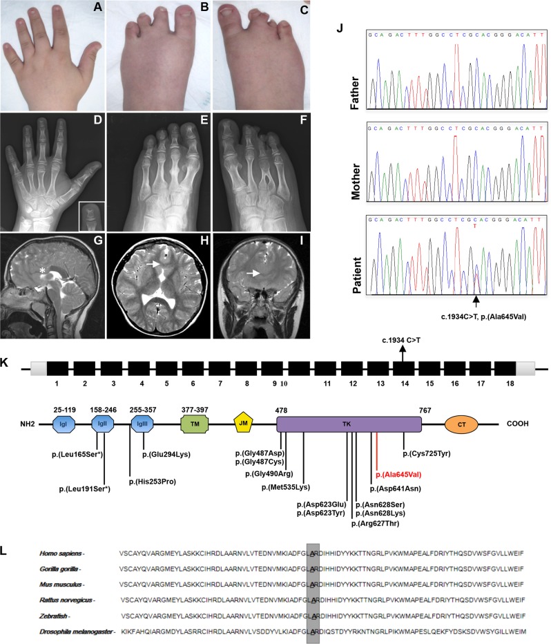
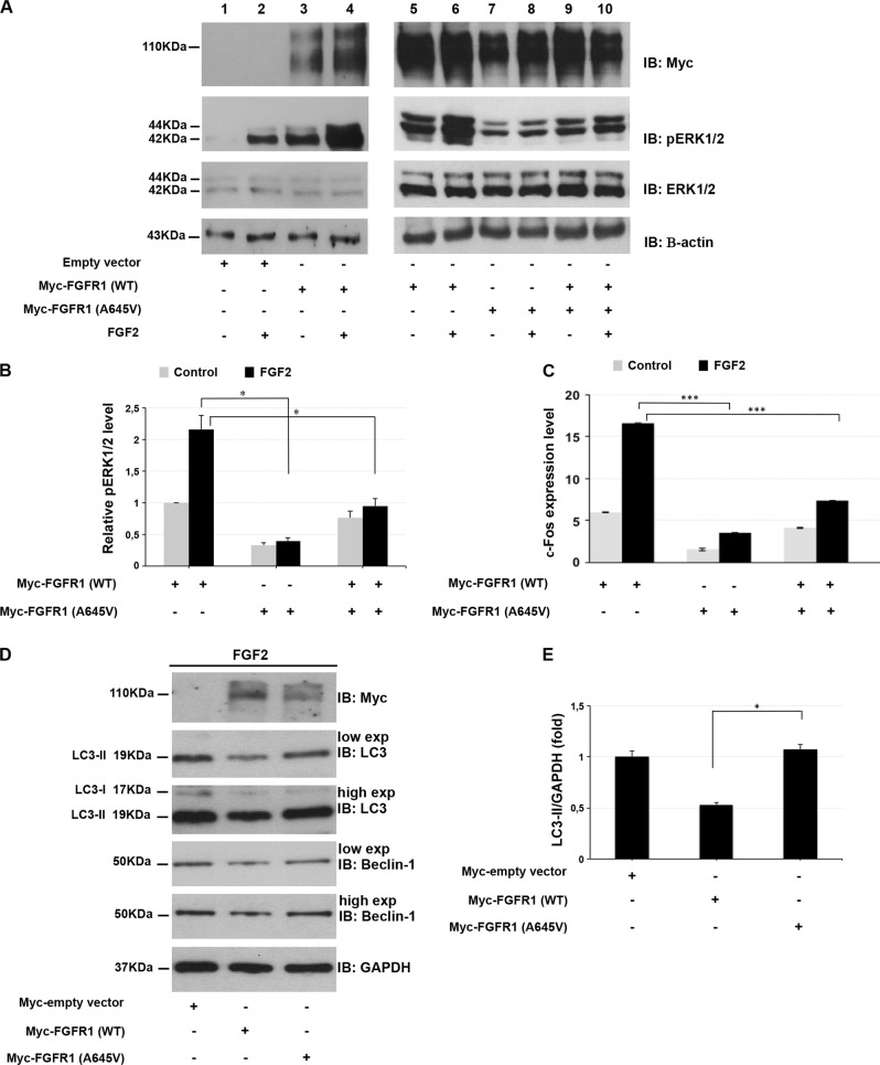
Hartsfield syndrome (HS) is an ultrarare developmental disorder mainly featuring holoprosencephaly and ectrodactyly. It is caused by heterozygous or biallelic variants in FGFR1. Recently, a dominant-negative effect was suggested for FGFR1 variants associated with HS. Here, exome sequencing analysis in a 12-year-old boy with HS disclosed a novel de novo heterozygous variant c.1934C>T in FGFR1 predicted to cause the p.(Ala645Val) amino-acid substitution. In order to evaluate whether the variant, changing a highly conserved residue of the kinase domain, affects FGFR1 function, biochemical studies were employed. We measured the FGFR1 receptor activity in FGF2-treated cell lines exogenously expressing wild-type or Ala645Val FGFR1 by monitoring the activation status of FGF2/FGFR1 downstream pathways. Our analysis highlighted that RAS/ERK1/2 signaling was significantly perturbed in cells expressing mutated FGFR1, in comparison with control cells. We also provided preliminary evidence showing a modulation of the autophagic process in cells expressing mutated FGFR1. This study expands the FGFR1 mutational spectrum associated with HS, provides functional evidence further supporting a dominant-negative effect of this category of FGFR1 variants and offers initial insights on dysregulation of autophagy in HS.


Conflict of interest statement

The authors declare that they have no conflict of interest.

Figures

Fig. 1
Fig. 1
Clinical features and molecular findings. a Brachydactyly with broad fingertips and short nails. b Left foot with four toes, brachydactyly II–IV and partial cutaneous syndactyly II–III. c Right foot presenting cutaneous syndactyly and severe brachydactyly II–IV. d Radiograph of the left hand showing short metacarpals, shortened middle phalanx of the fifth finger, and bifid distal phalanx of the third finger. e Radiograph of the left foot with four rays, malformed metatarsal of the second toe, absence of the middle phalanx of the fifth toe, and partial synostosis of the middle and distal phalanges of the second toe. f Radiograph of the right foot with inverted “Y” synostosis of the third and fourth metatarsal, rudimentary bones of the third toe, and absence of the middle phalanx of the fifth toe. Brain MRI with absence of the anterior part of the corpus callosum on the sagittal scan (g; asterisk indicates the absence of the genu and rostrum of the corpus callosum), and partial fusion of the frontal lobes on axial (h) and coronal scans (i; arrows indicate midline fusion of the frontal lobes in both scans). j Sanger sequencing showing the c.1934C>T nucleotide change in FGFR1. k Diagram showing the structure of FGFR1 and the secondary structure of the protein. Coding regions are in black, UTR sequences are in gray, introns are not to scale. Previously identified and the novel variants associated with Hartsfield syndrome are located on the protein structure. The amino-acid change identified in this work is in red. CT C-terminal-tail, IgI–III, immunoglobulin-like domains, JM juxtamembrane domain, TD transmembrane domain, TK tyrosine kinase domain. l Conservation of the involved amino-acid (Ala645) among species (color figure online)
Fig. 2
Fig. 2
Ala645Val FGFR1 impairs ERK1/2 signaling pathway and autophagy flux. a HEK293 cells were transfected with empty vector (lanes 1, 2), wild-type FGFR1 (lanes 3–6) and Ala645Val FGFR1-expressing plasmids (lanes 7, 8) or wild-type plus Ala645Val FGFR1 plasmids (lanes 9, 10). At 24 h after transfection, cells were grown in serum-free medium for 24 h, and then incubated in the absence (lanes 1, 3, 5, 7, 9) or presence of 1 nM FGF2 (lanes 2, 4, 6, 8, 10) for 15 min. After incubation with FGF2, the phosphorylated ERK1/2 was analyzed by SDS-polyacrylamide gel electrophoresis using indicated antibodies. Actin was used as loading control. b Quantitative analysis of ERK1/2 phosphorylation in cells transfected with indicated vectors. Protein levels were quantified by densitometry. The relative ERK1/2 phosphorylation level in cells transfected with the vector expressing wild-type FGFR1 was set as 1. Bar represents the average of three independent experiments and scale bars represent standard errors. *P < 0.05. c qPCR was performed to measure the c-Fos endogenous expression in HEK293 cells transfected with indicated plasmids and cultured in the absence or presence of 0.5 nM FGF2 for 1 h. The relative c-Fos expression in cells transfected with the empty vector was set as 1. Scale bars represent standard errors. ***P < 0.01. d Whole protein lysates of HEK293 cells transfected with empty vector, wild-type or Ala645Val FGFR1-expressing plasmids and cultured in the absence or presence of 25 ng/ml FGF2 for 2 h, were separated on 12% SDS-gel and subjected to immunoblotting with LC3 and Beclin-1 antibodies (low and high exposure). The autophagy flux was monitored by the conversion of LC3-I to its lipidated form, LC3-II. GAPDH was used as loading control. The same lysates were separated on 7.5% SDS-gel for immunoblotting with anti-Myc. e Quantification of LC3-II levels. Graph shows averages calculated on two different experiments and scale bars represent standard errors. *P < 0.05

References

    1. Dhamija R, Babovic-Vuksanovic D. Hartsfield syndrome. In: Adam MP, Ardinger HH, Pagon RA, Wallace SE, Bean LJH, Stephens K, Amemiya A, editors. GeneReviews® [Internet]. Seattle (WA): University of Washington, Seattle; 1993–2018 (last updated: 3 Mar 2016).
    1. Simonis N, Migeotte I, Lambert N. FGFR1 mutations cause Hartsfield syndrome, the unique association of holoprosencephaly and ectrodactyly. J Med Genet. 2013;50:585–92. doi: 10.1136/jmedgenet-2013-101603. - DOI - PMC - PubMed
    1. Hong S, Hu P, Marino J, et al. Dominant-negative kinase domain mutations in FGFR1 can explain the clinical severity of Hartsfield syndrome. Hum Mol Genet. 2016;25:1912–22. doi: 10.1093/hmg/ddw064. - DOI - PMC - PubMed
    1. Ornitz DM, Itoh N. The fibroblast growth factor signaling pathway. Wiley Interdisciplinary Rev Dev Biol. 2015;4:215–66. doi: 10.1002/wdev.176. - DOI - PMC - PubMed
    1. Teven CM, Farina EM, Rivas J, et al. Fibroblast growth factor (FGF) signaling in development and skeletal diseases. Genes Dis. 2014;1:199–213. doi: 10.1016/j.gendis.2014.09.005. - DOI - PMC - PubMed

Publication types

MeSH terms

Substances

Supplementary concepts