Psammomatoid Ossifying Fibroma of the Ethmoid Sinus with Secondary Intracranial Aneurymal Bone Cyst: A Case Report and Literature Review
- PMID: 30787712
- PMCID: PMC6298325
- DOI: 10.4103/1658-631X.178350
Psammomatoid Ossifying Fibroma of the Ethmoid Sinus with Secondary Intracranial Aneurymal Bone Cyst: A Case Report and Literature Review
Abstract
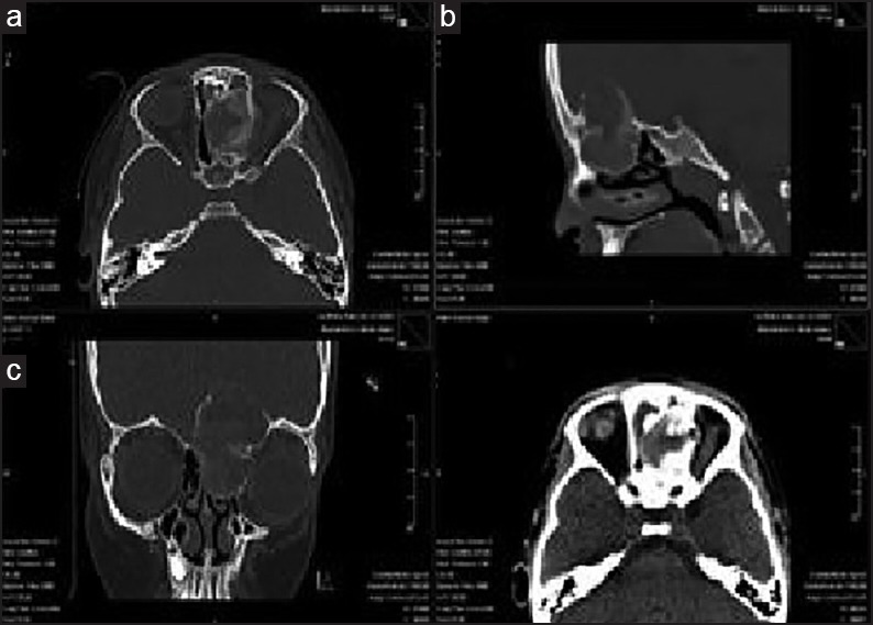
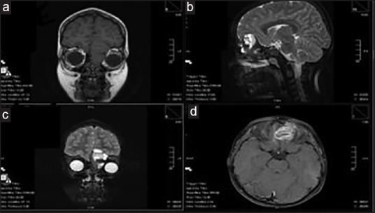
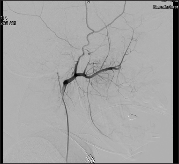
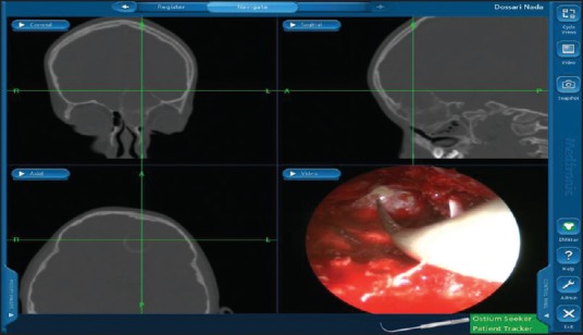
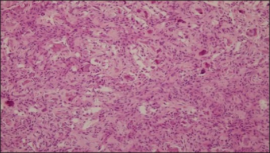
Juvenile psammomatoid ossifying fibroma (JPOF) is a rare, slowly progressive tumor of the extragnathic craniofacial bones, with a tendency toward locally aggressive behavior and recurrence. The pathognomonic histopathologic feature is the presence of spherical ossicles, which are similar to psammoma bodies. Very few cases in association with secondary aneurysmal bone cyst (ABC) formation have been reported in literature. Treatment consists of complete surgical removal. However, incomplete excision has been associated with a high local recurrence rate. The prognosis is good because malignant change and metastasis have not been reported. The authors are reporting a case of JPOF of the ethmoid bones with secondary ABC in a 7-year-old female patient.
ملخص البحث : الورم الليفي الرملي العظمي من الحالات النادرة وهو ورم بطئ النمو داخل الجمجمة وذو طبيعة متكررة. يعرض الباحثون حالة لطفلة تبلغ من العمر7 سنوات تم تشخيصها بهذا الورم في الجيب ألغربالي داخل الجمجمة. وتم علاجها جراحيا باستئصال الورم كاملاً باستخدام المنظار.
Keywords: Aneurysmal bone cyst; endoscopic management; paranasal sinuses; psammomatoid ossifying fibroma.
Conflict of interest statement
There are no conflicts of interest.
Figures
Similar articles
-
Juvenile psammomatoid ossifying fibroma of the calcaneus.BMJ Case Rep. 2020 Aug 18;13(8):e234555. doi: 10.1136/bcr-2020-234555. BMJ Case Rep. 2020. PMID: 32816930 Free PMC article.
-
Recurrent Psammomatoid Juvenile Ossifying Fibroma with Aneurysmal Bone Cyst: An Unusual Case Presentation.Iran J Med Sci. 2017 Nov;42(6):603-606. Iran J Med Sci. 2017. PMID: 29184270 Free PMC article.
-
Psammomatoid ossifying fibroma with secondary aneurysmal bone cyst of frontal sinus.Childs Nerv Syst. 2009 Nov;25(11):1513-6. doi: 10.1007/s00381-009-0906-7. Epub 2009 May 30. Childs Nerv Syst. 2009. PMID: 19484249 Free PMC article. Review.
-
Juvenile Psammomatoid Ossifying Fibroma (JPOF) of Proximal Radius: A Rare Entity.Open Orthop J. 2017 Jul 28;11:583-588. doi: 10.2174/1874325001711010583. eCollection 2017. Open Orthop J. 2017. PMID: 28932332 Free PMC article.
-
Juvenile psammomatoid ossifying fibroma of maxillary sinus: case report with review of literature.J Maxillofac Oral Surg. 2014 Jun;13(2):109-14. doi: 10.1007/s12663-013-0479-6. Epub 2013 Feb 7. J Maxillofac Oral Surg. 2014. PMID: 24822000 Free PMC article. Review.
Cited by
-
Synchronous Occurrence of Colloid Cyst With Intracranial Ossifying Fibromyxoid Tumor Masquerading as Meningioma.Cureus. 2020 Sep 26;12(9):e10662. doi: 10.7759/cureus.10662. Cureus. 2020. PMID: 33014664 Free PMC article.
-
Juvenile psammomatoid ossifying fibroma of the calcaneus.BMJ Case Rep. 2020 Aug 18;13(8):e234555. doi: 10.1136/bcr-2020-234555. BMJ Case Rep. 2020. PMID: 32816930 Free PMC article.
References
-
- Waknisa P, Sarodeb SC, Dolasa RS. Psammomatoid juvenile ossifying fibroma of the mandible with secondary aneurysmal bone cyst: A case report. Asian J Oral Maxillofac Surg. 2011;2:83–6.
-
- El-Mofty S. Psammomatoid and trabecular juvenile ossifying fibroma of the craniofacial skeleton: Two distinct clinicopathologic entities. Oral Surg Oral Med Oral Pathol Oral Radiol Endod. 2002;93:296–304. - PubMed
-
- Slootweg PJ, Panders AK, Koopmans R, Nikkels PG. Juvenile ossifying fibroma: An analysis of 33 cases with emphasis on histopathological aspects. J Oral Pathol Med. 1940;23:385–8. - PubMed
-
- Sarode SC, Sarode GS, Waknis P, Patil A, Jashika M. Juvenile psammom`: A review. Oral Oncol. 2011;47:1110–6. - PubMed
-
- Johnson LC, Yousefi M, Vinh TN, Heffner DK, Hyams VJ, Hartman KS. Juvenile active ossifying fibroma. Its nature, dynamics and origin. Acta Otolaryngol Suppl. 1991;488:1–40. - PubMed