. 2019 Feb 6:10:143.
doi: 10.3389/fimmu.2019.00143. eCollection 2019.

Faecalibacterium prausnitzii Skews Human DC to Prime IL10-Producing T Cells Through TLR2/6/JNK Signaling and IL-10, IL-27, CD39, and IDO-1 Induction

Affiliations

Faecalibacterium prausnitzii Skews Human DC to Prime IL10-Producing T Cells Through TLR2/6/JNK Signaling and IL-10, IL-27, CD39, and IDO-1 Induction

Joudy Alameddine et al. Front Immunol. .

Abstract

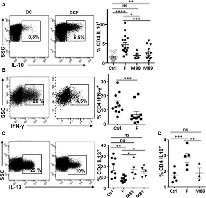
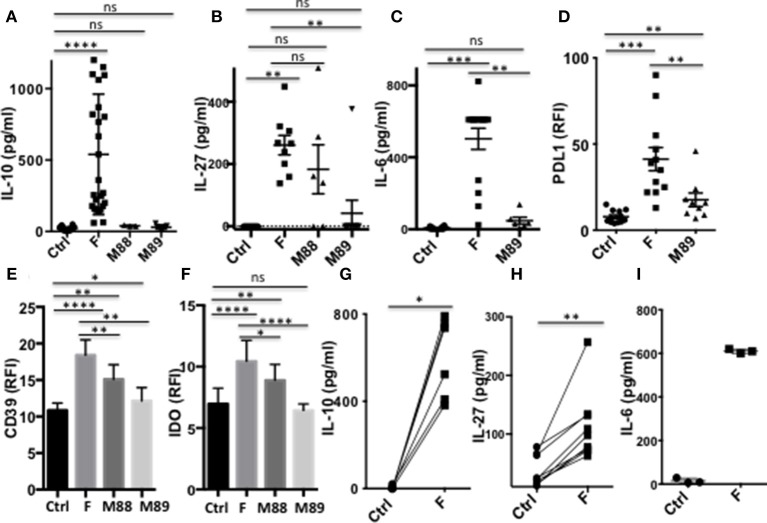
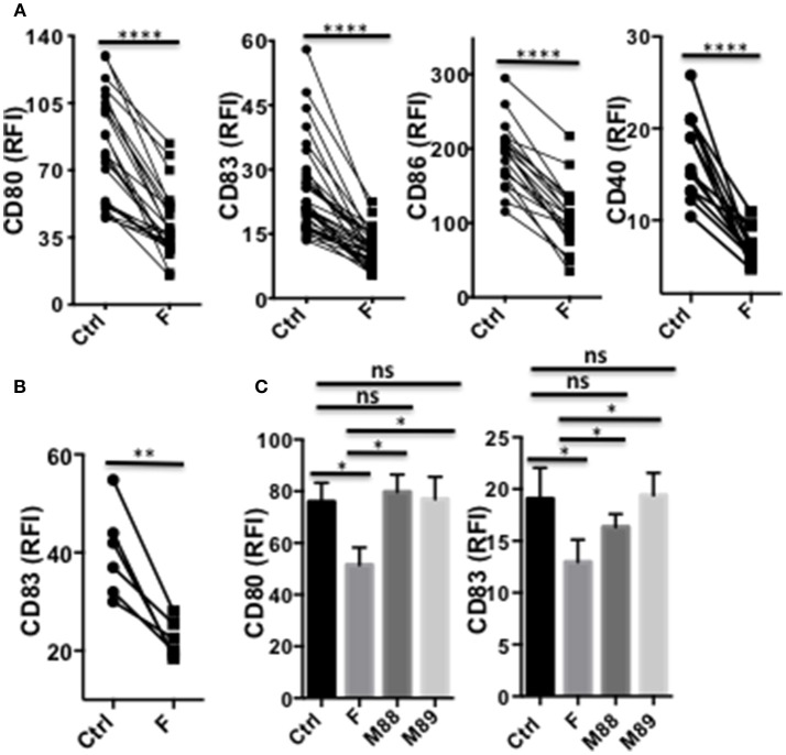
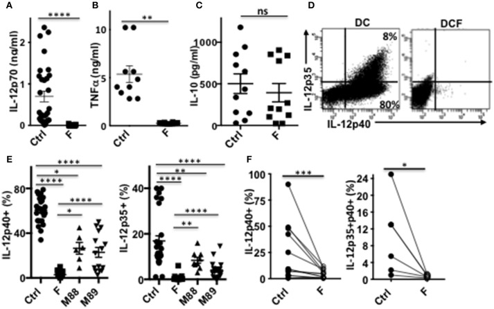
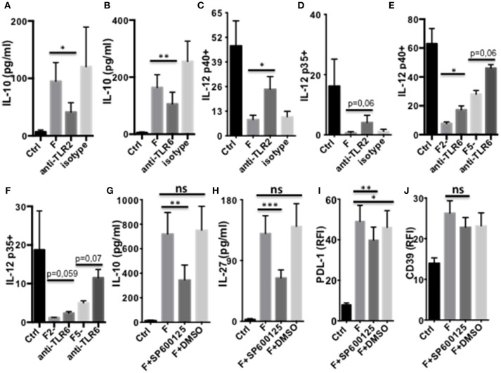
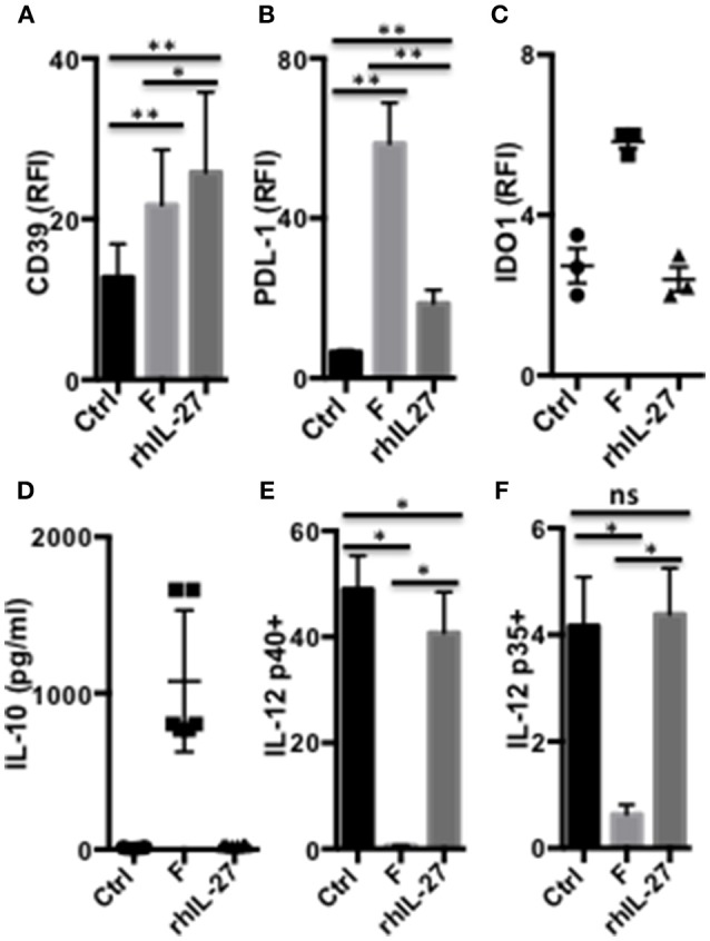
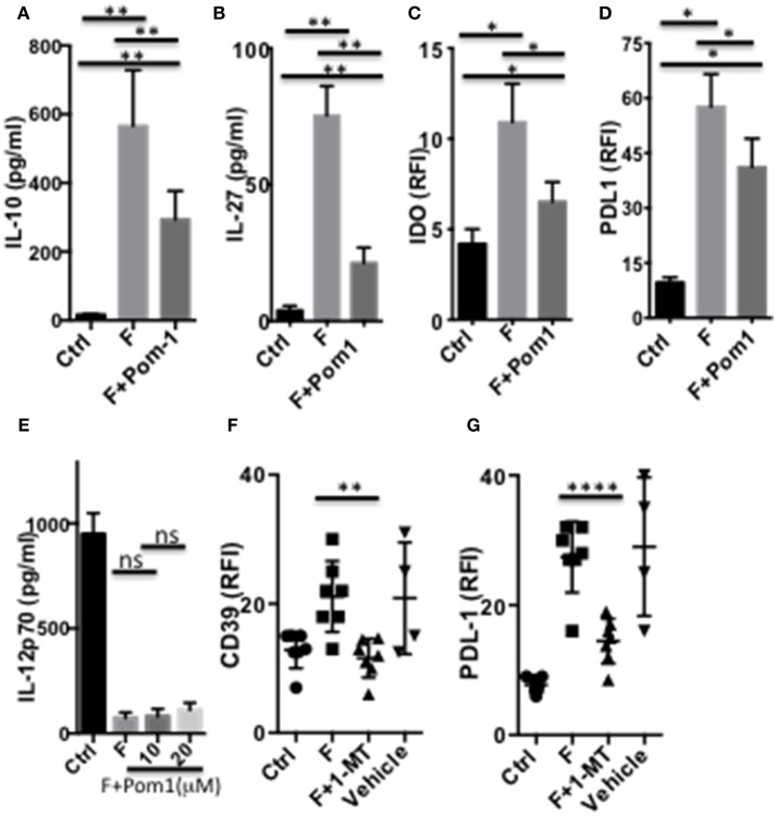
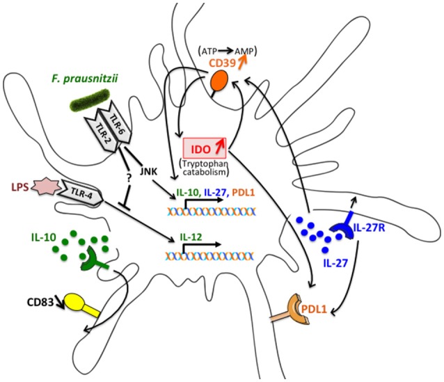
The human colonic mucosa contains regulatory type 1-like (Tr1-like, i.e., IL-10-secreting and Foxp3-negative) T cells specific for the gut Clostridium Faecalibacterium prausnitzii (F. prausnitzii), which are both decreased in Crohn's disease patients. These data, together with the demonstration, in mice, that colonic regulatory T cells (Treg) induced by Clostridium bacteria are key players in colon homeostasis, support a similar role for F. prausnitzii-specific Treg in the human colon. Here we assessed the mechanisms whereby F. prausnitzii induces human colonic Treg. We demonstrated that F. prausnitzii, but not related Clostridia, skewed human dendritic cells to prime IL-10-secreting T cells. Accordingly, F. prausnitzii induced dendritic cells to express a unique array of potent Tr1/Treg polarizing molecules: IL-10, IL-27, CD39, IDO-1, and PDL-1 and, following TLR4 stimulation, inhibited their up-regulation of costimulation molecules as well as their production of pro-inflammatory cytokines IL-12 (p35 and p40) and TNFα. We further showed that these potent tolerogenic effects relied on F. prausnitzii-induced TLR2/6 triggering, JNK signaling and CD39 ectonucleotidase activity, which was induced by IDO-1 and IL-27. These data, together with the presence of F. prausnitzii-specific Tr1-like Treg in the human colon, point out to dendritic cells polarization by F. prausnitzii as the first described cellular mechanism whereby the microbiota composition may affect human colon homeostasis. Identification of F. prausnitzii-induced mediators involved in Tr1-like Treg induction by dendritic cells opens therapeutic avenues for the treatment of inflammatory bowel diseases.

Keywords: CD39; IDO-1; JNK; TLR2/6; Tr1-like Treg; colon homeostasis; dendritic cells; gut microbiota.


Figures

Figure 1
Figure 1
Exposure to F. prausnitzii (F) promotes DC ability to prime IL-10-secreting T cells. VPD-stained naïve CD4+ T cells were stimulated by LPS-stimulated DC, pre-exposed or not to indicated bacteria at day 0 [(A) n = 6–18, (B) n = 11, (C) n = 5–7] or from day 4 to 6 of their differentiation [(D) n = 5], and were re-stimulated by CD3/CD28 beads. Representative examples and mean percentages ± sem of CD4 T cells expressing indicated cytokines. Paired t-test: *p < 0.05, **p < 0.005, ***p < 0.0005, and ****p < 0.00005.
Figure 2
Figure 2
DC exposure to F. prausnitzii promotes the production of immunoregulatory molecules. (A): IL-10 (Ctrl and F n = 26, M88 n = 3, M89 n = 9), (B): IL-27 (Ctrl, F and M89 n = 9, M88 n = 6) and (C): IL-6 (Ctrl and F n = 12, M89 n = 6) -secretion by DC exposed or not to indicated bacteria during the last 24 h. (D–F): expression of PDL-1 and CD39 (n = 15–29) or IDO-1 (n = 18–25) by DC exposed or not to indicated bacteria for the last 24 or 48 h. (G–I): IL-10 (n = 6), IL-27 (n = 9), and IL-6 (n = 3) secretion by myeloid DC maintained for 24 h in culture with or without F. prausnitzii. Results as mean ± sem. Wilcoxon test. *p < 0.05, **p < 0.005, ***p < 0.0005, ****p < 0.00005.
Figure 3
Figure 3
A single exposure to F. prausnitzii inhibits DC maturation. (A) Expression of CD80, CD83, CD86 or CD40 by DC exposed or not to F. prausnitzii at the beginning of their differentiation or (B) at day 5 of their differentiation (n = 6), and stimulated by LPS at day 6 during 48 h; (C) Expression of CD80 or CD83 (n = 7) by bacteria-exposed DC. Marker expression was measured by flow cytometry using a Canto II FACS Scan and expressed as relative fluorescence intensity (RFI) ± sem. Paired t-test. *p < 0.05, **p < 0.005, ****p < 0.00005.
Figure 4
Figure 4
DC exposure to F. prausnitzii switches their cytokine profile from pro-inflammatory to anti-inflammatory. IL-12p70 (A), TNF-a (B), and IL-10 levels (C) secreted in response to LPS by DC exposed or not (Ctrl) to Fprau at the beginning of their differentiation. (D) Representative dot plot of intracellular IL-12p35 and p40 co-labeling in LPS-stimulated DC exposed or not to F. prausnitzii. (E) LPS-induced IL-12p40 (n = 6–24) and IL-12p35 (n = 9–27) expression by DC exposed or not (Ctrl) to indicated bacteria (ratio:1:1) during the last 48 h. (F) IL-12p40 (n = 12) and p35 (n = 6) expression by myeloid DC maintained for 24 h in culture with or without F. prausnitzii, and then stimulated 12 h with LPS. Wilcoxon test. *p < 0.05, **p < 0.005, ***p < 0.0005, ****p < 0.00005.
Figure 5
Figure 5
TLR2/6- and JNK-dependent modulation of DC function by F. prausnitzii. (A,B):IL-10-secretion by d5-DC exposed or not to F. prausnitzii for the last 24 h, in the presence or not of neutralizing anti-TLR2 [(A) n = 3] or anti-TLR6 Ab [(B) n = 8]. (C–F): LPS-induced IL-12p40 or p35 expression by DC exposed or not to F. prausnitzii at DC:bacterium ratios 1:1 (F or F1), 2:1 (F2-) or 5:1 (F5-), during the last 48 h in the presence or not of neutralizing anti-TLR2 [(C) n = 6, (D) n = 5] or -TLR6 Ab [(E) n = 3, (F) n = 3]. Secretion of IL-10 [(G) n = 9), IL-27 [(H) n = 14] or expression of PDL-1 [(I) n = 9] or CD39 [(J) n = 12] by DC exposed or not (Ctrl) to F. prausnitzii for the last 24 h, in the presence or not of the JNK inhibitor or its vehicle control DMSO. Paired t-test for n = 3, Wilcoxon test otherwise. *p < 0.05, **p < 0.005, ***p < 0.0005.
Figure 6
Figure 6
IL-27-dependent modulation of DC functions by F. prausnitzii. Expression of CD39 [(A) n = 8], PDL-1 [(B) n = 9] or IDO1 [(C) n = 3], and IL-10 secretion [(D) n = 6] by DC incubated or not for the last 24 h with F. prausnitzii or with rhIL-27. IL-12p40 [(E) n = 6] or p35 [(F) n = 6] expression by DC incubated or not for the last 24 h with F. prausnitzii or with rhIL-27 and then stimulated overnight by LPS. Wilcoxon test. *p < 0.05, **p < 0.005.
Figure 7
Figure 7
CD39 and IDO1-dependent modulation of DC functions by F. prausnitzii. Secretion of IL-10 [(A) n = 8] or IL-27 [(B) n = 10] or expression of IDO1 [(C) n = 4] or PDL-1 [(D) n = 6] by DC incubated or not for the last 24 h with F. prausnitzii, following or not a 45 min-incubation with the CD39 inhibitor Pom1 (10 μM). (E) (n = 3) LPS-induced IL-12-secretion by DC incubated, or not (Ctrl) with F. prausnitzii for 48 h, following or not a 45 min-incubation with the Pom1 inhibitor. (F,G): (n = 5) CD39 and PDL-1 expression by DC incubated (F), or not (Ctrl) with F. prausnitzii for 24 h, following or not a 45 min-incubation with the IDO1 inhibitor 1-MT. (A,B): Wilcoxon test, (E): Mann Whitney, (C,D,F,G) Paired t-test. *p < 0.05, **p < 0.005, ****p < 0.00005.
Figure 8
Figure 8
Schematic representation of the interactions involved in the alteration of DC function following exposure to F. prausnitzii. IL-27 secretion induced by F. prausnitzii in a JNK dependent manner up-regulates CD39 expression. The ENTPDase activity of CD39 then contributes to IL-10 and IL-27 induction and up-regulates IDO1, in a feed-forward loop. IL-10 secretion also induced via the TLR2/6/JNK pathway limits the LPS-induced expression of CD83, which is affected by TLR2/6 triggering but not JNK blocking. Finally, PDL-1 expression relies on IDO1 enzymatic activity and, partly, on JNK and IL-27 signaling.

References

    1. Atarashi K, Tanoue T, Shima T, Imaoka A, Kuwahara T, Momose Y, et al. . Induction of colonic regulatory T cells by indigenous Clostridium species. Science (2011) 331:337–41. 10.1126/science.1198469 - DOI - PMC - PubMed
    1. Tanoue T, Atarashi K, Honda K. Development and maintenance of intestinal regulatory T cells. Nat Rev Immunol. (2016) 16:295–309. 10.1038/nri.2016.36 - DOI - PubMed
    1. Sarrabayrouse G, Bossard C, Chauvin JM, Jarry A, Meurette G, Quevrain E, et al. . CD4CD8alphaalpha lymphocytes, a novel human regulatory T cell subset induced by colonic bacteria and deficient in patients with inflammatory bowel disease. PLoS Biol. (2014) 12:e1001833. 10.1371/journal.pbio.1001833 - DOI - PMC - PubMed
    1. Godefroy E, Alameddine J, Montassier E, Mathe J, Desfrancois-Noel J, Marec N, et al. . Expression of CCR6 and CXCR6 by Gut-derived CD4(+)/CD8alpha(+) T-regulatory cells which are decreased in blood samples from patients with inflammatory bowel diseases. Gastroenterology (2018) 155:1205–17. 10.1053/j.gastro.2018.06.078 - DOI - PubMed
    1. Sokol H, Pigneur B, Watterlot L, Lakhdari O, Bermudez-Humaran LG, Gratadoux JJ, et al. . Faecalibacterium prausnitzii is an anti-inflammatory commensal bacterium identified by gut microbiota analysis of Crohn disease patients. Proc Natl Acad Sci USA. (2008) 105:16731–6. 10.1073/pnas.0804812105 - DOI - PMC - PubMed

Publication types

MeSH terms