Review
. 2019 Feb 6:10:156.
doi: 10.3389/fimmu.2019.00156. eCollection 2019.

Molecular Players in Hematologic Tumor Cell Trafficking

Affiliations
Review

Molecular Players in Hematologic Tumor Cell Trafficking

Javier Redondo-Muñoz et al. Front Immunol. .

Abstract

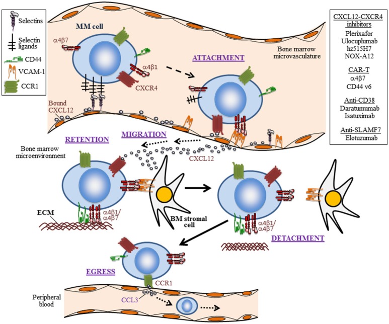
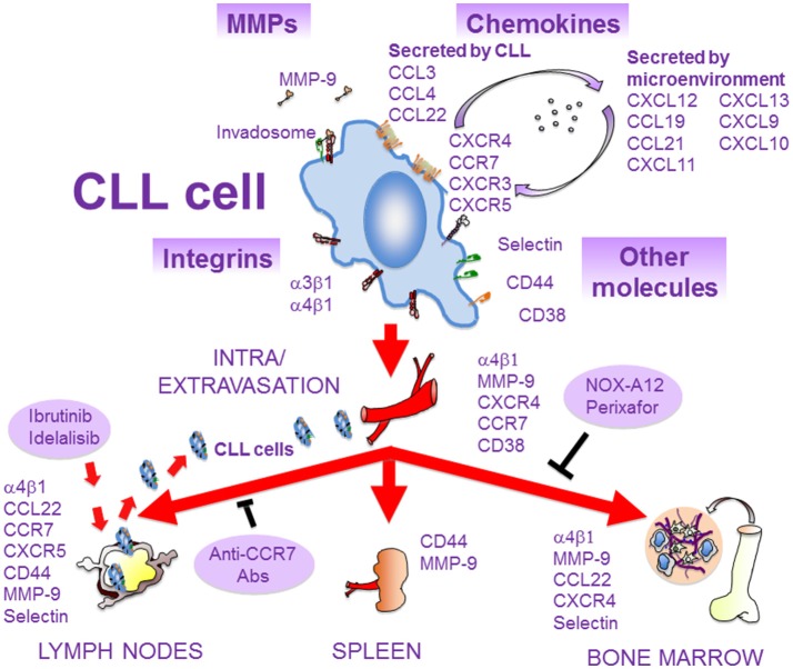
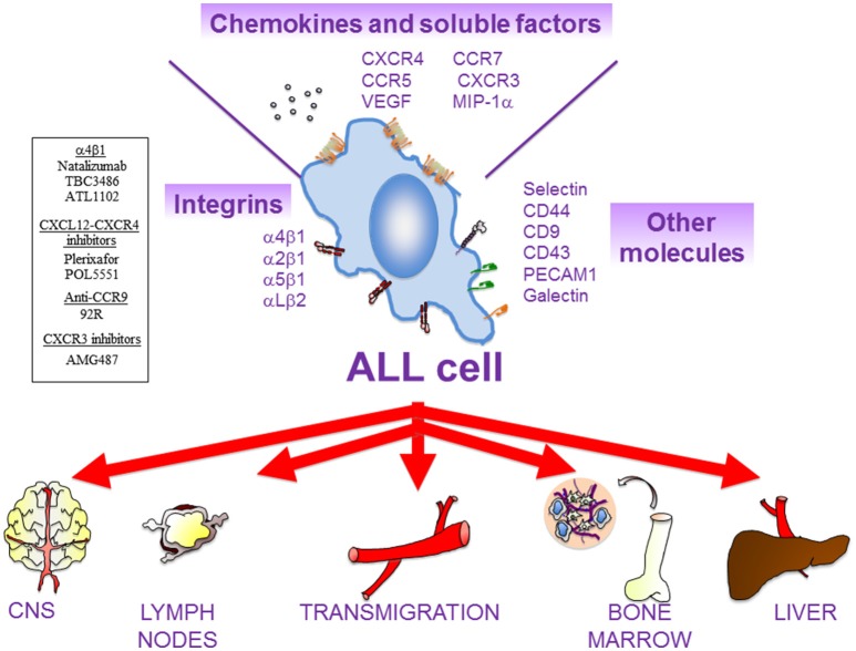
The trafficking of neoplastic cells represents a key process that contributes to progression of hematologic malignancies. Diapedesis of neoplastic cells across endothelium and perivascular cells is facilitated by adhesion molecules and chemokines, which act in concert to tightly regulate directional motility. Intravital microscopy provides spatio-temporal views of neoplastic cell trafficking, and is crucial for testing and developing therapies against hematologic cancers. Multiple myeloma (MM), chronic lymphocytic leukemia (CLL), and acute lymphoblastic leukemia (ALL) are hematologic malignancies characterized by continuous neoplastic cell trafficking during disease progression. A common feature of these neoplasias is the homing and infiltration of blood cancer cells into the bone marrow (BM), which favors growth and survival of the malignant cells. MM cells traffic between different BM niches and egress from BM at late disease stages. Besides the BM, CLL cells commonly home to lymph nodes (LNs) and spleen. Likewise, ALL cells also infiltrate extramedullary organs, such as the central nervous system, spleen, liver, and testicles. The α4β1 integrin and the chemokine receptor CXCR4 are key molecules for MM, ALL, and CLL cell trafficking into and out of the BM. In addition, the chemokine receptor CCR7 controls CLL cell homing to LNs, and CXCR4, CCR7, and CXCR3 contribute to ALL cell migration across endothelia and the blood brain barrier. Some of these receptors are used as diagnostic markers for relapse and survival in ALL patients, and their level of expression allows clinicians to choose the appropriate treatments. In CLL, elevated α4β1 expression is an established adverse prognostic marker, reinforcing its role in the disease expansion. Combining current chemotherapies with inhibitors of malignant cell trafficking could represent a useful therapy against these neoplasias. Moreover, immunotherapy using humanized antibodies, CAR-T cells, or immune check-point inhibitors together with agents targeting the migration of tumor cells could also restrict their survival. In this review, we provide a view of the molecular players that regulate the trafficking of neoplastic cells during development and progression of MM, CLL, and ALL, together with current therapies that target the malignant cells.

Keywords: adhesion molecule; cell trafficking; chemokines (CK); hematological cancer; immunotherapy.


Figures

Figure 1
Figure 1
The trafficking life of MM cells. Depicted are five steps (attachment, migration, retention, detachment, and egress) in the trafficking of myeloma cells into the BM and their escape to the periphery, as well as some of the involved trafficking receptors and their ligands. The initial attachment of MM cells to the BM microvasculature is controlled by selectins, the CXCL12-CXCR4 axis, and the α4β1 integrin interaction with VCAM-1. Importantly, CXCL12-CXCR4 leads to upregulation of α4β1-dependent MM cell adhesion. The migration and retention of MM cells in the BM is contributed by the CXCL12-CXCR4 chemoattraction module, by adhesion mediated by the integrins α4β1 and α4β7, as well as by CD44. Ligands for these adhesion receptors are components of the extracellular matrix (ECM), such as fibronectin and hialuronic acid. Weakening or disrupting these adhesive interactions causes MM cell detachment from the BM microenvironment and egress to peripheral blood. The homing of MM cells to the BM can be inhibited by the CXCL12 inhibitor NOX-A12, and neutralizing CXCL12 binding to CXCR4 with the plerixafor blocks MM cell interaction with the BM microenvironment. A putative active cell egress mechanism is proposed as depicted by the CCL3-CCR1 interaction. Not shown is the trafficking of circulating MM cells to extramedullary colonization sites. In addition to therapies targeting the CXCL12-CXCR4 axis, the use of CAR T cells addressing α4β7 and CD44, and humanized monoclonal antibodies against CD38 and SLAMF7, which are currently being tested in MM, are shown.
Figure 2
Figure 2
Molecules involved in the migration of CLL cells. As CLL progresses, malignant cells extravasate and infiltrate the BM and secondary lymphoid organs. This process is mediated by several molecules, expressed and/or secreted by CLL cells or by the microenvironment of lymphoid tissues. Some of these molecules interact physically and/or functionally and may constitute the CLL cell invadosome. The α4β1 integrin is critical for CLL cell homing to BM and LNs, and blocking BCR signaling with ibrutinib or idelalisib inhibits α4β1 function and results in lymphocytosis. The chemokine receptors CCR7 and CXCR4 are crucial for CLL cell traffic to LNs and BM, respectively. These receptors can be blocked by specific antibodies or inhibitors (NOX-A12, perixafor). MMP-9, CD44, and CD38 are also important for CLL cell migration and localization in lymphoid niches. Adhesive interactions with stromal cells in these niches, mediated by some of the depicted molecules, favor survival and chemoresistance of CLL cells and contribute to disease progression.
Figure 3
Figure 3
Main actors of ALL cell migration. There are multiple cell surface receptors and soluble molecules that drive ALL cell movement. ALL cells might remain in the BM or colonize multiple extramedullary organs, such as central nervous system (CNS), lymph nodes (LNs), liver, and testes. In general, β1 integrins (α4β1, α2β1, α5β1) and the integrin αLβ2 are critical for the homing of ALL cells into the BM and for their infiltration in most of the extramedullary organs. The chemokine receptor CXCR4 mediates ALL cell infiltration into BM, liver, lung, and CNS, whilst CCR7 controls ALL infiltration into CNS and LNs. CXCR3 is also critical for CNS infiltration. Other molecules that might contribute to ALL migration includes MIP-1α, VEGF, PECAM-1, VE-Cadherin (for CNS infiltration); IL-7 (for LNs); and CD44 (for BM). Several compounds targeting molecular players in ALL cell migration that are currently being tested in preclinical trials are shown on the left.

References

    1. Anderson KC, Carrasco RD. Pathogenesis of myeloma. Annu Rev Pathol. (2011) 6:249–74. 10.1146/annurev-pathol-011110-130249 - DOI - PubMed
    1. Kuehl WM, Bergsagel PL. Molecular pathogenesis of multiple myeloma and its premalignant precursor. J Clin Invest. (2012) 122:3456–63. 10.1172/JCI61188 - DOI - PMC - PubMed
    1. Bianchi G, Munshi NC. Pathogenesis beyond the cancer clone(s) in multiple myeloma. Blood (2015) 125:3049–58. 10.1182/blood-2014-11-568881 - DOI - PMC - PubMed
    1. van Nieuwenhuijzen N, Spaan I, Raymakers R, Peperzak V. From MGUS to multiple myeloma, a paradigm for clonal evolution of premalignant cells. Cancer Res. (2018) 78:2449–56. 10.1158/0008-5472.can-17-3115 - DOI - PubMed
    1. Pawlyn C, Morgan GJ. Evolutionary biology of high-risk multiple myeloma. Nat Rev Cancer (2017) 17:543–56. 10.1038/nrc.2017.63 - DOI - PubMed

Publication types