. 2019 Jan 15;11(1):31-44.
eCollection 2019.

lncRNA GHET1 knockdown suppresses breast cancer activity in vitro and in vivo

Affiliations

lncRNA GHET1 knockdown suppresses breast cancer activity in vitro and in vivo

Mingli Han et al. Am J Transl Res. .

Abstract

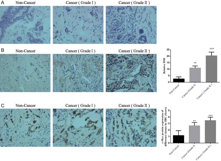
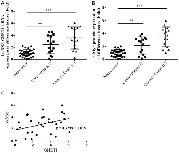
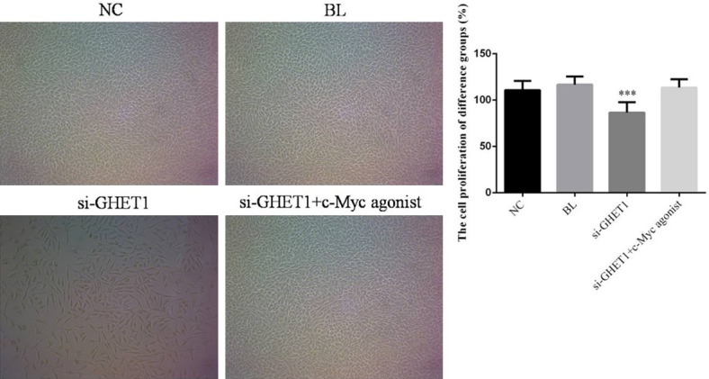
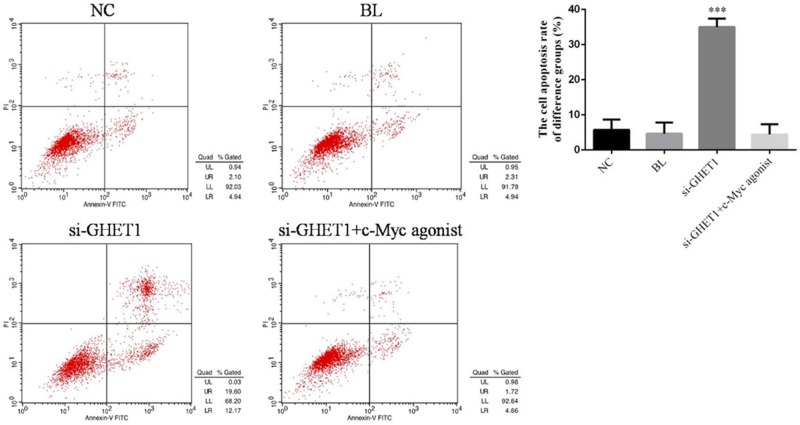
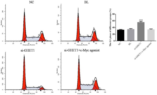
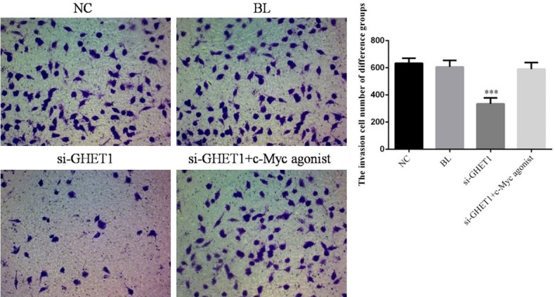
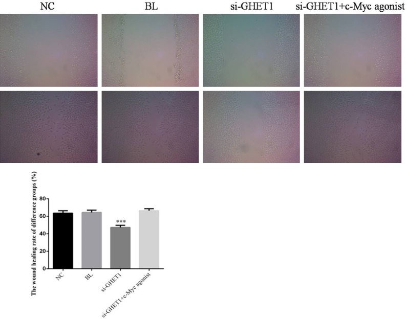
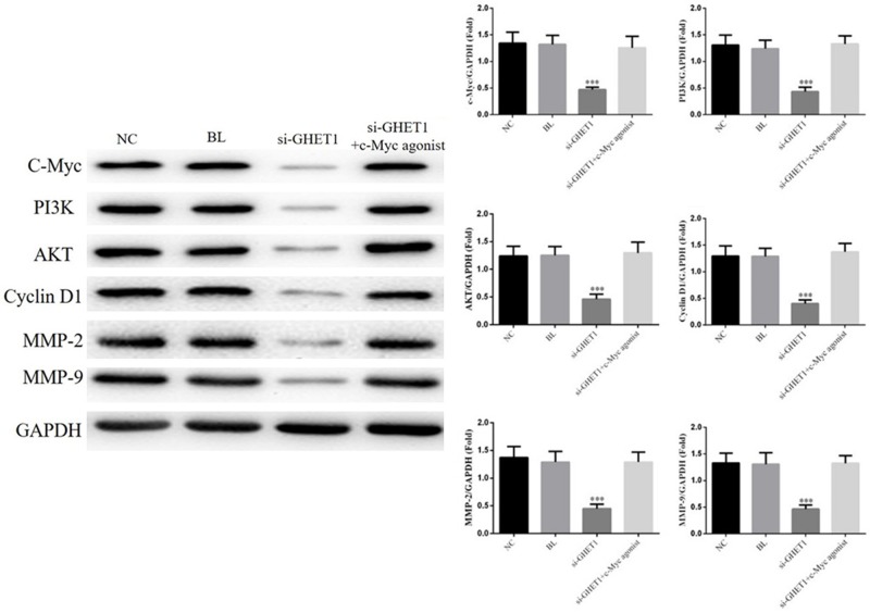
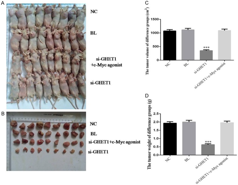
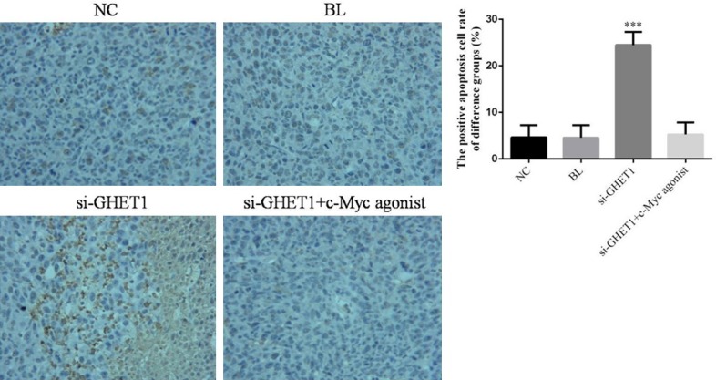
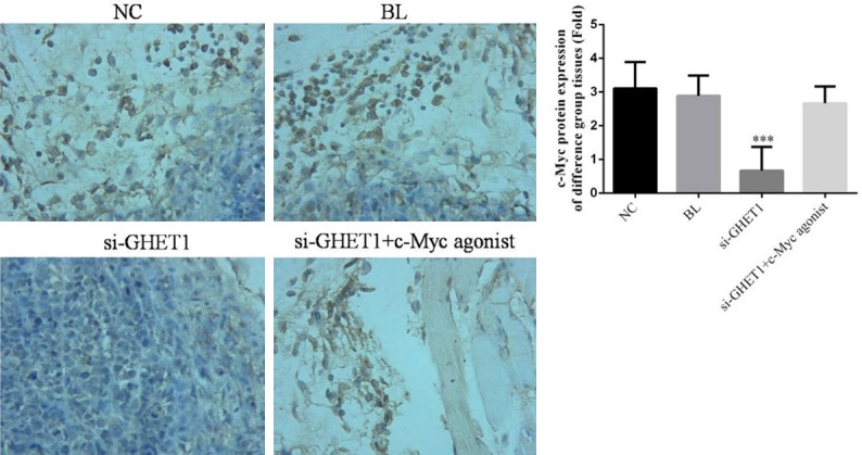
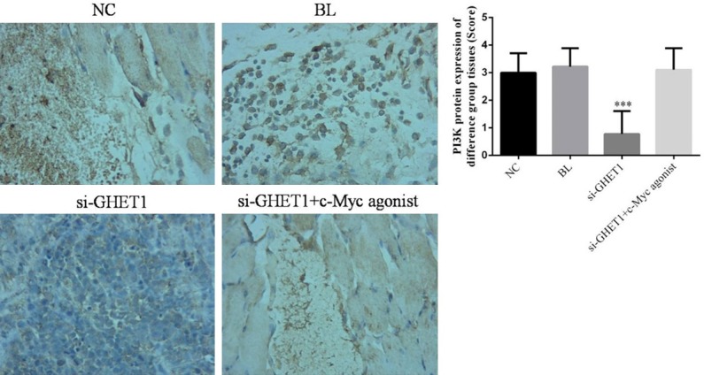
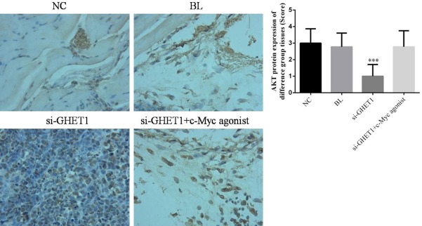
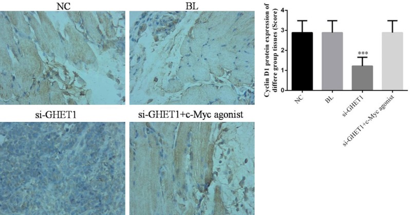
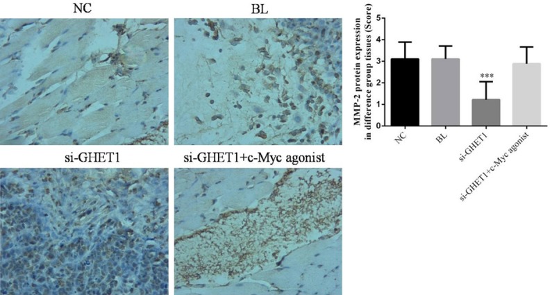
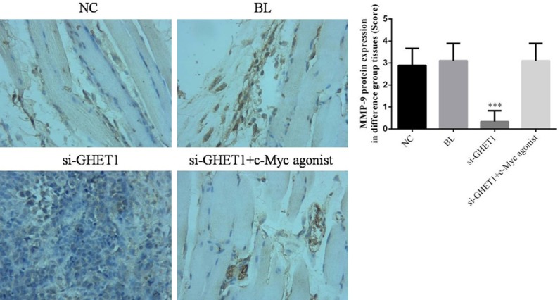
Long non-coding RNA gastric carcinoma high-expressed transcript 1 (lncRNA GHET1) is highly expressed in many tumors. The aim of the present study was to determine whether GHET1 inhibition decreases growth and metastasis of MCF-7 breast cancer cells by modulating epidermal growth factor receptor (EGFR) expression. In vitro, lncRNA GHET1 knockdown suppressed cell proliferation, migration, and invasion and enhanced cell apoptosis by maintaining MCF-7 cells in the G1 phase of the cell cycle. Furthermore, lncRNA GHET1 knockdown reduced the expression of EGFR and related proteins. Treatment of mice with a GHET1 inhibitor prevented tumor growth in vivo. The results indicate that lncRNA GHET1 inhibition directly suppresses EGFR expression, significantly inhibiting the downstream PI3K/AKT/Cyclin D1/MMP2/9 pathway. This mechanism may underlie the suppression of breast cancer cell activities including proliferation, migration, and invasion. In conclusion, lncRNA GHET1 knockdown suppresses tumor growth and metastasis by suppressing the activity of EGFR and downstream pathways.

Keywords: EGFR; GHET1; MCF-7; apoptosis; invasion; migration; proliferation.


Conflict of interest statement

None.

Figures

Figure 1
Figure 1
Tumor characteristics. A. Hematoxylin and eosin staining of non-cancer and Grade I and II breast cancer tissues (× 200). B. GHET1 expression in non-cancer and Grade I and II cancer tissues analyzed with ISH (× 200). **P < 0.05 and ***P < 0.001 vs. NC. C. c-Myc expression in non-cancer and Grade I and II cancer tissues analyzed with ISH (× 200). **P < 0.05 and ***P < 0.001 vs. NC.
Figure 2
Figure 2
Correlation between c-Myc and GHET1 expression. A. GHET1 expression in non-cancer and Grade I and II cancer tissues analyzed with RT-PCR. **P < 0.05 and ***P < 0.001 vs. NC. B. c-Myc expression in non-cancer and Grade I and II cancer tissues analyzed with RT-PCR. C. Correlation between GHET1 and c-Myc in cancer tissues (r = 0.325).
Figure 3
Figure 3
Cell proliferation rate in different groups analyzed with MTT assays. ***P < 0.001 vs. NC.
Figure 4
Figure 4
Apoptosis rates in different treatment groups. Flow cytometry was performed to measure cell death in control, cells transfected with empty vector (BL), and cells treated with shRNA against (si-GHET1) with or without c-Myc agonist. ***P < 0.001 vs. NC.
Figure 5
Figure 5
Cell cycle analyzed with flow cytometry in control, cells transfected with empty vector (BL), and cells treated with shRNA against (si-GHET1) with or without c-Myc agonist. ***P < 0.001 vs. NC.
Figure 6
Figure 6
Cell invasion analyzed with Transwell assays in control, cells transfected with empty vector (BL), and cells treated with shRNA against (si-GHET1) with or without c-Myc agonist. ***P < 0.001 vs. NC.
Figure 7
Figure 7
Wound healing rate in control, cells transfected with empty vector (BL), and cells treated with shRNA against (si-GHET1) with or without c-Myc agonist. ***P < 0.001 vs. NC.
Figure 8
Figure 8
Relative protein expressions analyzed with WB assays in control, cells transfected with empty vector (BL), and cells treated with shRNA against (si-GHET1) with or without c-Myc agonist. ***P < 0.001 vs. NC.
Figure 9
Figure 9
Tumor characteristics in each group. The NC, BL, si-GHET1, and si-GHET1+c-Myc groups were injected with normal saline, empty vector, si-GHET1, and si-GHET1+c-Myc agonist, respectively. ***P < 0.001 vs. NC. A. Tumor tissues in each group. B. Resected tumor tissues in each group. C. Analysis of tumor volumes. ***P < 0.001 vs. NC. D. Analysis of tumor weights. ***P < 0.001 vs. NC group.
Figure 10
Figure 10
Apoptosis analyzed with TUNEL assays (× 200) in control, cells transfected with empty vector (BL), and cells treated with shRNA against (si-GHET1) with or without c-Myc agonist. ***P < 0.001 vs. NC.
Figure 11
Figure 11
c-Myc expression analyzed with IHC (× 200) in control, cells transfected with empty vector (BL), and cells treated with shRNA against (si-GHET1) with or without c-Myc agonist. ***P < 0.001 vs. NC.
Figure 12
Figure 12
PI3K expression analyzed with IHC (× 200) in control, cells transfected with empty vector (BL), and cells treated with shRNA against (si-GHET1) with or without c-Myc agonist. ***P < 0.001 vs. NC.
Figure 13
Figure 13
AKT expression analyzed with IHC (× 200) in control, cells transfected with empty vector (BL), and cells treated with shRNA against (si-GHET1) with or without c-Myc agonist. ***P < 0.001 vs. NC.
Figure 14
Figure 14
Cyclin D1 expression analyzed with IHC (× 200) in control, cells transfected with empty vector (BL), and cells treated with shRNA against (si-GHET1) with or without c-Myc agonist. ***P < 0.001 vs. NC.
Figure 15
Figure 15
MMP-2 expression analyzed with IHC (× 200) in control, cells transfected with empty vector (BL), and cells treated with shRNA against (si-GHET1) with or without c-Myc agonist. ***P < 0.001 vs. NC.
Figure 16
Figure 16
MMP-9 expression analyzed with IHC (× 200) in control, cells transfected with empty vector (BL), and cells treated with shRNA against (si-GHET1) with or without c-Myc agonist. ***P < 0.001 vs. NC.

Similar articles

Cited by

References

    1. Santa-Maria CA, Gradishar WJ. Changing treatment paradigms in metastatic breast cancer: lessons learned. JAMA Oncol. 2015;1:528–534. - PubMed
    1. Fountzilas G, Dafni U, Papadimitriou C, Timotheadou E, Gogas H, Eleftheraki AG, Xanthakis I, Christodoulou C, Koutras A, Papandreou CN, Papakostas P, Miliaras S, Markopoulos C, Dimitrakakis C, Korantzopoulos P, Karanikiotis C, Bafaloukos D, Kosmidis P, Samantas E, Varthalitis I, Pavlidis N, Pectasides D, Dimopoulos MA. Dose-dense sequential adjuvant chemotherapy followed, as indicated, by trastuzumab for one year in patients with early breast cancer: first report at 5-year median follow-up of a Hellenic cooperative oncology group randomized phase III trial. BMC Cancer. 2014;14:515. - PMC - PubMed
    1. Liu Q, Huang J, Zhou N, Zhang Z, Zhang A, Lu Z, Wu F, Mo YY. LncRNA loc285194 is a p53-regulated tumor suppressor. Nucleic Acids Res. 2013;41:4976–4987. - PMC - PubMed
    1. Kallen AN, Zhou XB, Xu J, Qiao C, Ma J, Yan L, Lu L, Liu C, Yi JS, Zhang H, Min W, Bennett AM, Gregory RI, Ding Y, Huang Y. The imprinted H19 lncRNA antagonizes let-7 microRNA. Mol Cell. 2013;52:101–12. - PMC - PubMed
    1. Yang F, Xue X, Zheng L, Bi J, Zhou Y, Zhi K, Gu Y, Fang G. Long non-coding RNA GHET1 promotes gastric carcinoma cell proliferation by increasing c-Myc mRNA stability. FEBS J. 2014;281:802–813. - PubMed

LinkOut - more resources