. 2019 Jan 15;11(1):314-326.
eCollection 2019.

Downregulation of angiopoietin-like protein 2 inhibits cementoblast differentiation partially by activating the ERK1/2 signaling pathway

Affiliations

Downregulation of angiopoietin-like protein 2 inhibits cementoblast differentiation partially by activating the ERK1/2 signaling pathway

Chenxi Jiang et al. Am J Transl Res. .

Abstract

Angiopoietin-like protein 2 (ANGPTL2) is abundantly expressed in adipose tissue, is associated with tissue homeostasis, and promotes osteoblast and chondrocyte differentiation. In teeth, cementum, a thin layer of mineralized tissue that is formed by cementoblasts, covers the entire root surface and is a vital component of periodontium. The cementoblasts regulate the deposition and mineralization of the cementum matrix. However, the effects of ANGPTL2 on cementoblast differentiation have not been studied. The objective of this study was to elucidate the role of ANGPTL2 during cementoblast differentiation and determine its underlying mechanisms. Our results showed that the expression levels of ANGPTL2 gradually increased during cementoblast differentiation. After ANGPTL2 was knocked down using short-hairpin RNA, the levels of the osteogenic markers osterix (OSX), alkaline phosphatase (ALP), bone sialoprotein (BSP), and osteocalcin (OCN) decreased. In addition, ALP activity and the number of calcified nodules were dramatically reduced compared with those in the negative control. Interestingly, the ERK1/2 signaling pathway was activated after ANGPTL2 knockdown. Treatment with PD98059, the inhibitor of the ERK1/2 signaling pathway, partially rescued the decreased differentiation capability of cementoblast caused by ANGPTL2 downregulation. Collectively, ANGPTL2 knockdown inhibited cementoblast differentiation partially by activating the ERK1/2 signaling pathway. These findings suggest that ANGPTL2 was indispensable in cementoblast differentiation.

Keywords: ANGPTL2; ERK1/2 signaling pathway; cementoblast differentiation; cementogenesis; dental cementum; periodontal reconstruction.


Conflict of interest statement

None.

Figures
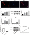
Figure 1
Figure 1
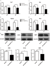
Immunofluorescence staining for ANGPTL2 expression and lentivirus infection of OCCM-30 cells. The effect of ANGPTL2 downregulation on cell proliferation. A. Immunofluorescence assay demonstrated that ANGPTL2 was located in the cytoplasm of OCCM-30 cells. B, C. The mRNA and protein levels of ANGPTL2 were examined by qRT-PCR and Western blotting on days 0, 4, 7, and 11. The expression level of ANGPTL2 gradually increased during osteogenic differentiation of OCCM-30 cells. The 0 day group was set as the control. *P < 0.05, **P < 0.01 (one-way ANOVA with post hoc Dunnett test). D. mRNA levels of osteogenic markers Bsp and Bglap were examined by qRT-PCR on days 0, 4, 7 and 11 during osteogenic differentiation of OCCM-30 cells. ***P < 0.001 (one-way ANOVA with post hoc Dunnett test). E, F. After infection of the cells with lentivirus and selection with puromycin (2 μg/mL) for 7 days, the mRNA and protein levels of ANGPTL2 significantly decreased in the SH-ANGPTL2 group compared with SH-NC group. **P < 0.01 (Student’s t-test). G. Knockdown of ANGPTL2 inhibited cell proliferation as seen using the CCK-8 assay. **P < 0.01 vs SH-NC (Student’s t-test). All data were based on three independent experiments. Values are presented as the mean ± SD.
Figure 2
Figure 2
ANGPL2 knockdown suppressed cementoblast differentiation. A-D. mRNA levels of Sp7, Alp, Bglap, and Bsp were examined by qRT-PCR on days 4 and 7, and the expression levels in the knockdown group were lower than those in the SH-NC group during osteogenic differentiation of OCCM-30 cells. *P < 0.05, **P < 0.01 vs SH-NC (Student’s t-test). E. The protein levels of osterix (OSX), bone sialoprotein (BSP), and osteocalcin (OCN) were detected by Western blotting on day 7 during osteogenic differentiation of OCCM-30 cells. F. Histograms representing the quantification of OSX, OCN, and BSP protein levels were obtained using Image J. *P < 0.05, **P < 0.01 vs SH-NC (Student’s t-test). All data were based on at least three independent experiments. Values are presented as the mean ± SD.
Figure 3
Figure 3
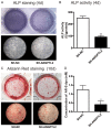
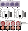
ANGPL2 knockdown suppressed ALP activity, ALP staining, and mineralized nodules. A. ALP staining was conducted on day 4 during osteogenic differentiation of OCCM-30 cells. The cells were stained with nitro-blue tetrazolium and 5-bromo-4-chloro-3’-indolyphosphate (NBT/BCIP) for 15 min and photographed. Additionally, the attenuated ALP staining intensity was observed in the knockdown group compared to that in the SH-NC group. B. A kit for ALP activity assay was used and the data were normalized to total protein level. Downregulation of ANGPTL2 had an evident inhibitory effect on the ALP activity of OCCM-30 cells. **P < 0.01 vs SH-NC (Student’s t-test). C. Alizarin Red staining of the OCCM-30 cells was detected on day 10. There were fewer calcified nodules in the knockdown group than in the SH-NC group. D. Mineralized nodules were desorbed with 10% cetylpyridinium chloride, and the absorbance of the solution was read at 562 nm. *P < 0.05 vs SH-NC (Student’s t-test). Values are presented as the mean ± SD.
Figure 4
Figure 4
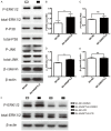
Activation of the MAPK signaling pathway by ANGPTL2 downregulation. A. Four different signaling pathways were detected by Western blotting-ERK1/2, P38, JNK, and β-catenin after ANGPTL2 inhibition. B-E. Histograms representing quantification of (phospho-ERK1/2)/(total ERK1/2), (phospho-P38)/(total P38), (phospho-JNK)/(total JNK), and (β-catenin)/(β-actin) ratios obtained by Image J. The results show that phospho-ERK1/2 was significantly enhanced after ANGPTL2 silencing. *P < 0.01 vs SH-NC (Student’s t-test). F. The protein levels of (P-ERK1/2)/(total ERK1/2) significantly decreased after the cells were treated with PD98059 (10 μM) in dimethyl sulfoxide (DMSO) for 6 h compared with those treated with DMSO alone. Values are presented as the mean ± SD.
Figure 5
Figure 5
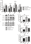
Decreased differentiation capability of cementoblast could be partially rescued by inhibition of ERK1/2 signaling pathway. A. The mRNA expression levels of Alp, Sp7, Bglap, and Bsp were measured by qRT-PCR in SH-NC and SH-ANGPTL2 cementoblast after PD98059 or DMSO treatment on day 4. B, C. The protein levels of OSX, OCN, and BSP were examined by Western blotting on day 7 in SH-NC and SH-ANGPTL2 cementoblast after PD98059 or DMSO treatment. The results demonstrated that the repressed levels of osteogenic markers were partially rescued in the PD98059-treated SH-ANGPTL2 group as compared with those in the DMSO-treated SH-ANGPTL2 group. D-F. Histograms representing the quantification of OSX, OCN, and BSP protein levels were obtained from Image J. All data are based on at least three independent experiments. Values are presented as the mean ± SD. *P < 0.05, **P < 0.01, ***P < 0.001 vs DMSO-treated SH-ANGPTL2 (one-way ANOVA with post hoc Tukey test).
Figure 6
Figure 6
Decreased ALP activity and mineralized nodules could be partially rescued by inhibiting the ERK1/2 signaling pathway. A. SH-NC and SH-ANGPTL2 cementoblast were stained with ALP after PD98059 or DMSO treatment on day 4. The cells were stained with NBT/BCIP for 15 min and photographed. B. ALP activity was measured in SH-NC and SH-ANGPTL2 cementoblast after PD98059 or DMSO treatment on day 4. The results were normalized to total protein levels. The ALP activity increased after inhibiting the ERK1/2 signaling pathway using PD98059. C. Four groups were stained with Alizarin Red on day 10, and there were more calcified nodules in the PD98059-treated ANGPTL2-SH group than in the DMSO-treated ANGPTL2-SH group. D. Mineralized nodules in the four groups were desorbed with 10% cetylpyridinium chloride, respectively, and absorbance of the solution was read at 562 nm. All data were based on at least three independent experiments. Values are presented as the mean ± SD. ***P < 0.001 vs DMSO-treated SH-ANGPTL2 (one-way ANOVA with post hoc Tukey test).

Similar articles

References

    1. Kassebaum NJ, Bernabe E, Dahiya M, Bhandari B, Murray CJ, Marcenes W. Global burden of severe periodontitis in 1990-2010: a systematic review and meta-regression. J Dent Res. 2014;93:1045–1053. - PMC - PubMed
    1. Yang H, Xiao L, Zhang L, Deepal S, Ye G, Zhang X. Epidemic trend of periodontal disease in elderly Chinese population, 1987-2015: a systematic review and meta-analysis. Sci Rep. 2017;7:45000. - PMC - PubMed
    1. Pihlstrom BL, Michalowicz BS, Johnson NW. Periodontal diseases. Lancet. 2005;366:1809–1820. - PubMed
    1. Bosshardt DD, Selvig KA. Dental cementum: the dynamic tissue covering of the root. Periodontol 2000. 1997;13:41–75. - PubMed
    1. Zeichner-David M. Regeneration of periodontal tissues: cementogenesis revisited. Periodontol 2000. 2006;41:196–217. - PubMed

LinkOut - more resources