. 2019 Jan 15;11(1):442-452.
eCollection 2019.

High CCL7 expression is associated with migration, invasion and bone metastasis of non-small cell lung cancer cells

Affiliations

High CCL7 expression is associated with migration, invasion and bone metastasis of non-small cell lung cancer cells

Shuai Han et al. Am J Transl Res. .

Abstract

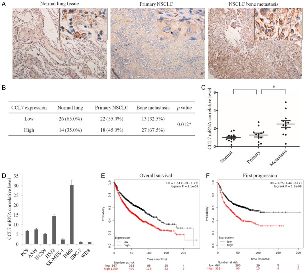
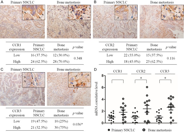
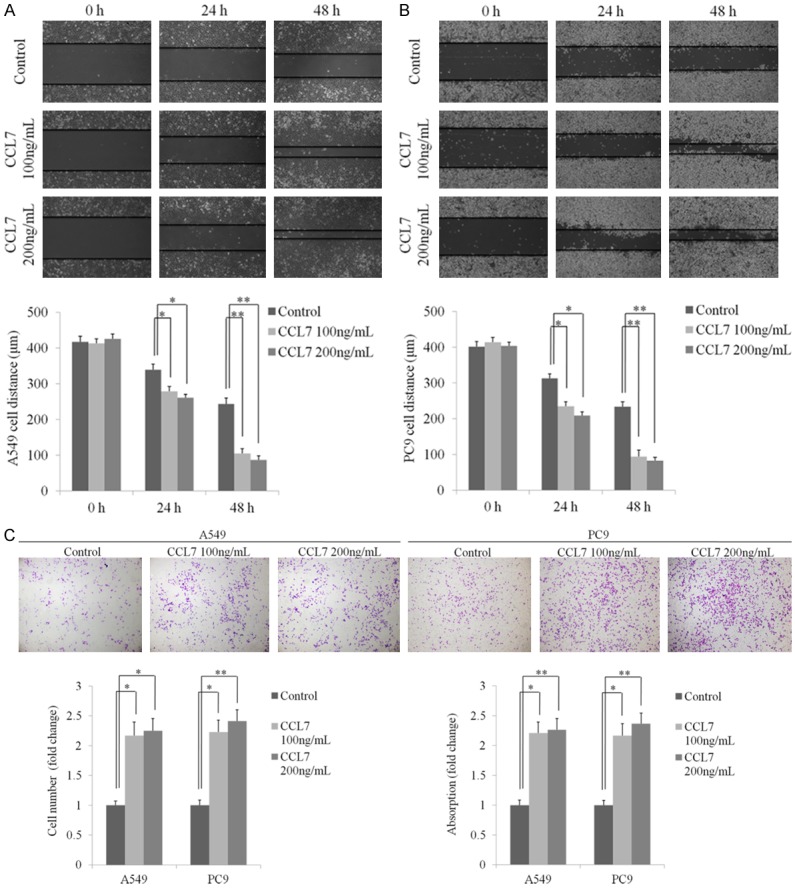
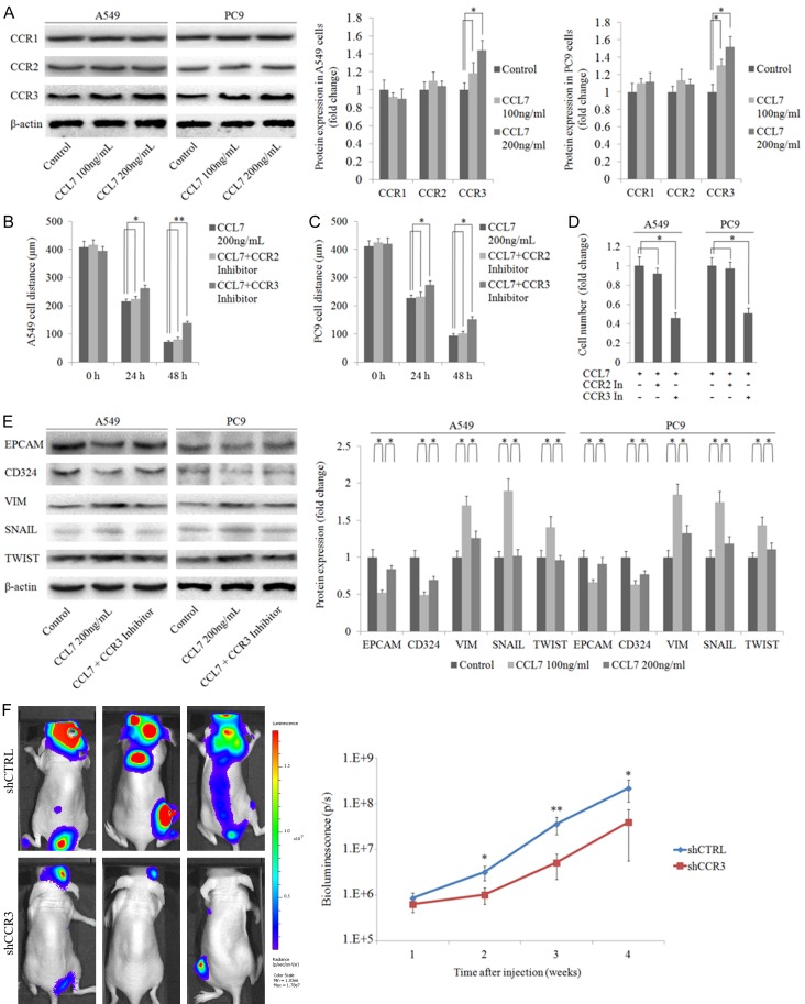
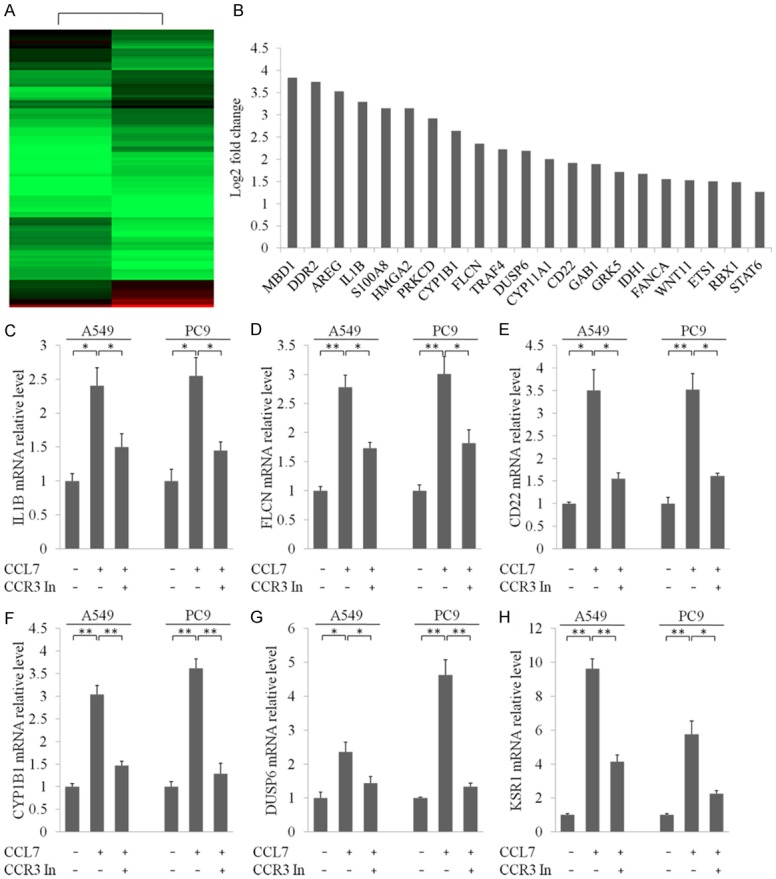
Distant metastasis is the main cause of death for non-small cell lung cancer (NSCLC) patients, with the bone as a more frequent metastatic site. The expression of various chemokines and their receptors may contribute to the predilection of organ specific metastasis. In this study, we demonstrated that CC chemokine ligand 7 (CCL7) and its receptors CCR1, CCR2 and CCR3 were up-regulated markedly in lung cancer bone metastasis. In addition, CCL7 promoted migration and invasion of lung cancer cells in a dose-dependent pattern but played an insignificant role in cell proliferation mainly through CCR3. Finally, we identified a cohort of critical downstream genes of CCL7 related to cancer metastasis using Illumina deep mRNA sequencing. These findings may help better understand the molecular aspects of bone metastasis in lung cancer, and prefigure a potential clinical value for the prevention of bone recurrences.

Keywords: CCL7; bone metastasis; invasion; migration; non-small cell lung cancer (NSCLC).


Conflict of interest statement

None.

Figures

Figure 1
Figure 1
CCL7 expression is negatively correlated with the clinical outcome of non-small cell lung cancer (NSCLC) patients. A. Immunohistochemical detection of CCL7 expression in NSCLC bone metastasis, primary NSCLC and normal lung tissues (n = 40). B. Statistics of all samples associated with CCL7 staining. C. qRT-PCR assay of mRNA level of CCL7 in NSCLC bone metastasis, primary NSCLC and normal lung tissues. D. qRT-PCR assay of mRNA level of CCL7 in various lung cancer cell lines. E, F. High CCL7 mRNA expression is positively correlated with poor overall survival (OS) and progression-free survival (FPS) in NSCLC patients. *: P < 0.05.
Figure 2
Figure 2
Expression of possible target chemokine receptors of CCL7 in primary NSCLC and bone metastasis tissues. A-C. IHC detection of CCR1, CCR2 and CCR3 expression in primary NSCLC and bone metastasis tissues. CCR1, CCR2 and CCR3 were mainly expressed on the cell membrane and in cytoplasm (arrow). D. qRT-PCR measurement of mRNA levels of CCR1, CCR2, CCR3 in primary NSCLC and bone metastasis tissues. *: P < 0.05.
Figure 3
Figure 3
CCL7 promotes migration and invasion of NSCLC cells. A, B. Representative images and statistical analysis of wound healing assay of A549 and PC9 cells incubated with recombinant CCL7 (100 ng/ml, 200 ng/ml) at 24 and 48 h. C. Pictures and bar graphs of transwell invasion assay showing increased invasion of A549 and PC9 cells after treatment with recombinant CCL7 (100 ng/ml, 200 ng/ml). *: P < 0.05; **: P < 0.001.
Figure 4
Figure 4
CCL7 induces EMT in A549 and PC9 cells via interacting with CCR3. A. Protein expression of CCR1, CCR2 and CCR3 in A549 and PC9 cells with different doses of recombinant CCL7 (100 ng/ml, 200 ng/ml); the result showed that CCL7 had no effect on the expression of CCR. B-D. Wound healing assay and transwell invasion assay of A549 and PC9 cells after co-incubation with recombinant CCL7 and CCR inhibitors respectively; interestingly, inhibition of CCR3 significantly suppressed cell invasion. E. CCL7 increased the expression of mesenchymal markers and decreased the expression of epithelial markers in A549 and PC9 cells via CCR3. F. Bioluminescence imaging showed that CCR3 knockdown inhibited A549 cell metastasis in vivo. *: P < 0.05; **: P < 0.001.
Figure 5
Figure 5
RNA-seq profiling of PC9 cells after incubation with recombinant CCL7. A. Heatmap differentially expressed genes in PC9 cells treated with recombinant CCL7 and negative control. B. List of genes that were involved in multi organ metastasis and underwent significant alterations after treatment with recombinant CCL7. C-H. qRT-PCR measurement to confirm the effect of CCL7 with or without co-incubation with CCR3 inhibitor. *: P < 0.05; **: P < 0.001.

References

    1. Siegel RL, Miller KD, Jemal A. Cancer statistics, 2015. CA Cancer J Clin. 2015;65:5–29. - PubMed
    1. Howlader N, Noone AM, Yu M, Cronin KA. Use of imputed population-based cancer registry data as a method of accounting for missing information: application to estrogen receptor status for breast cancer. Am J Epidemiol. 2012;176:347–356. - PMC - PubMed
    1. Hoffman PC, Mauer AM, Vokes EE. Lung cancer. Lancet. 2000;355:479–485. - PubMed
    1. DeSantis CE, Lin CC, Mariotto AB, Siegel RL, Stein KD, Kramer JL. Cancer treatment and survivorship statistics, 2014. CA Cancer J Clin. 2014;64:252–271. - PubMed
    1. Popper HH. Progression and metastasis of lung cancer. Cancer Metastasis Rev. 2016;35:75–91. - PMC - PubMed

LinkOut - more resources