. 2019 Jan 26:17:151-159.
doi: 10.1016/j.csbj.2019.01.004. eCollection 2019.

Divaricoside Exerts Antitumor Effects, in Part, by Modulating Mcl-1 in Human Oral Squamous Cell Carcinoma Cells

Affiliations

Divaricoside Exerts Antitumor Effects, in Part, by Modulating Mcl-1 in Human Oral Squamous Cell Carcinoma Cells

Jing-Ru Weng et al. Comput Struct Biotechnol J. .

Abstract

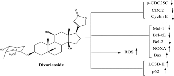
Cardiac glycosides (CGs), prescribed to treat congestive heart failure and arrhythmias, exert potent antitumor activity. In this study, divaricoside (DIV), a CG isolated from Strophanthus divaricatus was examined for its antitumor potency in oral squamous cell carcinoma (OSCC) cells. Cell growth was inhibited by DIV in a dose- and time-dependent manner in SCC2095 and OECM-1 OSCC cells using MTT assays. DIV induced S and G2/M phase arrest accompanied by downregulation of phosphorylated CDC25C, CDC25C, and CDC2 in SCC2095 cells. In addition, DIV induced apoptosis by activating caspase-3 and downregulating the expression of Mcl-1. Furthermore, overexpression of Mcl-1 partially reversed DIV-induced death in SCC2095 cells. Additionally, western blot and transmission electron microscopy analyses also indicated that DIV induced autophagy in SCC2095 cells. However, the combination of autophagy inhibitor did not affect DIV-mediated apoptosis in SCC2095 cells. Together, these findings suggest that translational potential of DIV to be developed as a therapeutic agent for OSCC treatment.

Keywords: Apoptosis; Autophagy; Cardiac glycoside; Divaricoside; Mcl-1; Oral squamous cell carcinoma.


Figures

Unlabelled Image
Graphical abstract
Fig. 1
Fig. 1
Effect of divaricoside (DIV) on the viability of oral cancer cells (SCC2095 and OECM-1) and DOK cells. (A) The chemical structure of DIV. (B) SCC2095, (C) OECM-1, and (D) DOK cells. Cells were seeded into 96-well plates, after 24 h of incubation, cells were treated with DIV for 24 h or 48 h, and cell viability was detected by MTT assays. Points represent means; bars represent S.D. (n = 3–6). *P < 0.05, **P < 0.01 compared to the control group. (E) SCC2095 cells were treated with DIV and detected for the protein expression by Western blot analysis.
Fig. 2
Fig. 2
Effect of divaricoside (DIV) on cell cycle and cell cycle-regulating proteins. (A) Cell cycle analysis showed an increase in S and G2/M phase population after treatment with DIV at the indicated concentrations for 48 h in SCC2095 cells, followed by propidium iodide (PI) staining. Three independent experiments were performed; and data are presented in (B) as mean ± S.D. (C) Western blot analysis of DIV-treated SCC2095 cells indicating the phosphorylation and expression of cyclin E, CDC25C, and CDC2. Cells were treated with DIV in 5% FBS-supplemented DMEM/F12 medium for 48 h, and cell lysates were immunoblotted as described in the Materials and Methods section. The values in percentage denote the relative intensity of protein bands of drug-treated samples to that of the respective DMSO vehicle-treated control after being normalized to the respective internal reference (total respective protein or β-actin).
Fig. 3
Fig. 3
Effect of divaricoside (DIV) treatment on apoptosis. (A) SCC2095 cells were treated with DMSO or DIV in 5% FBS-supplemented DMEM/F12 medium for 48 h and stained with propidium iodide (PI)/annexin V. (B) The percentage of apoptotic cells (Q2 + Q4) after the treatment with DMSO or DIV for 48 h. Cells were analyzed by flow cytometry after staining with fluorescein-conjugated annexin V and PI. Columns represent means; bars represent S.D. (n = 3). *P < 0.05, **P < 0.01 compared to the control group. (C) Concentration-dependent effect of DIV on caspase-3 activation and PARP cleavage in SCC2095 cells after 48 h treatment in 5% FBS-supplemented DMEM/F12 medium.
Fig. 4
Fig. 4
Reactive oxygen species (ROS) analysis in SCC2095 cells. (A) Left panel, cells were treated with DMSO or divaricoside (DIV) at the indicated concentration for 24 h and were stained with carboxy-DCF-DA. Right panel, data are presented as the mean ± S.D. (n = 3). *P < 0.05, **P < 0.01. (B) Upper panel, cells were treated with DMSO or 500 nM DIV for 24 h and were stained with carboxy-DCF-DA. N-acetylcysteine (NAC) or glutathione (GSH) was used to inhibit ROS production. Lower panel, data are presented as the mean ± S.D. (n = 4). *P < 0.05, **P < 0.01.
Fig. 5
Fig. 5
Divaricoside (DIV) induces autophagy. (A) Electron microscopy analysis of autophagosome formation in DMSO- or DIV-treated SCC2095 cells as described in the Materials and Methods section; arrows indicate autophagosomes. (B) SCC2095 cells were treated with DMSO or DIV or rapamycin (RAP) for 48 h. Cells were harvested and stained with acridine orange to determine the ratio of autophagy using flow cytometry; arrows indicate autophagosomes. (C) The expression levels of LC3B-II and p62 after DIV treatment for 48 h in SCC2095 cells. (D) Left, SCC2095 cells were treated with 250 nM DIV alone or in combination with 20 μM 3-methyladenine (3-MA) or 10 μM chloroquine (CQ) for 48 h, and dual staining with propidium iodide (PI)/annexin V-FITC was performed. Percentages in the graphs are representative of cell percentage in the respective quadrants (n = 3). Columns represent means; bars represent S.D. **P < 0.01. NS, indicates not significant when comparing DIV alone treatment and the combined treatment of DIV and 3-MA or CQ.
Fig. 6
Fig. 6
(A) Effect of DIV treatment on Bcl-2 family of proteins. (A) Effect of DIV treatment on the expression levels of Mcl-1, Bcl-xL, Bak, Bcl-2, NOXA, and Bax in SCC2095 cells. Cells were treated with DIV in 5% FBS-supplemented DMEM/F12 medium for 48 h, and cell lysates were immunoblotted as described in the Material and Methods section. (B) Time-dependent effect of DIV treatment on the expression of Mcl-1 and Bcl-xL. (C) Effect of ectopic Mcl-1 expression after DIV treatment on apoptosis-associated proteins. SCC2095 cells were transfected with control vector or Mcl-1 plasmid for 18 h and then treated with DIV for 24 h. Whole cell extracts were subjected to western blot analysis. (D) Effect of Mcl-1 overexpression on the viability of SCC2095 cells treated with 50 nM DIV for 24 h. After incubation, cells were analyzed using MTT assay. Columns represent means; bars represent S.D. *P < 0.05.

Similar articles

Cited by

References

    1. Alkire B.C., Bergmark R.W., Chambers K., Lin D.T., Deschler D.G. Head and neck cancer in South Asia: macroeconomic consequences and the role of the head and neck surgeon. Head Neck. 2016;38:1242–1247. - PubMed
    1. Mehrtash H., Duncan K., Parascandola M., David A., Gritz E.R. Defining a global research and policy agenda for betel quid and areca nut. Lancet Oncol. 2017;18:e767–e775. - PubMed
    1. Choi S., Myers J.N. Molecular pathogenesis of oral squamous cell carcinoma: implications for therapy. J Dent Res. 2008;87:14–32. - PubMed
    1. Seki S., Fujiwara M., Matsuura M., Fujita S., Ikeda H. Prediction of outcome of patients with oral squamous cell carcinoma using vascular invasion and the strongly positive expression of vascular endothelial growth factors. Oral Oncol. 2011;47:588–593. - PubMed
    1. Kaur V., Kumar M., Kumar A., Kaur K., Dhillon V.S. Pharmacotherapeutic potential of phytochemicals: Implications in cancer chemoprevention and future perspectives. Biomed Pharmacother. 2018;97:564–586. - PubMed