Review
. 2019 Feb;10(1):144-154.
doi: 10.21037/jgo.2018.08.20.

Gastrointestinal stromal tumors: a comprehensive review

Affiliations
Review

Gastrointestinal stromal tumors: a comprehensive review

Trisha M Parab et al. J Gastrointest Oncol. 2019 Feb.

Abstract

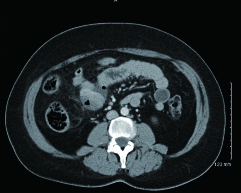
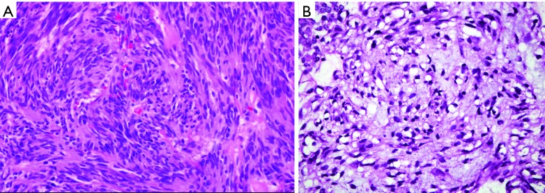
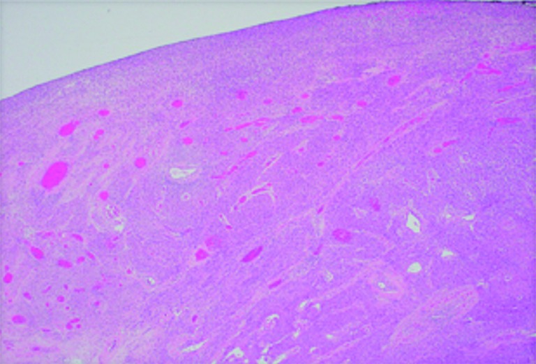
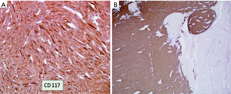
Gastrointestinal stromal tumors (GISTs) are rare neoplasms of the gastrointestinal tract associated with high rates of malignant transformation. Most GISTs present asymptomatically. They are best identified by computed tomography (CT) scan and most stain positive for CD117 (C-Kit), CD34, and/or DOG-1. There have been many risk stratification classifications systems which are calculated based on tumor size, mitotic rate, location, and perforation. The approaches to treating GISTs are to resect primary low-risk tumors, resect high-risk primary or metastatic tumors with imatinib 400 mg daily for 12 months, or if the tumor is unresectable, neoadjuvant imatinib 400 mg daily followed by surgical resection is recommended. Sunitinib is required for KIT exon 9, 13, and 14 mutations, while ponatinib is used for exon 17 mutations and regorafenib for highly refractory tumors. High-risk tumors should be monitored for recurrence with serial abdominal CT scans. Radiofrequency ablation has shown to be effective when surgery is not suitable. Newer therapies of ipilimumab, nivolumab, and endoscopic ultrasound alcohol ablation have shown promising results. This report addresses the epidemiology, clinical presentation, diagnostic imaging, histologic diagnosis, classification and risk stratification, staging and grading, surgical treatment, adjuvant treatment, and metastasis of GISTs.

Keywords: Gastrointestinal stromal tumor (GIST); outcome; prognosis; risk classification; tyrosine kinase receptor inhibitor (TKI).


Conflict of interest statement

Conflicts of Interest: The authors have no conflicts of interest to declare.

Figures

Figure 1
Figure 1
Gross photo of a fresh total gastrectomy specimen showing a large polypoid mass near the GE junction at lesser curvature and very close to the proximal resection margin. The specimen is opened along the greater curvature. The tumor is about 7 cm, in size with surface ulceration, hemorrhage, deep fissuring, and tumor fragmentation.
Figure 2
Figure 2
Gross photo. A large, bulky, intraluminal mass in resected small bowel. The cut surface of the tumor has a fish-flesh appearance with hemorrhage, necrosis, and cystic softening.
Figure 3
Figure 3
Gross photo showing a firm polypoid mass (green arrow) on the colon mucosal surface.
Figure 4
Figure 4
Microscopic photo. H&E stain. 4× objective magnification showing normal gastric mucosa on the left with intraluminal growth of the tumor on the right.
Figure 5
Figure 5
CT with oral contrast showing a mass compressing the mid-jejunal lumen with perforation.
Figure 6
Figure 6
Microscopic photo, H&E stain. (A) 40× objective magnification image of a GIST spindle cell type; (B) 100× objective magnification image of a GIST epithelioid type. GIST, gastrointestinal stromal tumor.
Figure 7
Figure 7
Microscopic photo, H&E stain. 4× objective magnification image of a GIST showing microabscesses. GIST, gastrointestinal stromal tumor.
Figure 8
Figure 8
Microscopic photo. (A) 40× objective magnification of GIST that stains CD117 positive; (B) 40× objective magnification of GIST that stains DOG-1 positive. GIST, gastrointestinal stromal tumor.
Figure 9
Figure 9
CT Imaging correlating with histology. (A) Abdominal CT transverse cut showing extraluminal mass with contrast filling area of necrosis (arrow) (9); (B) microscopic photo, H&E stain. 4× objective magnification showing a GIST with ischemic and coagulative necrosis. GIST, gastrointestinal stromal tumor.
Figure 10
Figure 10
Microscopic photo, H&E stain. 10× objective magnification of calcifications.

References

    1. Mazur MT, Clark HB. Gastric stromal tumors. Reappraisal of histogenesis. Am J Surg Pathol 1983;7:507-19. 10.1097/00000478-198309000-00001 - DOI - PubMed
    1. Rammohan A, Sathyanesan J, Rajendran K, et al. A gist of gastrointestinal stromal tumors: A review. World J Gastrointest Oncol 2013;5:102-12. 10.4251/wjgo.v5.i6.102 - DOI - PMC - PubMed
    1. Kindblom LG, Remotti HE, Aldenborg F, et al. Gastrointestinal pacemaker cell tumor (GIPACT): gastrointestinal stromal tumors show phenotypic characteristics of the interstitial cells of Cajal. Am J Pathol 1998;152:1259-69. - PMC - PubMed
    1. Hirota S, Isozaki K, Moriyama Y, et al. Gain-of-function mutations of c-kit in human gastrointestinal stromal tumors. Science 1998;279:577-80. 10.1126/science.279.5350.577 - DOI - PubMed
    1. Corbin KS, Kindler HL, Liauw SL. Considering the role of radiation therapy for gastrointestinal stromal tumor. Onco Targets Ther 2014;7:713-8. - PMC - PubMed