. 2019 Feb 6:9:43.
doi: 10.3389/fonc.2019.00043. eCollection 2019.

Differential Oxygenation in Tumor Microenvironment Modulates Macrophage and Cancer Cell Crosstalk: Novel Experimental Setting and Proof of Concept

Affiliations

Differential Oxygenation in Tumor Microenvironment Modulates Macrophage and Cancer Cell Crosstalk: Novel Experimental Setting and Proof of Concept

Noelia Campillo et al. Front Oncol. .

Abstract

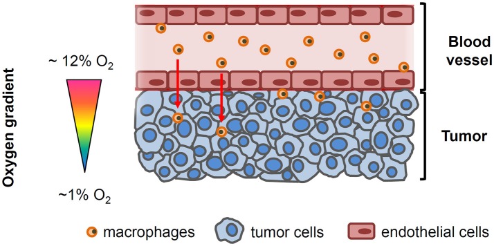
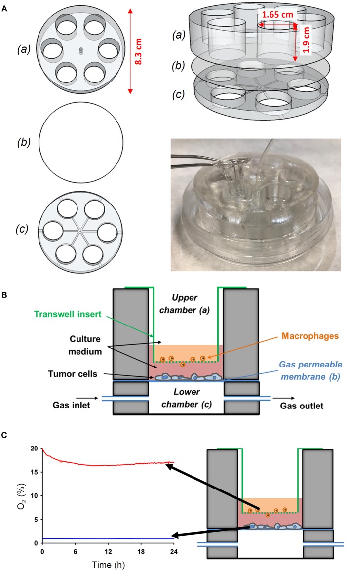
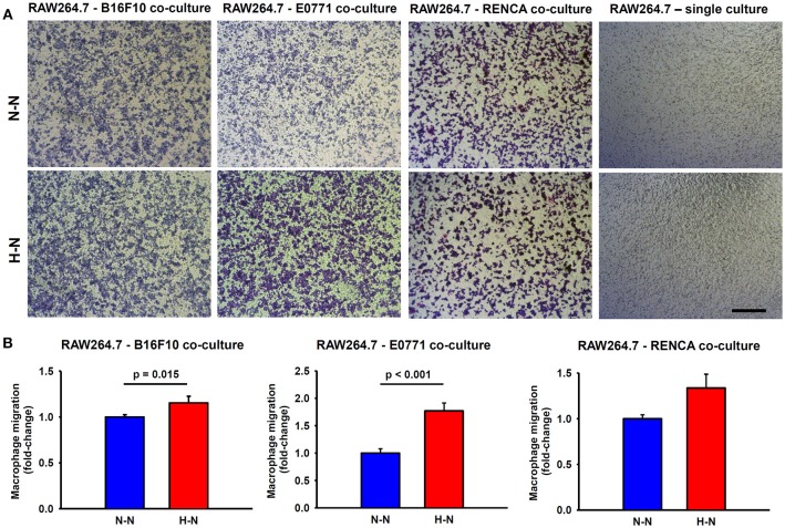
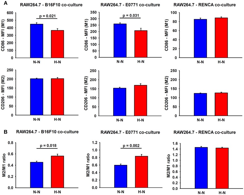
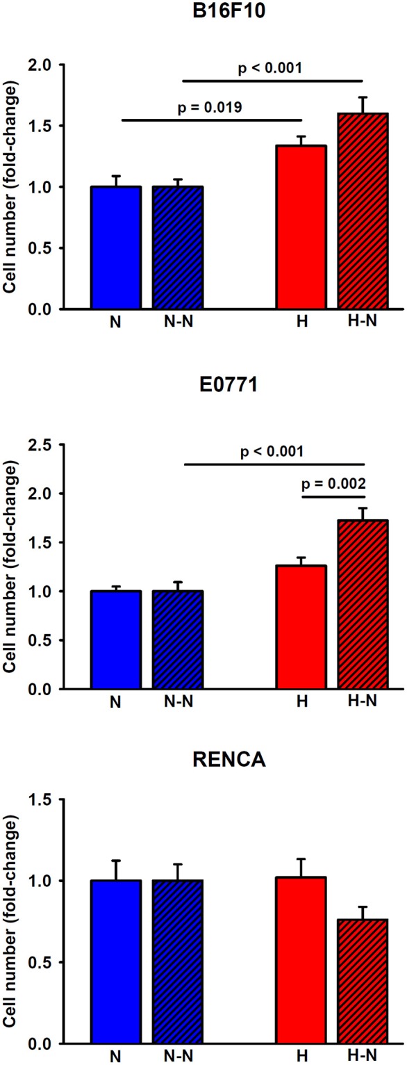
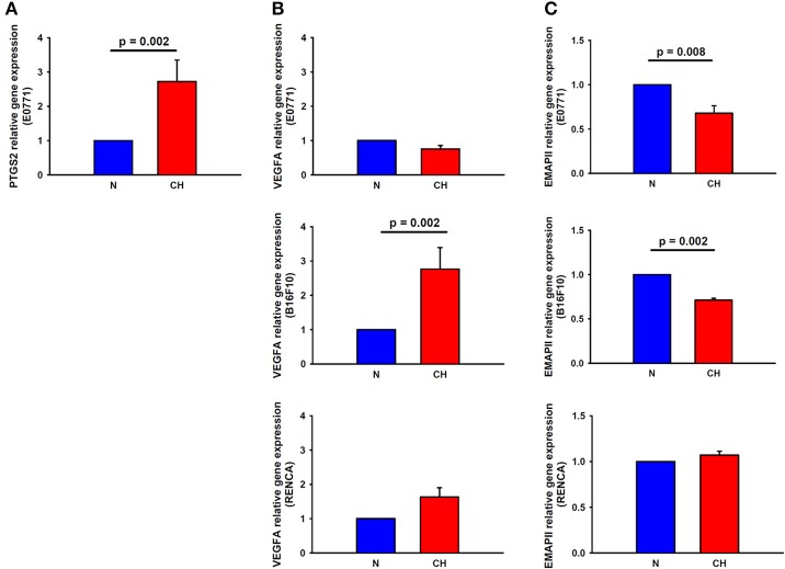
Hypoxia is a common characteristic of many solid tumors that has been associated with tumor aggressiveness. Limited diffusion of oxygen generates a gradient of oxygen availability from the blood vessel to the interstitial space and may underlie the recruitment of macrophages fostering cancer progression. However, the available data based on the recruitment of circulating cells to the tumor microenvironment has been so far carried out by conventional co-culture systems which ignore the hypoxic gradient between the vessel to the tumor interstitium. Here, we have designed a novel easy-to-build cell culture device that enables evaluation of cellular cross-talk and cell migration while they are being simultaneously exposed to different oxygenation environments. As a proof-of-concept of the potential role of differential oxygenation among interacting cells we have evaluated the activation and recruitment of macrophages in response to hypoxic melanoma, breast, and kidney cancer cells. We found that hypoxic melanoma and breast cancer cells co-cultured with normoxic macrophages enhanced their directional migration. By contrast, hypoxic kidney cells were not able to increase their recruitment. We also identified well-described hypoxia-induced pathways which could contribute in the immune cell recruitment (VEGFA and PTGS2 genes). Moreover, melanoma and breast cancer increased their proliferation. However, oxygenation levels affected neither kidney cancer cell proliferation nor gene expression, which in turn resulted in no significant changes in macrophage migration and polarization. Therefore, the cell culture device presented here provides an excellent opportunity for researchers to reproduce the in vivo hypoxic gradients in solid tumors and to study their role in recruiting circulating cells to the tumor in specific types of cancer.

Keywords: hypoxia gradient; macrophage motility; models of host-tumor interactions; novel assay technology; tumor progression.


Figures

Figure 1
Figure 1
Simplified drawing showing the oxygenation gradient from the blood vessel to tumor cells. Circulating macrophages and endothelial cells lining the tumor vessel are exposed to the physiological oxygen concentrations in blood (~12 O2) while oxygen levels within the tumor decline as the distance from the functional blood vessel increases.
Figure 2
Figure 2
Novel experimental in vitro setting to reproduce different oxygenation patterns between the tumor-vessel interface. (A) The device was performed by assembling an upper (a) and a lower (c) chamber with a PDMS membrane (b) between both chambers. The wells of the upper chamber where adjusted to fit to a conventional transwell insert. (B) Scheme of the novel in vitro experimental setting developed to study the interactions between tumor cells and macrophages simultaneously exposed to different oxygen levels mimicking the pathophysiological conditions in the solid tumor microenvironment. (C) The novel setting was able to produce an oxygen gradient as measured by an oxygen optical sensor.
Figure 3
Figure 3
Macrophage migration through a permeable membrane in co-culture with mouse melanoma (B16F10), breast (E0771), or renal (RENCA) cancer cells exposed to different oxygen levels. (A) Representative bright field images showing stained macrophages which migrated to the lower side of the transwell membrane after 24 h in co-culture with melanoma, breast, or renal cancer cells cultured under normoxic conditions (N-N) or under a physiological gradient of oxygen (H-N). In the absence of tumor cells (right), macrophages did not migrate through the transwell membrane. Scale bar = 250 μm. (B) The number of macrophages (RAW 264.7) that migrated to the lower side of the membrane was significantly higher when B16F10 (left) or E0771 (center) were selectively exposed to hypoxia (H-N) in comparison to normoxia (N-N), while no differences were found between treatments in the migration of macrophages co-cultured with RENCA cells (right). Data were normalized to the control group (20% O2).
Figure 4
Figure 4
Assessment of macrophage polarity. (A) Representative graphs obtained during FACS analyses which depict the median fluorescence intensities (MFI) for M1 and M2 markers in macrophages co-cultured with melanoma (left), breast (center) and renal (right) cancer cells exposed to hypoxia (H-N) or normoxia (N-N). Hypoxia-treated melanoma and breast tumor cells reduced the M1-like polarization of macrophages (A), as reflected by an increase in the M2/M1 ratio compared to that observed when both cell types were exposed to normoxia (N-N) No significant changes in the phenotype of macrophages were observed when co-cultured with renal cancer cells exposed to different oxygen levels (B). The M2/M1 ratio was calculated as the ratio of the median fluorescence intensity (MFI) of CD206/CD86 surface markers.
Figure 5
Figure 5
Proliferation of melanoma, breast, and renal cancer cells. Melanoma (upper) and breast (middle) cancer cells increased their proliferative rates when cultured under low levels of oxygen either in single culture (H) or in co-culture with normoxic macrophages (H-N) when compared to N and (N-N) treatment groups, respectively. The hypoxia-induced proliferation observed in single culture (H) was further enhanced when tumor cells were co-cultured with normoxic macrophages (H-N). Renal cancer cell proliferation remained similar among the different treatment groups (right).
Figure 6
Figure 6
Expression of macrophage recruitment-related genes in melanoma, breast, and cancer cells in response to hypoxia. PTGS2 (A) and VEGFA (B, center) gene expression was upregulated in hypoxia-treated breast and melanoma cancer cells, respectively, in comparison to normoxia. (C) EMAPII gene expression was reduced by hypoxia in breast (upper) and melanoma (center) tumor cells, while remained invariable in renal cancer cells (lower).

References

    1. Herman AB, Savage VM, West GB. A quantitative theory of solid tumor growth, metabolic rate and vascularization. PLoS ONE (2011) 6:e22973. 10.1371/journal.pone.0022973 - DOI - PMC - PubMed
    1. Muz B, de la Puente P, Azab F, Azab AK. The role of hypoxia in cancer progression, angiogenesis, metastasis, and resistance to therapy. Hypoxia (2015) 3:83–92. 10.2147/HP.S93413 - DOI - PMC - PubMed
    1. Rofstad EK, Galappathi K, Mathiesen BS. Tumor interstitial fluid pressure-a link between tumor hypoxia, microvascular density, and lymph node metastasis. Neoplasia (2014) 16:586–94. 10.1016/j.neo.2014.07.003 - DOI - PMC - PubMed
    1. Lewis DM, Park KM, Tang V, Xu Y, Pak K, Eisinger-Mathason TS, et al. . Intratumoral oxygen gradients mediate sarcoma cell invasion. Proc Natl Acad Sci USA. (2016) 113:9292–7. 10.1073/pnas.1605317113 - DOI - PMC - PubMed
    1. Quail DF, Joyce JA. Microenvironmental regulation of tumor progression and metastasis. Nat Med. (2013) 19:1423–37. 10.1038/nm.3394 - DOI - PMC - PubMed