Review
. 2019 Feb 5:6:49-69.
doi: 10.2147/JHC.S186239. eCollection 2019.

LI-RADS: a conceptual and historical review from its beginning to its recent integration into AASLD clinical practice guidance

Affiliations
Review

LI-RADS: a conceptual and historical review from its beginning to its recent integration into AASLD clinical practice guidance

Khaled M Elsayes et al. J Hepatocell Carcinoma. .

Abstract

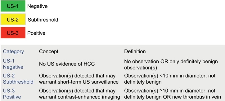
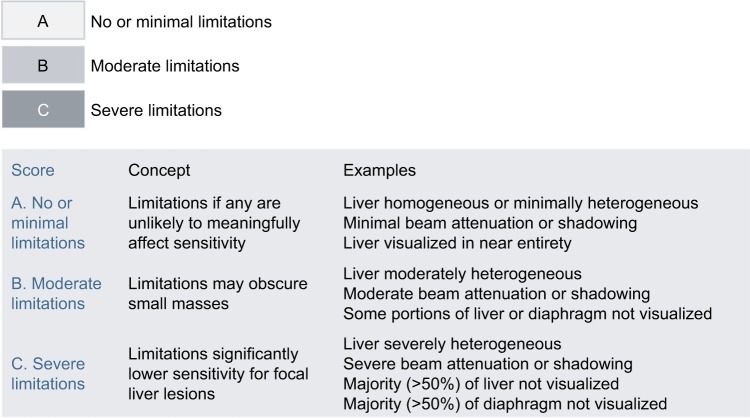
The Liver Imaging Reporting and Data System (LI-RADS®) is a comprehensive system for standardizing the terminology, technique, interpretation, reporting, and data collection of liver observations in individuals at high risk for hepatocellular carcinoma (HCC). LI-RADS is supported and endorsed by the American College of Radiology (ACR). Upon its initial release in 2011, LI-RADS applied only to liver observations identified at CT or MRI. It has since been refined and expanded over multiple updates to now also address ultrasound-based surveillance, contrast-enhanced ultrasound for HCC diagnosis, and CT/MRI for assessing treatment response after locoregional therapy. The LI-RADS 2018 version was integrated into the HCC diagnosis, staging, and management practice guidance of the American Association for the Study of Liver Diseases (AASLD). This article reviews the major LI-RADS updates since its 2011 inception and provides an overview of the currently published LI-RADS algorithms.

Keywords: CEUS; CT; HCC; LI-RADS; MRI; US; cirrhosis; liver imaging; reporting; v2018.


Conflict of interest statement

Disclosure The authors report no conflicts of interest in this work. The views expressed in this work are those of the authors and do not necessarily reflect the official policy or position of the Department of the Navy, Department of Defense or the United States Government. Dr Robert M Marks is a military service member. This work was prepared as part of official duties. Title 17 U.S.C. 105 provides that ‘Copyright protection under this title is not available for any work of the United States Government.

Figures

Figure 1
Figure 1
LI-RADS CT/MRI diagnostic algorithm and table v2018. Notes: Reproduced with permission from American College of Radiology. Liver Imaging Reporting and Data System version 2018 core. Available from: https://www.acr.org/Clinical-Resources/Reporting-and-Data-Systems/LI-RADS. Abbreviations: ACR, American College of Radiology; APHE, Arterial phase hyperenhancement; HCC, hepatocellular carcinoma; LI-RADS, Liver Imaging Reporting and Data System; LR-NC, LR-noncategorizable; TIV, tumor in vein.
Figure 2
Figure 2
US LI-RADS US category. Notes: Reproduced with permission from American College of Radiology. Ultrasound LI-RADS v2017. https://www.acr.org/Clinical-Resources/Reporting-and-Data-Systems/LI-RADS/Ultrasound-LI-RADS-v2017. Abbreviations: ACR, American College of Radiology; HCC, hepatocellular carcinoma; LI-RADS, Liver Imaging Reporting and Data System; US, ultrasound.
Figure 3
Figure 3
US LI-RADS visualization score. Notes: Reproduced with permission from American College of Radiology. Ultrasound LI-RADS v2017. https://www.acr.org/Clinical-Resources/Reporting-and-Data-Systems/LI-RADS/Ultrasound-LI-RADS-v2017. Abbreviations: ACR, American College of Radiology; LI-RADS, Liver Imaging Reporting and Data System.
Figure 4
Figure 4
US LI-RADS Category 3 observation in a 59-year-old male with hepatitis C cirrhosis undergoing US surveillance. Notes: Sagittal US image shows a 3.6 cm solid hypoechoic observation with lobulated margins in segment 6. This patient requires contrast-based studies; CEUS, CECT, or CEMRI to further characterize the lesion. Abbreviations: CEUS, contrast-enhanced ultrasound; LI-RADS, Liver Imaging Reporting and Data System; US, ultrasound.
Figure 5
Figure 5
US LI-RADS Category 3 observation in a 70-year-old female with cryptogenic cirrhosis undergoing US surveillance. Notes: Transverse US image shows a large area of heterogeneity (arrows) distinctive from background liver, shown to represent an HCC with infiltrative appearance on a diagnostic CEUS (not shown). Abbreviations: CEUS, contrast-enhanced ultrasound; HCC, hepatocellular carcinoma; LI-RADS, Liver Imaging Reporting and Data System; US, ultrasound.
Figure 6
Figure 6
Seventy-one-year-old female demonstrating nonrim arterial phase hyperenhancement. Notes: Contrast-enhanced CT shows a large mass (arrows) in the left hepatic lobe, partially exophytic, demonstrating heterogeneous arterial phase hyperenhancement. Posthepatectomy pathology confirmed well-differentiated hepatocellular carcinoma.
Figure 7
Figure 7
Ancillary imaging features used in LI-RADS CT/MRI. Note: These ancillary features are supportive, and their use is optional. Reproduced with permission from American College of Radiology. Liver Imaging Reporting and Data System version 2018 core. Available from: https://www.acr.org/Clinical-Resources/Reporting-and-Data-Systems/LI-RADS. Abbreviations: ACR, American College of Radiology; HCC, hepatocellular carcinoma; LI-RADS, Liver Imaging Reporting and Data System; US, ultrasound.
Figure 8
Figure 8
Fifty-eight-year-old man with cirrhosis (AFP=285 ng/mL) tumor in vein (TIV), probably due to hepatocellular carcinoma. Notes: Gadobenate dimeglumine-enhanced MRI shows unequivocal enhancement of tissue (arrow) within the left portal vein, which meets the criterion for LR-TIV. Notice that the tissue expands the lumen of the left portal vein, (A) hyperenhances in the arterial phase, (B) appears to washout in the portal venous phase and has high signal intensity on (C) T2-weighted and (D) diffusion-weighted images. Involvement of the parenchyma by the tumor is more conspicuous on diffusion-weighted images (arrowhead in D).
Figure 9
Figure 9
Sixty-one-year-old man with chronic hepatitis C and cirrhosis. Notes: A 2 cm observation in hepatic segment 5 shows (A) rim arterial phase hyperenhancement, (B) progressive delayed central enhancement on portal venous, and (C) delayed phase, corresponding to a targetoid appearance (LR-M). Biopsy confirmed intrahepatic cholangiocarcinoma. Arrow shows the enhancement pattern.
Figure 10
Figure 10
Diagnostic categories for CEUS LI-RADS. Notes: Reproduced with permission from American College of Radiology. CEUS LI-RADS v2017. Available from: https://www.acr.org/Clinical-Resources/Reporting-and-Data-Systems/LI-RADS/CEUS-LI-RADS-v2017. Abbreviations: ACR, American College of Radiology; CEUS, contrast-enhanced ultrasound; HCC, hepatocellular carcinoma; LI-RADS, Liver Imaging Reporting and Data System; LR-NC, LR-noncategorizable; TIV, tumor in vein.
Figure 11
Figure 11
CEUS LI-RADS TIV. Notes: (A) Soft tissue in the portal vein (arrows) contiguous with a parenchymal mass (star) on B-mode ultrasound. (B, C) Both soft tissue within the portal vein (arrows) and the mass (star) show arterial phase hyperenhancement and (D) mild washout in the late phase. CEUS LI-RADS TIV Criteria: • Unequivocal arterial phase hyperenhancement and washout of soft tissue within the lumen of portal and/or hepatic veins. • Must correspond on dual screen with mass in vein. • Most LR-TIVs are HCC. Some are iCCA or cHCC-CCA. • CEUS has been shown to have high sensitivity and specificity to diagnose TIV. Abbreviations: CEUS, contrast-enhanced ultrasound; HCC, hepatocellular carcinoma; iCCA, intrahepatic cholangiocarcinoma; TIV, tumor in vein.
Figure 12
Figure 12
CEUS LI-RADS M criteria. Notes: Reproduced with permission from American College of Radiology. CEUS LI-RADS v2017. Available from: https://www.acr.org/Clinical-Resources/Reporting-and-Data-Systems/LI-RADS/CEUS-LI-RADS-v2017. Abbreviations: ACR, American College of Radiology; CEUS, contrast-enhanced ultrasound; LI-RADS, Liver Imaging Reporting and Data System.
Figure 13
Figure 13
Washout. Notes: (A) Early weak washout seen within 1 minute after contrast injection (CEUS LR-M feature). (B) Marked washout seen within 2 minutes after contrast injection (CEUS LR-M feature). (C) Late and mild washout seen >1 minute after contrast injection (CEUS LR-5 feature). Abbreviation: CEUS, contrast-enhanced ultrasound.
Figure 14
Figure 14
AFs used in CEUS LI-RADS. Notes: Reproduced with permission from American College of Radiology. CEUS LI-RADS v2017. Available from: https://www.acr.org/Clinical-Resources/Reporting-and-Data-Systems/LI-RADS/CEUS-LI-RADS-v2017. Abbreviations: ACR, American College of Radiology; AF, ancillary feature; CEUS, contrast-enhanced ultrasound; HCC, hepatocellular carcinoma; LI-RADS, Liver Imaging Reporting and Data System.
Figure 15
Figure 15
CEUS LI-RADS diagnostic algorithm and table. Notes: Reproduced with permission from American College of Radiology. CEUS LI-RADS v2017. Available from: https://www.acr.org/Clinical-Resources/Reporting-and-Data-Systems/LI-RADS/CEUS-LI-RADS-v2017. Abbreviations: ACR, American College of Radiology; APHE, arterial phase hyperenhancement; CEUS, contrast-enhanced ultrasound; HCC, hepatocellular carcinoma; LI-RADS, Liver Imaging Reporting and Data System; ; LR-NC, LR-noncategorizable; TIV, tumor in vein; US, ultrasound.
Figure 16
Figure 16
CEUS LR-5. Notes: (A) A 17 mm hypoechoic nodule on B-mode ultrasound. (B) The entire nodule shows hyperenhancement in the arterial phase. (C) At 1 minute, the nodule is isoenhancing compared with the surrounding liver parenchyma. (D) The nodule shows mild but definite hypoenhancement compared with the surrounding liver parenchyma at 2 minutes. This is late and mild washout. Arrows show the outline of the nodule. Abbreviation: CEUS, contrast-enhanced ultrasound.
Figure 17
Figure 17
CEUS LR-4. Notes: (A) A 12 mm hypoechoic nodule on B-mode ultrasound. (B) The entire nodule shows hyperenhancement in the arterial phase. (C) At 2 minutes, the nodule is isoenhancing compared with the surrounding liver parenchyma. (D) The nodule remains isoenhancing at 5.5 minutes without washout. Arrows show to the outline of the nodule. Abbreviation: CEUS, contrast-enhanced ultrasound.
Figure 18
Figure 18
CEUS LI-RADS AFs upgrade and downgrade of categories. Notes: Reproduced with permission from American College of Radiology. CEUS LI-RADS v2017. Available from: https://www.acr.org/Clinical-Resources/Reporting-and-Data-Systems/LI-RADS/CEUS-LI-RADS-v2017. Abbreviations: ACR, American College of Radiology; AF, ancillary feature; CEUS, contrast-enhanced ultrasound; LI-RADS, Liver Imaging Reporting and Data System.
Figure 19
Figure 19
CEUS LI-RADS tiebreaking rules. Notes: Reproduced with permission from American College of Radiology. CEUS LI-RADS v2017. Available from: https://www.acr.org/Clinical-Resources/Reporting-and-Data-Systems/LI-RADS/CEUS-LI-RADS-v2017. Abbreviations: ACR, American College of Radiology; CEUS, contrast-enhanced ultrasound; LI-RADS, Liver Imaging Reporting and Data System; TIV, tumor in vein.
Figure 20
Figure 20
Treatment response categories with examples from a 70-year-old man who underwent transarterial radioembolization (Y90) followed by transarterial bland embolization and microwave ablation (MWA). Notes: Left column shows no evidence of enhancement after MWA, categorized as LR-TR nonviable. Center column shows ill-defined enhancement (arrows) 5 months after Y90, categorized as LR-TR equivocal. Right column shows mass-like arterial phase hyperenhancement in the treated lesion (arrowheads), categorized as LR-TR viable.

References

    1. Marrero JA, Kulik LM, Sirlin CB, et al. Diagnosis, staging, and management of hepatocellular carcinoma: 2018 practice guidance by the American Association for the Study of Liver Diseases. Hepatology. 2018;68(2):723–750. - PubMed
    1. Mittal S, El-Serag HB. Epidemiology of hepatocellular carcinoma: consider the population. J Clin Gastroenterol. 2013;47(Suppl):S2–S6. - PMC - PubMed
    1. Forner A, Reig ME, de Lope CR, Bruix J. Current strategy for staging and treatment: the BCLC update and future prospects. Semin Liver Dis. 2010;30(1):61–74. - PubMed
    1. Heimbach JK, Kulik LM, Finn RS, et al. AASLD guidelines for the treatment of hepatocellular carcinoma. Hepatology. 2018;67(1):358–380. - PubMed
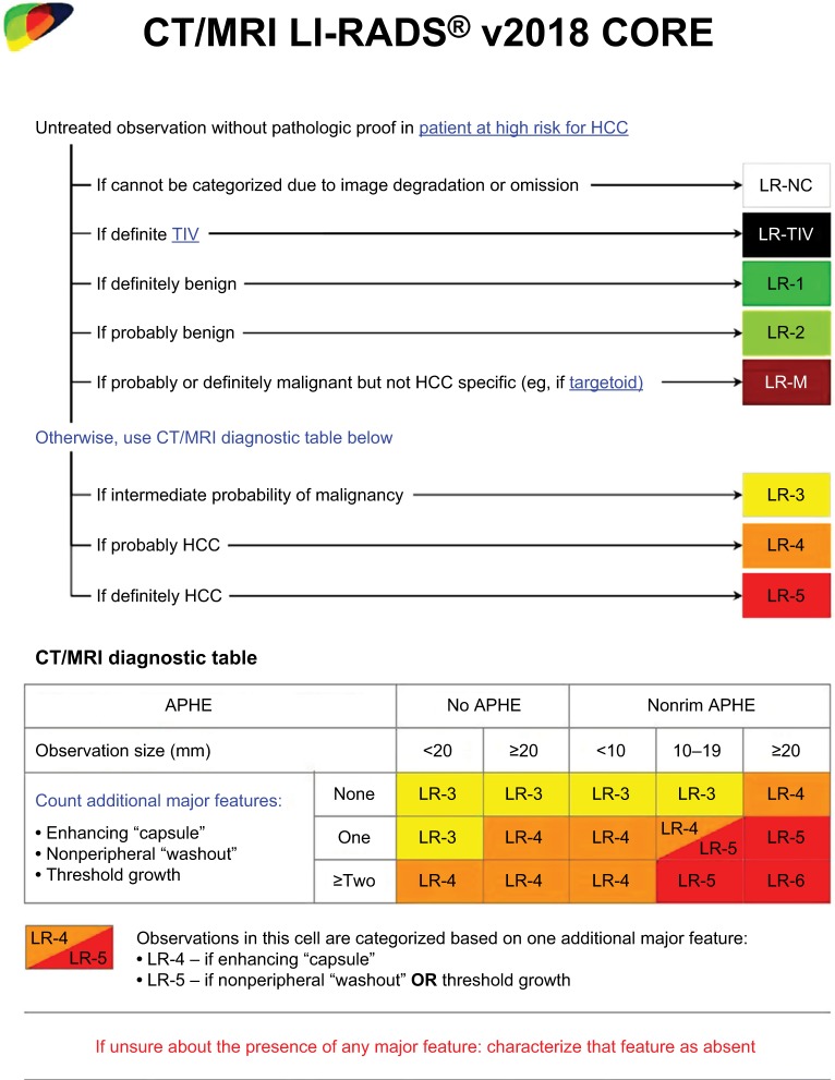
    1. Chernyak V, Santillan CS, Papadatos D, Sirlin CB. LI-RADS® algorithm: CT and MRI. Abdom Radiol (NY) 2018;43(1):111–126. - PubMed