. 2019 Jan 25:13:167-179.
doi: 10.1016/j.omtm.2019.01.007. eCollection 2019 Jun 14.

AAV9- TAZ Gene Replacement Ameliorates Cardiac TMT Proteomic Profiles in a Mouse Model of Barth Syndrome

Affiliations

AAV9- TAZ Gene Replacement Ameliorates Cardiac TMT Proteomic Profiles in a Mouse Model of Barth Syndrome

Silveli Suzuki-Hatano et al. Mol Ther Methods Clin Dev. .

Abstract

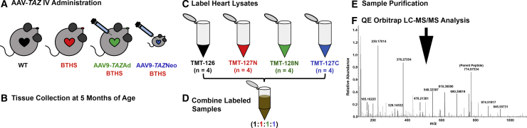
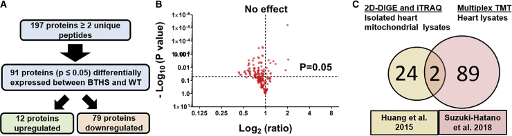
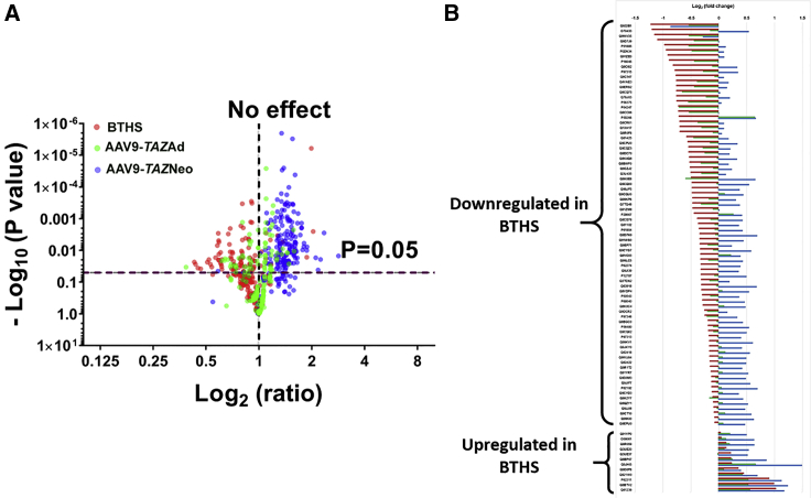
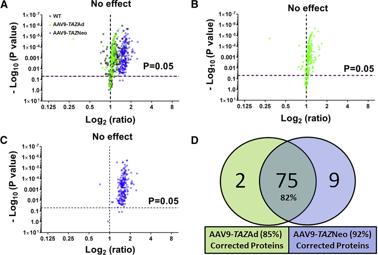
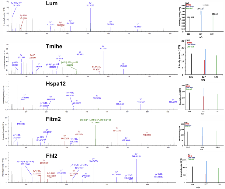
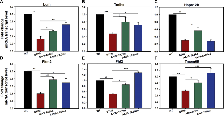
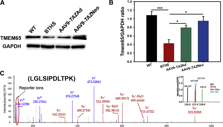
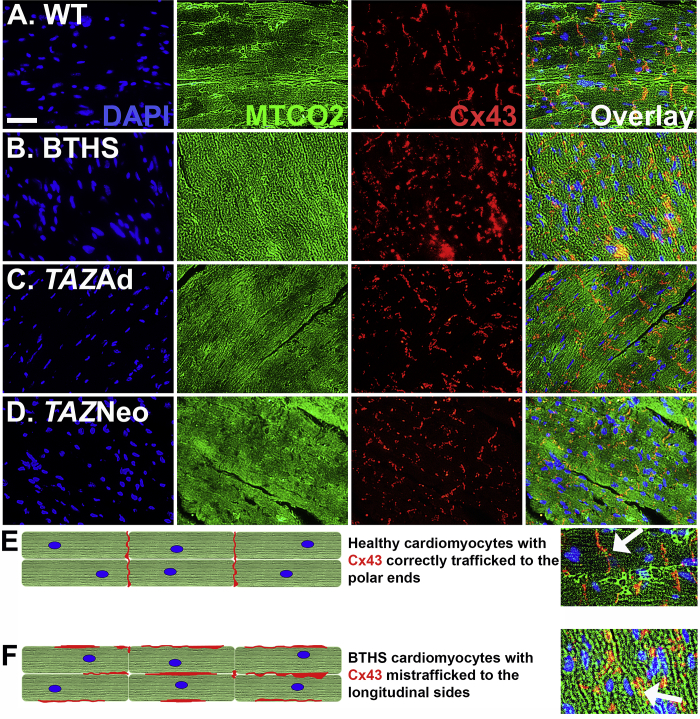
Barth syndrome (BTHS) is a rare mitochondrial disease that causes severe cardiomyopathy and has no disease-modifying therapy. It is caused by recessive mutations in the gene tafazzin (TAZ), which encodes tafazzin-an acyltransferase that remodels the inner mitochondrial membrane lipid cardiolipin. To identify novel mechanistic pathways involved in BTHS and evaluate the effects of gene therapy on proteomic profiles, we performed a multiplex tandem mass tagging (TMT) quantitative proteomics analysis to compare protein expression profiles from heart lysates isolated from BTHS, healthy wild-type (WT), and BTHS treated with adeno-associated virus serotype 9 (AAV9)-TAZ gene replacement as neonates or adults. 197 proteins with ≥2 unique peptides were identified. Of these, 91 proteins were significantly differentially expressed in BTHS compared to WT controls. Cause-effect relationships between tafazzin deficiency and altered protein profiles were confirmed through demonstrated significant improvements in expression levels following administration of AAV9-TAZ. The importance of TMEM65 in Cx43 localization to cardiac intercalated discs was revealed as a novel consequence of tafazzin deficiency that was improved following gene therapy. This study identifies novel mechanistic pathways involved in the pathophysiology of BTHS, demonstrates the ability of gene delivery to improve protein expression profiles, and provides support for clinical translation of AAV9-TAZ gene therapy.

Keywords: AAV9; BTHS; Barth syndrome; Cx43; TMEM65; TMT proteomics; cardiac gene therapy; gap junction localization; mitochondrial disease; tafazzin.


Figures

None
Graphical abstract
Figure 1
Figure 1
TMT Experimental Workflow (A) AAV9-TAZ was administered to BTHS mice as adults (3 months) or neonates (1 or 2 days). (B) Hearts from WT, untreated BTHS, and treated BTHS mice were collected at 5 months of age. (C) Heart lysates were labeled with unique isobaric labels. (D) The labeled samples were combined and purified (E). (F) LC-MS/MS was performed on the purified sample, and data were analyzed against the Mus musculus protein database for peptide identification.
Figure 2
Figure 2
Proteins Differentially Expressed in BTHS (A) Flowchart depicting the study screening process. (B) A volcano plot displaying log 2 fold change ratios of untreated BTHS heart proteins as compared to healthy WT controls. All proteins above the horizontal dashed line (p = 0.05) display significantly altered expression levels. (C) A Venn diagram comparison of proteins identified as being differentially expressed in a previous study using isolated heart mitochondrial lysates and this study using whole-heart lysates.
Figure 3
Figure 3
Relative Distribution of Proteins from BTHS, AAV9-TAZAd, and AAV9-TAZNeo Mouse Hearts (A) Volcano plot showing an overlay of log2 ratio fold changes in protein expression levels between untreated BTHS mice, AAV9-TAZneo, and AAV9-TAZad-treated cohorts as compared to healthy WT control levels clearly demonstrates improved expression in treatment groups. (B) Log2 fold change line diagram displaying relative expression levels for each protein identified as significantly differentially expressed in BTHS (red) and corresponding AAV9-TAZAd (green) and AAV9-TAZNeo (blue) levels as compared to that of healthy WT controls.
Figure 4
Figure 4
AAV9-TAZ Administration Improves Aberrant Protein Expression Profiles (A) Volcano plots of log2 fold change ratios of proteins from healthy WT (black), AAV9-TAZAd-treated (green), and AAV9-TAZNeo-treated (blue) heart lysate samples as compared to untreated BTHS. (B) The AAV9-TAZAd-treated profile is very similar to that of healthy WT controls. (C) The AAV9-TAZNeo-treated profile shows the overall highest expression levels. (D) A Venn diagram showing the total numbers of proteins found to be differentially expressed in BTHS that display significant improvement in expression levels in the treated groups.
Figure 5
Figure 5
MS2 Spectra for Representative Proteins Identified by TMT Reporter ions for each are located on the right-hand side of each spectra.
Figure 6
Figure 6
Relative Fold mRNA Transcript Levels for 6 Highly Interesting Proteins Identified by TMT as Being Significantly Differentially Expressed in BTHS (A) Lumican (Lum), (B) Trimethyllysine dioxygenase (Tmlhe), (C) heat shock 70 kDa protein 12B (Hspa12b), (D) fat-storage-inducing transmembrane protein 2 (Fitm2), (E) four and a half LIM domains protein 2 (Fhl2), and (F) transmembrane protein 65 (Tmem65) (all data are presented as ± SE; *p ≤ 0.05; **p ≤ 0.01; ***p ≤ 0.001).
Figure 7
Figure 7
Tmem65 Expression Levels (A) Western blot (WB) analysis confirms decreased TMEM65 expression in BTHS that is improved with AAV9-TAZ treatment. (B) Graphical representation of TMEM65 as compared to GAPDH expression by WB (n = 5). (C) MS2 spectra identified by TMT with reporter ions located on the right-hand side (all data are presented as ± SE; *p ≤ 0.05; ***p ≤ 0.001).
Figure 8
Figure 8
Cx43 Localization Is Impaired in Untreated BTHS Mice (A–D) IF staining showing DAPI staining of nuclei (blue), MTCO2 expression localized to mitochondria (green), and Cx43 expression (red) in (A) healthy WT control (scale bar = 25 μm), (B) untreated BTHS, (C) BTHS AAV9-TAZ adult, and (D) BTHS AAV9-TAZ neonatal treated heart tissues. (E) A depiction of how Cx43 properly localizes to the polar ends of healthy cardiomyocytes. (F) A depiction of how Cx43 is mislocalized to the longitudinal sides and more clumped in untreated BTHS cardiomyocytes.

References

    1. Houtkooper R.H., Turkenburg M., Poll-The B.T., Karall D., Pérez-Cerdá C., Morrone A., Malvagia S., Wanders R.J., Kulik W., Vaz F.M. The enigmatic role of tafazzin in cardiolipin metabolism. Biochim. Biophys. Acta. 2009;1788:2003–2014. - PubMed
    1. Neuwald A.F. Barth syndrome may be due to an acyltransferase deficiency. Curr. Biol. 1997;7:R465–R466. - PubMed
    1. Saini-Chohan H.K., Holmes M.G., Chicco A.J., Taylor W.A., Moore R.L., McCune S.A., Hickson-Bick D.L., Hatch G.M., Sparagna G.C. Cardiolipin biosynthesis and remodeling enzymes are altered during development of heart failure. J. Lipid Res. 2009;50:1600–1608. - PMC - PubMed
    1. Schlame M. Cardiolipin synthesis for the assembly of bacterial and mitochondrial membranes. J. Lipid Res. 2008;49:1607–1620. - PMC - PubMed
    1. Schlame M., Kelley R.I., Feigenbaum A., Towbin J.A., Heerdt P.M., Schieble T., Wanders R.J., DiMauro S., Blanck T.J. Phospholipid abnormalities in children with Barth syndrome. J. Am. Coll. Cardiol. 2003;42:1994–1999. - PubMed

LinkOut - more resources