. 2019 Jun;137(6):981-1001.
doi: 10.1007/s00401-019-01973-6. Epub 2019 Feb 20.

Demonstration of prion-like properties of mutant huntingtin fibrils in both in vitro and in vivo paradigms

Affiliations

Demonstration of prion-like properties of mutant huntingtin fibrils in both in vitro and in vivo paradigms

Maria Masnata et al. Acta Neuropathol. 2019 Jun.

Abstract

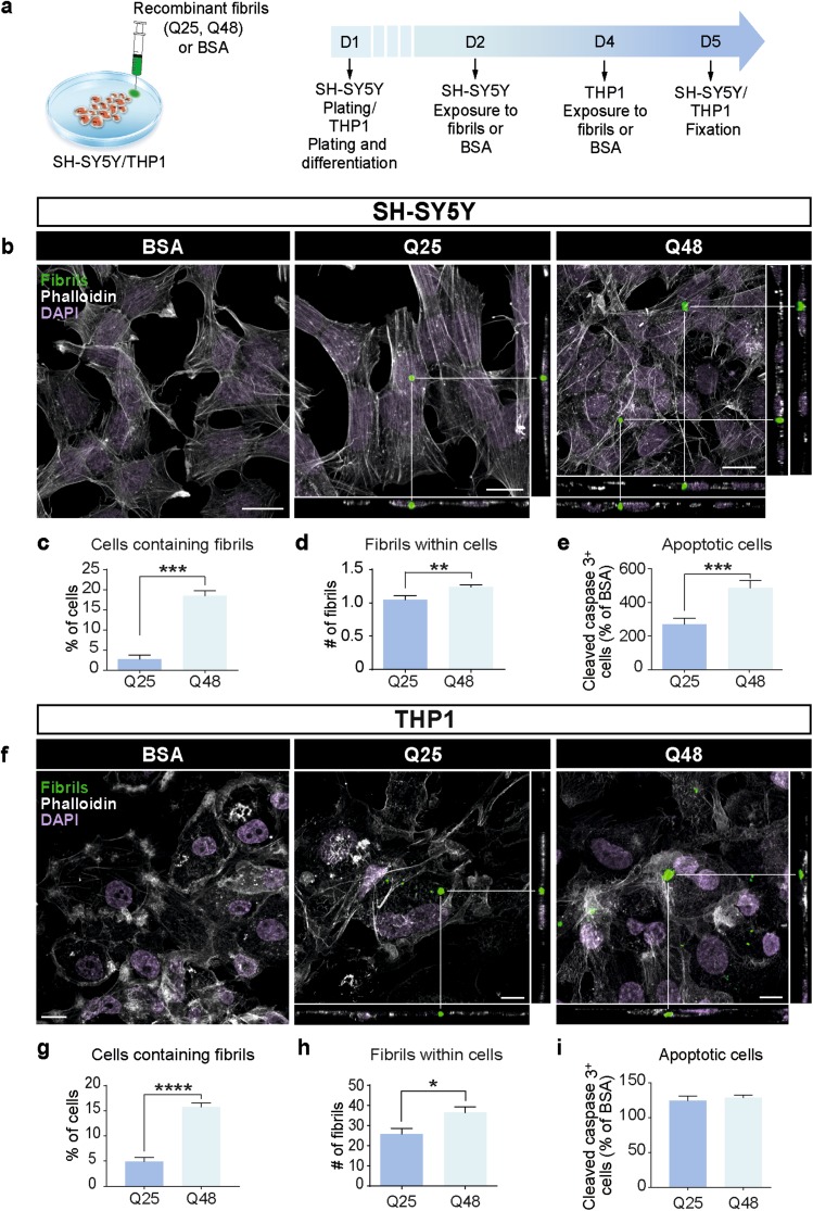
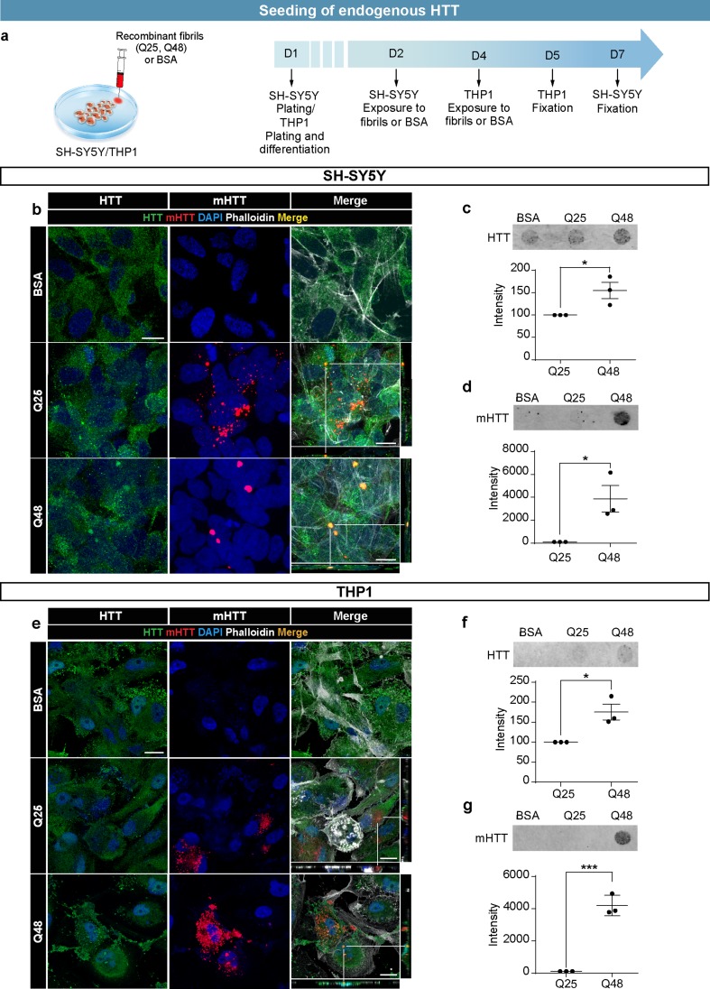
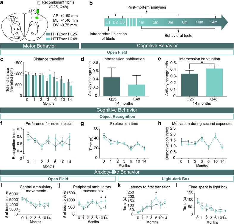
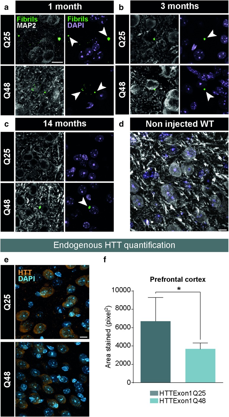
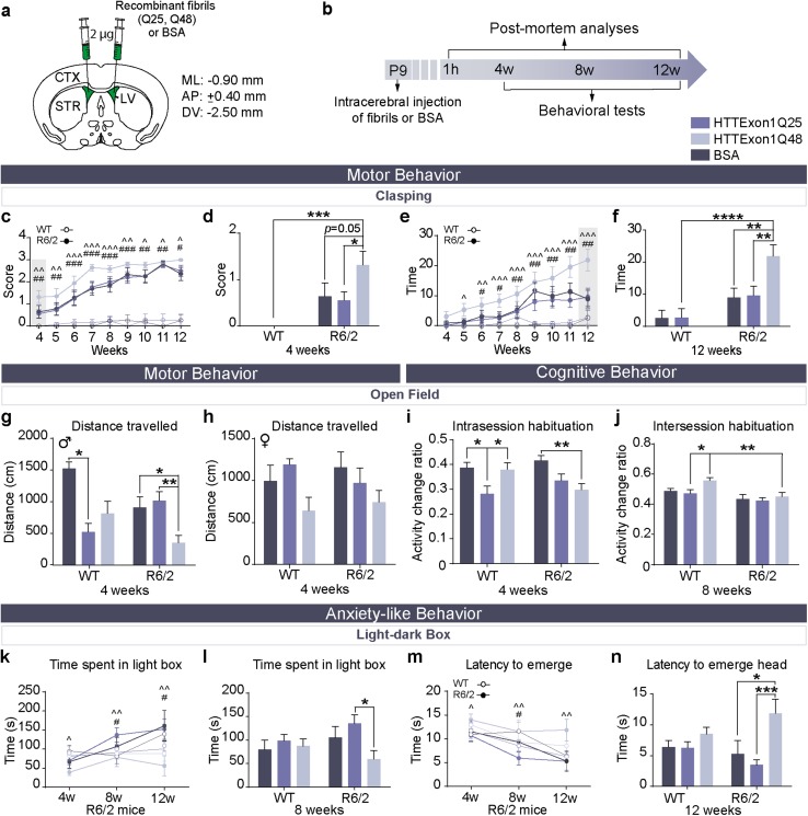
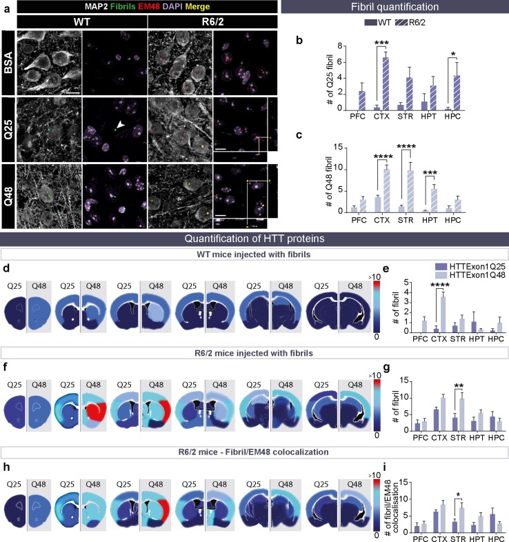
In recent years, evidence has accumulated to suggest that mutant huntingtin protein (mHTT) can spread into healthy tissue in a prion-like fashion. This theory, however, remains controversial. To fully address this concept and to understand the possible consequences of mHTT spreading to Huntington's disease pathology, we investigated the effects of exogenous human fibrillar mHTT (Q48) and huntingtin (HTT) (Q25) N-terminal fragments in three cellular models and three distinct animal paradigms. For in vitro experiments, human neuronal cells [induced pluripotent stem cell-derived GABA neurons (iGABA) and (SH-SY5Y)] as well as human THP1-derived macrophages, were incubated with recombinant mHTT fibrils. Recombinant mHTT and HTT fibrils were taken up by all cell types, inducing cell morphology changes and death. Variations in HTT aggregation were further observed following incubation with fibrils in both THP1 and SH-SY5Y cells. For in vivo experiments, adult wild-type (WT) mice received a unilateral intracerebral cortical injection and R6/2 and WT pups were administered fibrils via bilateral intraventricular injections. In both protocols, the injection of Q48 fibrils resulted in cognitive deficits and increased anxiety-like behavior. Post-mortem analysis of adult WT mice indicated that most fibrils had been degraded/cleared from the brain by 14 months post-surgery. Despite the absence of fibrils at these later time points, a change in the staining pattern of endogenous HTT was detected. A similar change was revealed in post-mortem analysis of the R6/2 mice. These effects were specific to central administration of fibrils, as mice receiving intravenous injections were not characterized by behavioral changes. In fact, peripheral administration resulted in an immune response mounting against the fibrils. Together, the in vitro and in vivo data indicate that exogenously administered mHTT is capable of both causing and exacerbating disease pathology.


Figures

Fig. 1
Fig. 1
Uptake of fibrillar HTTExon1Q25 and Q48 by different cell types. Schematic of experimental design (a). Representative confocal photomicrographs of human SH-SY5Y cells (b) and THP1-derived macrophages (f) demonstrating uptake of both ATTO488-labeled HTTExon1Q25 and Q48 fibrils (green), 72 (SH-SY5Y) and 24 h (THP1) post-exposure, respectively. For all photomicrographs, the cell membrane was labelled with phalloidin (white) and cell nuclei were stained with DAPI (purple). The percentage of SH-SY5Y (c) and THP1 (g) cells containing puncta, the number of puncta per SH-SY5Y (d) and THP1 (h) cell and the number of apoptotic SH-SY5Y (e) and THP1 (i) cells were all calculated. All graphs are the average of three independent experiments. Data are expressed as mean ± SEM. Statistical analysis was performed using a students’ unpaired t test. *p < 0.05, **p < 0.01, ***p < 0.001, ****p < 0.0001. Scale bars b = 25 µm, f = 10 µm. BSA bovine serum albumin
Fig. 2
Fig. 2
Seeding of endogenous HTT by exogenous HTTExon1Q48 fibrils. Schematic of experimental design for SH-SY5Y cells and THP1-derived macrophages (a). Representative confocal photomicrographs of cells exposed to ATTO550-labeled HTTExon1Q25/Q48 fibrils for 5 days (SH-SY5Y—b) and 24 h (THP1—e). Filter retardation assay and quantification of HTT aggregation immunodetected with anti-WT HTT antibody MAB2166 and anti-aggregated HTT antibody EM48 for SH-SY5Y (c, d) and THP1 (f, g) cells. For all filter retardation assay quantifications, the Q48 intensity is shown as percentage of the intensity of Q25. For immunofluorescence, endogenous HTT was detected with MAB2170 (green), HTTExon1Q25 and Q48 (red) and cell nuclei were stained with DAPI (blue). All graphs are the average of three independent experiments. Data are expressed as mean ± SEM. Statistical analysis was performed using a students’ unpaired t test (f). *p < 0.05, ***p < 0.001. Scale bars = 10 µm. GFP green fluorescent protein, HTT huntingtin, BSA bovine serum albumin
Fig. 3
Fig. 3
Manifestation of behavioral impairments in adult WT mice injected with HTTExon1Q48 fibrils. Atlas coordinates for intracerebral injections, treatment legend (a) and experimental timeline (b). Animals underwent motor, cognitive and anxiety-like behavioral tests from 1 to 14 months post-injection. Motor behavior was assessed using total distance traveled in 60 min in the open field (c). At 14 months post-injection, short-term memory was evaluated by assessing the change in distance traveled between the first 5 and the last 5 min of testing (d) and long-term memory was assessed by calculating the change in distance traveled in the first 5 min of baseline to 14 months post-injection (e). Cognitive performance was assessed at each time point by measuring the preference for the novel object (f). The absence of confounding factors was determined by measuring the exploration time (g) and motivation during the exposure to the novel object (h). Anxiety-like behavior was assessed in the open field by quantifying the distance traveled in the center of the open field (i) and in the periphery (j) as well as in the light–dark box by latency to leave the dark box (k) and total time spent in the light box (l). Data are expressed as mean ± SEM. Baseline HTTExon1Q25 n = 12, HTTExon1Q48 n = 12; 1 month HTTExon1Q25 n = 11, HTTExon1Q48 n = 11; 2 months HTTExon1Q25 n = 10, HTTExon1Q48 n = 10; 3–14 months HTTExon1Q25 n = 9, HTTExon1Q48 n = 9. Statistical analysis was performed using a students’ unpaired t test for individual time points and a two-way ANOVA followed by Tukey’s post hoc test for across time graphs. *p < 0.05. ACB nucleus accumbens, AP antero-posterior, CTX M1 primary motor cortex, D day, DV dorso-ventral, m month, ML medio-lateral, STR striatum
Fig. 4
Fig. 4
Post-mortem identification of HTTExon1Q25 and Q48 fibrils in adult WT mice. Representative confocal photomicrographs of brain tissue of WT mice sacrificed 1 month (HTTExon1Q25 n = 2, HTTExon1Q48 n = 2) (a), 3 months (HTTExon1Q25 n = 2, HTTExon1Q48 n = 2) (b) and 14 months (HTTExon1Q25 n = 8, HTTExon1Q48 n = 8) post-injection and immunostained for HTTExon1 (green) and MAP2 (white). Arrowheads indicate the localization of fibrils. Specificity of anti-HTTExon1 antibody was assessed by staining a non injected WT mouse (d). Representative confocal photomicrographs of brain tissue of WT sacrificed at 14 months post-surgery and immunostained for endogenous HTT (orange) (e). Quantification of HTT intensity as measured by total area stained in the prefrontal cortex (f). Nuclei were detected with DAPI (purple ad; blue e). Data are expressed as mean ± SEM. WT Q25 n = 7, WT Q48 n = 7. Statistical analysis was performed using a students’ unpaired t test. *p < 0.05. Scale bars = 10 µm. MAP2 microtubule-associated protein 2, WT wild type. Arrowheads indicate the localization of fibrils
Fig. 5
Fig. 5
Precipitation of behavioral phenotype in R6/2 mice following injection of HTTExon1Q48 fibrils. Atlas coordinates for intraventricular injections, treatment legend (a) and experimental timeline (b). Clasping score in R6/2 mice at all tested time points (c) and at 4 weeks of age (d). Duration of full-clasping behavior at all tested time points (e) and at 12 weeks of age (f) WT BSA n = 13–14; WT HTTExon1Q25 n = 11–13; WT HTTExon1Q48 n = 12–15; R6/2 BSA n = 11–14; R6/2 HTTExon1Q25 n = 12–18; R6/2 HTTExon1Q48 n = 9–19. Motor endurance was measured by assessing the distance traveled in the last 5 min of the open field at 4 weeks post-injection for male (g) and female mice (h). WT BSA n = 9F, 4M; WT HTTExon1Q25 n = 3F, 10M; WT HTTExon1Q48 n = 8F, 9M; R6/2 BSA n = 7F, 9M; R6/2 HTTExon1Q25 n = 7F, 10M; R6/2 HTTExon1Q48 n = 11F, 8M. At 4 weeks post-injection, short-term memory was evaluated by assessing the change in distance traveled between the first 5 min and min 25–30 of testing (i) and at 8 weeks post-injection, long-term memory was measured by calculating the change in distance traveled between the first 10 min of testing at 4 and 8 weeks post-injection (j). WT BSA n = 12–13; WT HTTExon1Q25 n = 12–13; WT HTTExon1Q48 n = 14–15; R6/2 BSA n = 12–16 R6/2 HTTExon1Q25 n = 16–17; R6/2Q HTTExon148 n = 16–19. Anxiety-like behavior was assessed in the light–dark box using time spent in the light box at 4, 8 and 12 weeks of age (k) and at 8 weeks only (l) and latency to emerge head at 4, 8 and 12 weeks of age (m) and at 12 weeks only (n). WT HTTExon1Q25 n = 11–14; WT HTTExon1Q48 n = 11–17; R6/2 HTTExon1Q25 n = 8–14; R6/2 HTTExon1Q48 n = 8–17. Data are expressed as mean ± SEM. For c, i, k, and m, statistics were calculated using a linear mixed effects model. For all other graphs, statistics were performed using a two-way ANOVA with Tukey’s post hoc test. *p < 0.05, **p < 0.01. AP antero-posterior, CTX cortex, DV dorso-ventral, LV lateral ventricle, ML medio-lateral, STR striatum, w weeks, WT wild type, M male, F female
Fig. 6
Fig. 6
Colocalization of HTTExon1Q48 fibrils and endogenous mHTT in R6/2 mice. Representative confocal photomicrographs of fibrils in the brains of WT and R6/2 mice at 12 weeks post-injection (a). Arrowheads indicate the localization of fibrils. BSA-injected mice were used as negative controls to set the lasers on the confocal microscope. Direct comparison of puncta number between WT and R6/2 mice injected HTTExon1Q25 (b) and HTTExon1Q48 (c). Heat maps depicting the number of puncta detected in different brain regions by converting low numbers of puncta to dark blue, and high numbers to red, in WT (d) and R6/2 mice (f). The corresponding graphs are displayed for WT (e) and R6/2 mice (g). The colocalization between EM48 and HTT fibrils depicted as a heat map (h) and graph (i). Quadruple immunofluorescence of injected fibrils (green), endogenous aggregates EM48 (red), microtubule-associated protein MAP2 (white) and cell nuclei DAPI (purple). Scale bars = 10 µm. Data are expressed as mean ± SEM. WT HTTExon1Q25 n = 5, WT HTTExon1Q48 n = 5, R6/2 HTTExon1Q25 n = 5, R6/2 HTTExon1Q48 n = 5. Statistics are performed using a two-way ANOVA with Tukey’s post hoc tests. *p < 0.05, **p < 0.01, ***p < 0.001, ****p < 0.0001. BSA bovine serum albumin, CTX cortex, HPC hippocampus, HPT hypothalamus, MAP2 microtubule associated protein, PFC prefrontal cortex, STR striatum, WT wild type

References

    1. Akhtar S, Wenborn A, Brandner S, Collinge J, Lloyd SE. Sex effects in mouse prion disease incubation time. PLoS One. 2011;6:e28741. doi: 10.1371/journal.pone.0028741. - DOI - PMC - PubMed
    1. Allen E, Carlson KM, Zigmond MJ, Cavanaugh JE. L-DOPA reverses motor deficits associated with normal aging in mice. Neurosci Lett. 2011;489:1–4. doi: 10.1016/j.neulet.2010.11.054. - DOI - PMC - PubMed
    1. Antunes M, Biala G. The novel object recognition memory: neurobiology, test procedure, and its modifications. Cogn Process. 2012;13:93–110. doi: 10.1007/s10339-011-0430-z. - DOI - PMC - PubMed
    1. Ast A, Buntru A, Schindler F, Hasenkopf R, Schulz A, Brusendorf L, Klockmeier K, Grelle G, McMahon B, Niederlechner H, et al. mHTT seeding activity: a marker of disease progression and neurotoxicity in models of Huntington’s disease. Mol Cell. 2018;71(675–688):e676. - PubMed
    1. Bates G. Huntingtin aggregation and toxicity in Huntington’s disease. Lancet. 2003;361:1642–1644. doi: 10.1016/S0140-6736(03)13304-1. - DOI - PubMed

Publication types

MeSH terms

Grants and funding

LinkOut - more resources