

Validation of Alternative Methods for Detecting Meibomian Gland Dropout Without an Infrared Light System: Red Filter for Simple and Effective Meibography
- PMID: 30789441
- PMCID: PMC6455092
- DOI: 10.1097/ICO.0000000000001892
Validation of Alternative Methods for Detecting Meibomian Gland Dropout Without an Infrared Light System: Red Filter for Simple and Effective Meibography
Erratum in
-
Validation of Alternative Methods for Detecting Meibomian Gland Dropouts without an Infrared Light System: Red Filter for Simple and Effective Meibography-Erratum.Cornea. 2019 Oct;38(10):e49. doi: 10.1097/ICO.0000000000002018. Cornea. 2019. PMID: 31135494 Free PMC article. No abstract available.
Abstract
Purpose: To evaluate the validity and reliability of alternative methods for evaluating meibomian gland (MG) dropout without using an infrared light system: red-filtered or isolated red-channel images (RCIs) of the everted eyelid.
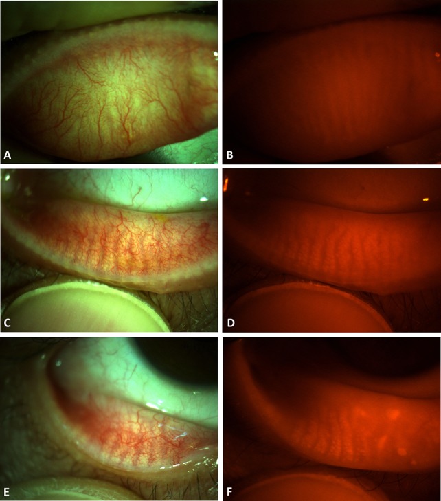
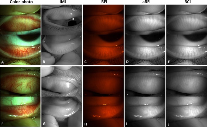
Methods: We evaluated MG dropout in the everted upper and lower eyelids of 125 eyes of 64 patients with good-quality infrared meibography images (IMIs) and color digital photographs with and without a red filter. Red-filtered images (RFIs) were converted to black and white and adjusted for contrast/brightness [adjusted red-filtered images (aRFIs)]. RCIs were computationally isolated from color digital photographs obtained without a red filter. After randomization, the total meiboscore (0-6) was evaluated by 2 independent evaluators (interobserver reliability) masked to the image origin, and again after a 30-day interval (intraobserver reliability).
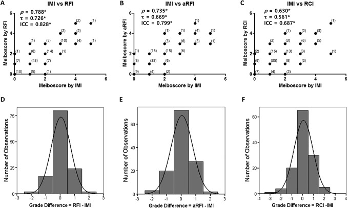
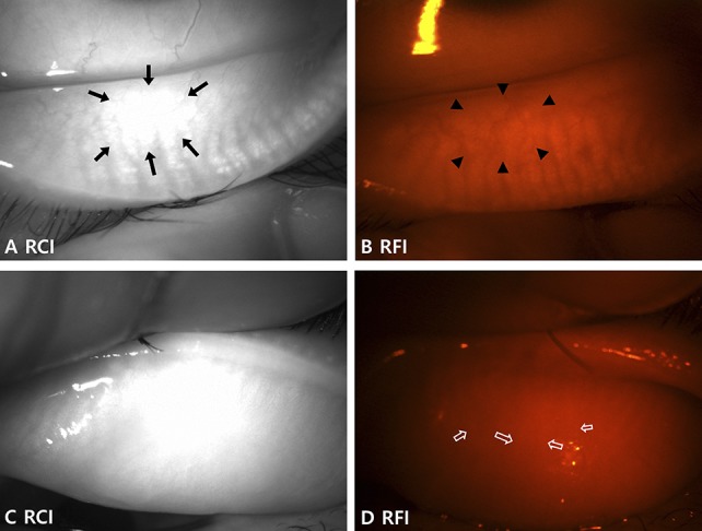
Results: The meiboscores evaluated using the RFI, aRFI, and RCI were strongly positively correlated with those evaluated using the IMI (RFI: ρ = 0.788; aRFI: ρ = 0.735; RCI: ρ = 0.630; all P < 0.001, Spearman correlation analysis). Linear-weighted κ-values (κw) showed substantial agreement between the RFI and IMI (κw = 0.676, 95% CI = 0.594-0.759). The RFI had substantial intraobserver reliability (κw = 0.735, 95% CI = 0.685-0.785) and moderate interobserver reliability (κw = 0.467, 95% CI = 0.371-0.563). Computational adjustment of RFIs did not enhance the validity or reliability, and RCIs had limitations in some cases.
Conclusions: MGs were successfully visualized using a red filter on a slit lamp and showed substantial agreement with visualization using the standard infrared method. Although interobserver reliability was only moderate, this alternative technique may be useful for evaluating MG dropout when an infrared meibography device is not available.
Conflict of interest statement
The authors have no conflicts of interest to disclose.
Figures
Comment in
-
Reply.Cornea. 2019 Aug;38(8):e34. doi: 10.1097/ICO.0000000000002009. Cornea. 2019. PMID: 31135492 No abstract available.
-
Evaluating a Simpler Method of Noncontact Infrared Meibography Using Still Photography.Cornea. 2019 Aug;38(8):e32-e34. doi: 10.1097/ICO.0000000000002010. Cornea. 2019. PMID: 31135495 No abstract available.
References
-
- Craig JP, Nichols KK, Akpek EK, et al. TFOS DEWS II definition and classification report. Ocul Surf. 2017;15:276–283. - PubMed
-
- Jester JV, Rife L, Nii D, et al. In vivo biomicroscopy and photography of meibomian glands in a rabbit model of meibomian gland dysfunction. Invest Ophthalmol Vis Sci. 1982;22:660–667. - PubMed
-
- Mathers WD, Daley T, Verdick R. Video imaging of the meibomian gland. Arch Ophthalmol. 1994;112:448–449. - PubMed
-
- Arita R, Itoh K, Inoue K, et al. Noncontact infrared meibography to document age-related changes of the meibomian glands in a normal population. Ophthalmology. 2008;115:911–915. - PubMed
Publication types
MeSH terms
LinkOut - more resources
Full Text Sources
Medical
