. 2019 Mar 26;58(12):1627-1647.
doi: 10.1021/acs.biochem.9b00044. Epub 2019 Mar 7.

A New Microbial Pathway for Organophosphonate Degradation Catalyzed by Two Previously Misannotated Non-Heme-Iron Oxygenases

A New Microbial Pathway for Organophosphonate Degradation Catalyzed by Two Previously Misannotated Non-Heme-Iron Oxygenases

Lauren J Rajakovich et al. Biochemistry. .

Abstract

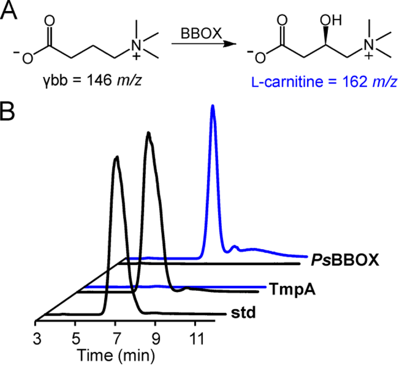
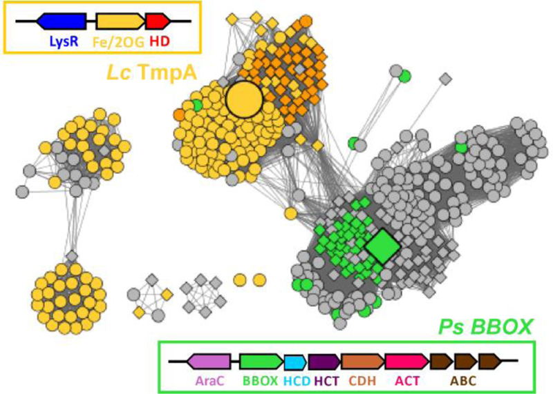
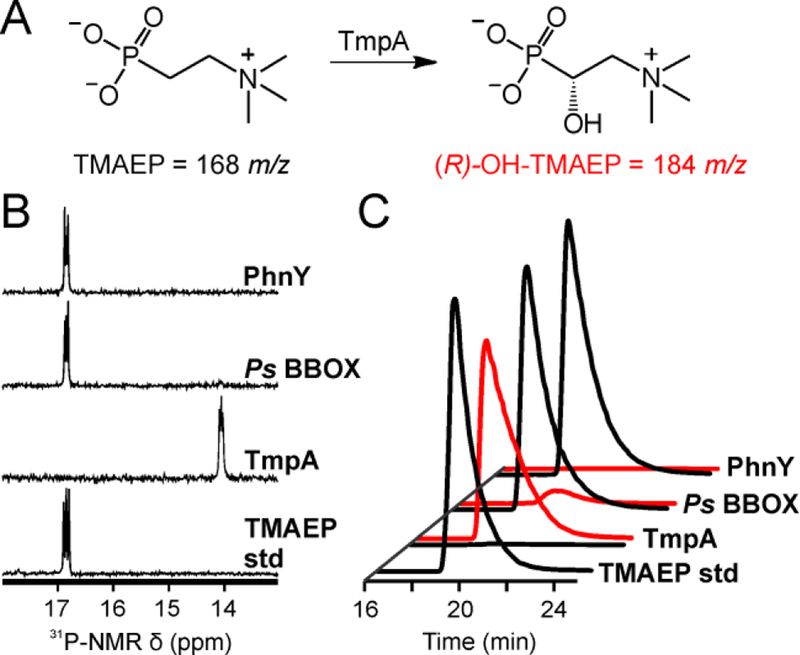
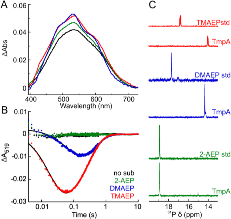
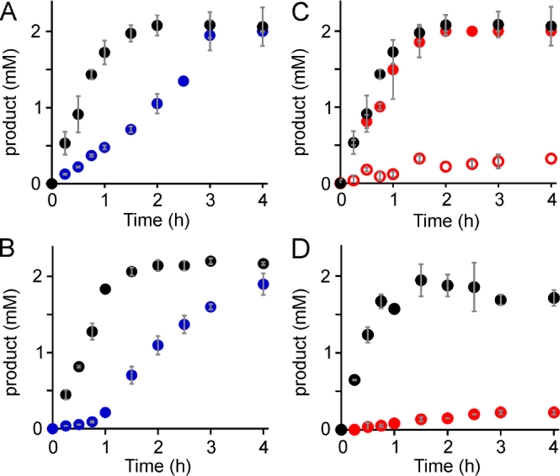
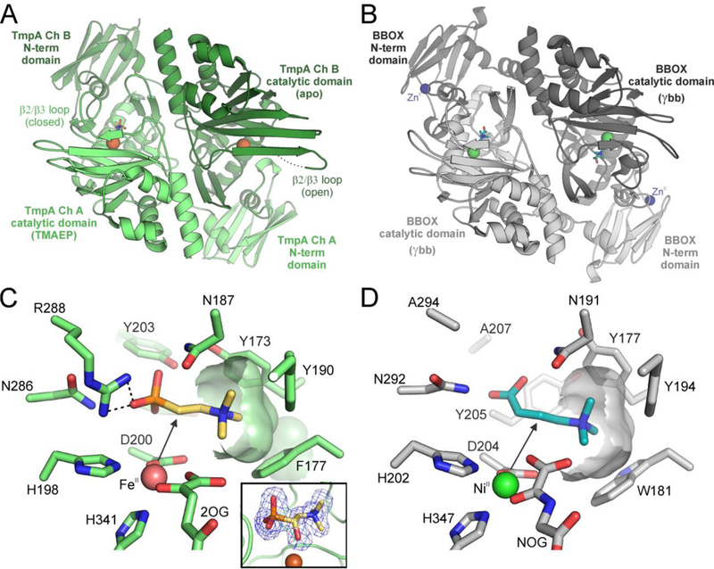
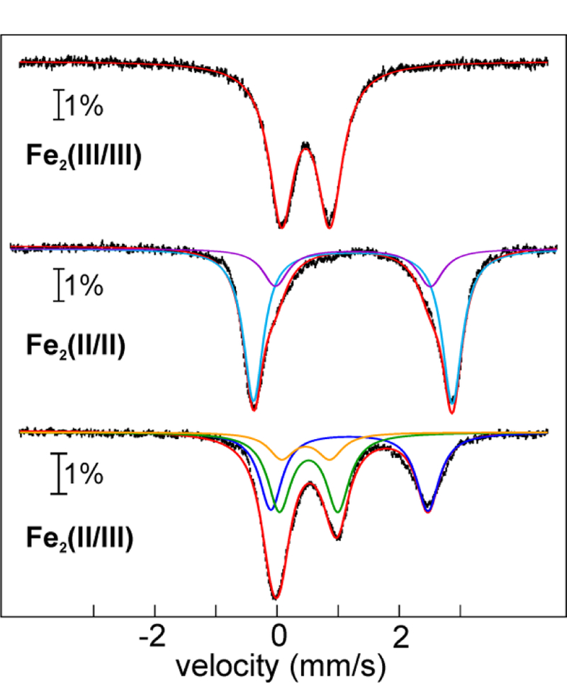
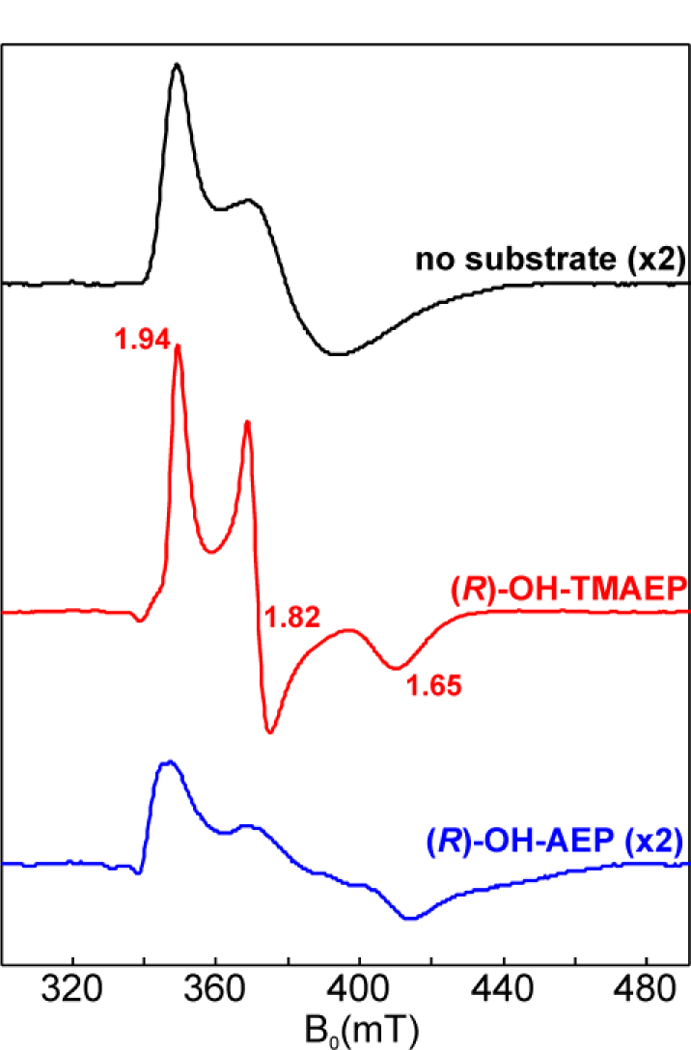
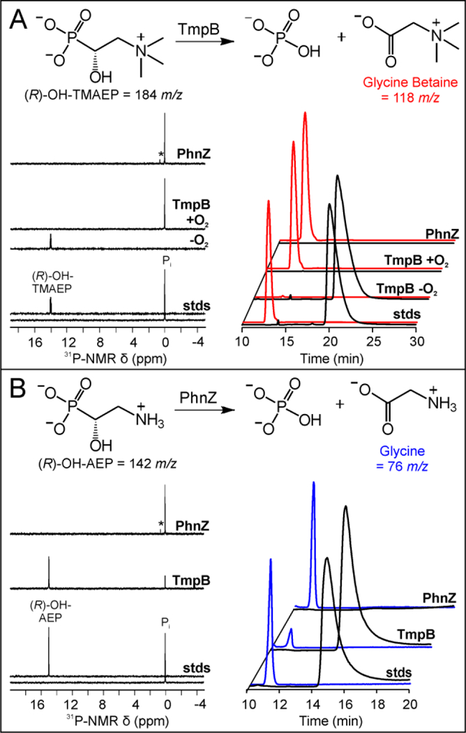
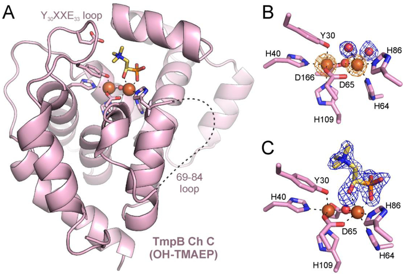
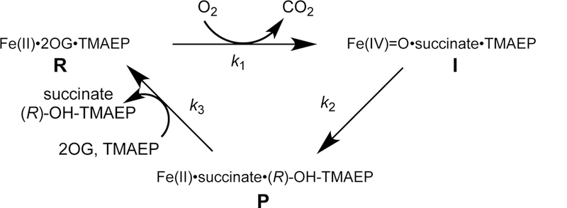
The assignment of biochemical functions to hypothetical proteins is challenged by functional diversification within many protein structural superfamilies. This diversification, which is particularly common for metalloenzymes, renders functional annotations that are founded solely on sequence and domain similarities unreliable and often erroneous. Definitive biochemical characterization to delineate functional subgroups within these superfamilies will aid in improving bioinformatic approaches for functional annotation. We describe here the structural and functional characterization of two non-heme-iron oxygenases, TmpA and TmpB, which are encoded by a genomically clustered pair of genes found in more than 350 species of bacteria. TmpA and TmpB are functional homologues of a pair of enzymes (PhnY and PhnZ) that degrade 2-aminoethylphosphonate but instead act on its naturally occurring, quaternary ammonium analogue, 2-(trimethylammonio)ethylphosphonate (TMAEP). TmpA, an iron(II)- and 2-(oxo)glutarate-dependent oxygenase misannotated as a γ-butyrobetaine (γbb) hydroxylase, shows no activity toward γbb but efficiently hydroxylates TMAEP. The product, ( R)-1-hydroxy-2-(trimethylammonio)ethylphosphonate [( R)-OH-TMAEP], then serves as the substrate for the second enzyme, TmpB. By contrast to its purported phosphohydrolytic activity, TmpB is an HD-domain oxygenase that uses a mixed-valent diiron cofactor to enact oxidative cleavage of the C-P bond of its substrate, yielding glycine betaine and phosphate. The high specificities of TmpA and TmpB for their N-trimethylated substrates suggest that they have evolved specifically to degrade TMAEP, which was not previously known to be subject to microbial catabolism. This study thus adds to the growing list of known pathways through which microbes break down organophosphonates to harvest phosphorus, carbon, and nitrogen in nutrient-limited niches.


Figures

Figure 1
Figure 1
Assessment of the γbb hydroxylase activities of TmpA and Ps BBOX. (A) The reaction catalyzed by BBOX. (B) LC-MS chromatograms monitoring γbb (146 m/z, black) and L-carnitine (162 m/z, blue) after a 4-h incubation of 0.01 mM TmpA or Ps BBOX, 0.02 mM (NH4)2Fe(SO4)2, 0.2 mM sodium L-ascorbate, 1 mM 2OG and 1 mM γbb.
Figure 2
Figure 2
Sequence similarity network (SSN) illustrating divergence of BBOX-like and TmpA-like proteins. The clusters of bacterial protein sequences shown here are derived from the IPR003819 SSN (Figures S1–3). The nodes represent protein sequences with > 90% identity. Edges between the nodes represent pairwise alignment scores of < 10−71 (corresponding to ~ 33% sequence identity). The large yellow circle and green diamond represent Lc TmpA and Ps BBOX, respectively. Orange nodes represent predicted fusion proteins with both TmpA- and TmpB-like domains. Diamond shaped nodes represent sequences with an N-terminal Zn(II)-binding motif (see discussion). The yellow and green boxes summarize the genomic context of proteins represented by the yellow and green nodes, respectively. Other designations: LysR, LysR-type transcription regulator; AraC, AraC-type transcription regulator; CDH, carnitine dehydrogenase; HCT, 4-hydroxybenzoyl-CoA thioesterase; HCD, 3-hydroxybutyryl-CoA dehydrogenase; ACT, Acetyl-CoA acetyltransferase/thiolase; ABC, ABC-type glycine/betaine transporter.
Figure 3
Figure 3
Activities of selected Fe/2OG oxygenases toward TMAEP. (A) The chemical transformation catalyzed by TmpA. (B) 31P-NMR spectra and (C) LC-MS chromatograms after a 4-h incubation of a solution containing 0.01 mM TmpA, PhnY or Ps BBOX with 0.02 mM (NH4)2Fe(SO4)2, 0.2 mM L-ascorbate, 3 mM 2OG and 2 mM TMAEP. The triplet splitting in the 1H-decoupled 31P-NMR spectra of the TMAEP substrate and (R)-OH-TMAEP product in panel B is presumed to result from the 14N nucleus on C1. The reason it is present in the spectrum of the N-trimethylated compound, but not those of the di- and unmethylated analogs, is not clear and is discussed more in the Supporting Information.
Figure 4
Figure 4
Ultraviolet-visible absorption data showing binding of TMAEP to TmpA•Fe(II)•2OG and triggering of O2 addition. (A) Difference absorption spectra associated with binding of 2OG (5 mM) to the TmpA•Fe(II) complex [0.6 mM TmpA, 0.5 mM (NH4)2Fe(SO4)2] in the absence of substrate (black) and presence of 5 mM TMAEP (red). (B) Stopped-flow absorption spectra acquired after mixing at 5 °C of a solution of 1.2 mM TmpA, 1 mM (NH4)2Fe(SO4)2, 10 mM 2OG and 10 mM TMAEP with an equal volume of air-saturated 50 mM sodium HEPES buffer, pH 7.5 (~ 0.4 mM O2 at 5 °C). The inset shows the absorption spectra at indicated time points after subtraction of the spectrum of the TmpA•Fe(II)•2OG•TMAEP complex. (C) Kinetic traces at 318 nm (blue dots) and 519 nm (red dots) extracted from the time-dependent spectra in panel B. The solid black lines are regression fits to the data, as described in the Experimental Section.
Figure 5
Figure 5
Activity of TmpA toward TMAEP analogues with varying degrees of N-methylation. (A) Absorption difference spectra caused by binding of 2OG (5 mM) to the TmpA•Fe(II) complex [0.6 mM TmpA, 0.5 mM (NH4)2Fe(SO4)2] in the absence of a substrate (black) and in the presence of 5 mM TMAEP (red), DMAEP (blue) or 2-AEP (green). (B) Kinetic traces at 519 nm after mixing of the solutions described in panel A with air-saturated 50 mM sodium HEPES buffer, pH 7.5 (~ 0.2 mM O2 final at 5 °C). The solid black lines are non-linear regression fits to the data, as described in the Experimental Section. (C) 31P-NMR spectra of reaction samples after a 4 h incubation of a solution of 0.01 mM TmpA, 0.02 mM (NH4)2Fe(SO4)2, 0.2 mM sodium ascorbate, 3 mM 2OG and 2 mM of either TMAEP (red), DMAEP (blue), or 2-AEP (green).
Figure 6
Figure 6
Evaluation of relative specificity of TmpA for TMAEP and either DMAEP or PC by direct competition. All reactions were performed at 3 °C and contained 0.02 mM TmpA, 0.03 mM (NH4)2Fe(SO4)2, 0.4 mM sodium ascorbate, 6 mM 2OG and 2 mM of each substrate. (A) Control reactions containing either TMAEP (black) or DMAEP (blue) in the absence of the other compound, monitoring corresponding hydroxylated products by LC-MS. (B) Competition reactions containing both TMAEP and DMAEP, monitoring corresponding hydroxylated products by LC-MS. (C) Control reactions containing either TMAEP (black) or PC (filled red), monitoring (R)-OH-TMAEP production and PC consumption, respectively, by LC-MS The open red circles are the concentration of PC consumed, detected by LC-MS, from a reaction containing PC and 2 mM (R)-OH-TMAEP. (D) Competition reactions containing both TMAEP and PC, monitoring (R)-OH-TMAEP and phosphate products by 31P-NMR because PC and (R)-OH-TMAEP have the same m/z and similar retention times.
Figure 7
Figure 7
Structural comparison of Lc TmpA and Hs BBOX. (A) Homodimeric quaternary structure of TmpA with chain A in light green and chain B in dark green. Fe(II) ions are shown as brown spheres. (B) Homodimeric quaternary structure of Hs BBOX (PDB accession code 3MS5) with chain A in dark gray and chain B in light gray. The Zn(II) and Ni(II) ions are shown as blue and green spheres, respectively. (C) Active site of chain A in the TmpA substrate-bound structure, showing the co-substrate (2OG), amino acid side chains, and TMAEP (yellow) in stick format. Electrostatic interactions are designated by black dashed lines and the black arrow identifies the position of hydroxylation. The inset shows the Fo-Fc omit map contoured at 3.0σ (blue mesh) and atomic model for (R)-OH-TMAEP (yellow sticks) in chain A of the TmpA product-bound structure. (D) Active site in the substrate-bound structure of Hs BBOX (PDB accession code 3MS5), showing the co-substrate analog, N-oxalylglycine (NOG), amino acids side chains, and γbb (blue) in stick format.
Figure 8
Figure 8
120-K/0-T Mössbauer spectra of 2 mM O2-free TmpA prepared in three oxidation states. All incubations were carried out for 45 min in an anoxic chamber. Experimental spectra are shown as black vertical bars. Overall simulations are shown as red lines. (Top) Fe2(III/III) state obtained by treatment of the as-isolated protein with 3 mM potassium ferricyanide. (Middle) Fe2(II/II) state obtained by treatment of the as-isolated protein with 20 mM sodium dithionite. The two quadrupole-doublet components of the simulated spectrum of the Fe2(II/II) complex are shown as purple and cyan lines; the parameters are provided in the main text. (Bottom) Fe2(II/III) state accumulated to ~ 65 % by treatment of the as-isolated protein with 20 mM sodium L-ascorbate. The simulated sub-spectra corresponding to the Fe2(II/III) species (orange) and the paired Fe(II) (blue) and Fe(III) (green) sub-sites are shown as solid lines; their parameters are provided in the main text. The contribution of the Fe2(II/II) species (22%) has already been removed (by subtraction from the experimental spectrum; Figure S20) for clarity.
Figure 9
Figure 9
X-band EPR spectra of O2-free 0.25 mM TmpB after treatment with 10 mM sodium L-ascorbate (black) followed by addition of either 10 mM (R)-OH-TMAEP (red) or (R)-OH-AEP (blue). Experimental conditions: temperature = 10 K, microwave power = 0.2 mW, microwave frequency = 9.479 GHz, modulation amplitude = 1 mT.
Figure 10
Figure 10
Activity of the HD-MVDOs against aminophosphonate compounds with and without N-methylation. The chemical transformations are depicted at the top of each panel. Aerobic reactions containing 0.01 mM TmpB or PhnZ, 0.2 mM L-ascorbate, and 2 mM of either (A) (R)-OH-TMAEP or (B) (R)-OH-AEP were incubated for 4 h at 3 °C. (Left) 31P-NMR spectra detecting the substrates, (R)-OH-TMAEP or (R)-OH-AEP, and the product phosphate. The asterisks mark a contaminant present in the PhnZ protein preparation. (Right) LC-MS chromatograms detecting the substrates, (R)-OH-TMAEP or (R)-OH-AEP, and products, glycine betaine or glycine.
Figure 11
Figure 11
Views of TmpB from the X-ray crystal structure solved in this study. (A) Cartoon depiction of chain C of the TmpB structure. Iron ions are shown as brown spheres and (R)-OH-TMAEP (yellow) is shown in stick format. A dashed line represents the unmodeled region of the structure. TmpB active site of (B) chain A lacking substrate and (C) chain D with (R)-OH-TMAEP (yellow sticks) bound. Water molecules are shown as red spheres and iron ions as orange spheres. Orange mesh depicts the anomalous Fourier density contoured at 10σ. Blue mesh depicts the FoFc omit map contoured at 3σ for either the coordinating water molecules or the substrate.
Scheme 1
Scheme 1

Similar articles

Cited by

References

    1. Sono M, Roach MP, Coulter ED, Dawson JH (1996) Heme-containing oxygenases. Chem. Rev 96, 2841–2888. - PubMed
    1. Hausinger RP (2015) 2-Oxoglutarate-Dependent Oxygenases, Hausinger RP, Schofield CJ, Ed., The Royal Society of Chemistry.
    1. Andrews SC (2010) The ferritin-like superfamily: Evolution of the biological iron storeman from a rubrerythrin-like ancestor. Biochim. Biophys. Acta 1800, 691–705. - PubMed
    1. Aravind L, Koonin EV (1998) The HD domain defines a new superfamily of metal-dependent phosphohydrolases. Trends Biochem. Sci 23, 469–472. - PubMed
    1. Baier F, Copp JN, Tokuriki N (2016) Evolution of enzyme superfamilies: Comprehensive exploration of sequence-function relationships. Biochemistry 55, 6375–6388. - PubMed

Publication types

MeSH terms

Supplementary concepts

LinkOut - more resources