. 2019 Apr 1;316(4):G527-G538.
doi: 10.1152/ajpgi.00148.2018. Epub 2019 Feb 21.

Inhibition of soluble epoxide hydrolase ameliorates hyperhomocysteinemia-induced hepatic steatosis by enhancing β-oxidation of fatty acid in mice

Affiliations

Inhibition of soluble epoxide hydrolase ameliorates hyperhomocysteinemia-induced hepatic steatosis by enhancing β-oxidation of fatty acid in mice

Liu Yao et al. Am J Physiol Gastrointest Liver Physiol. .

Abstract

Hepatic steatosis is the beginning phase of nonalcoholic fatty liver disease, and hyperhomocysteinemia (HHcy) is a significant risk factor. Soluble epoxide hydrolase (sEH) hydrolyzes epoxyeicosatrienoic acids (EETs) and other epoxy fatty acids, attenuating their cardiovascular protective effects. However, the involvement of sEH in HHcy-induced hepatic steatosis is unknown. The current study aimed to explore the role of sEH in HHcy-induced lipid disorder. We fed 6-wk-old male mice a chow diet or 2% (wt/wt) high-metnionine diet for 8 wk to establish the HHcy model. A high level of homocysteine induced lipid accumulation in vivo and in vitro, which was concomitant with the increased activity and expression of sEH. Treatment with a highly selective specific sEH inhibitor (0.8 mg·kg-1·day-1 for the animal model and 1 μM for cells) prevented HHcy-induced lipid accumulation in vivo and in vitro. Inhibition of sEH activated the peroxisome proliferator-activated receptor-α (PPAR-α), as evidenced by elevated β-oxidation of fatty acids and the expression of PPAR-α target genes in HHcy-induced hepatic steatosis. In primary cultured hepatocytes, the effect of sEH inhibition on PPAR-α activation was further confirmed by a marked increase in PPAR-response element luciferase activity, which was reversed by knock down of PPAR-α. Of note, 11,12-EET ligand dependently activated PPAR-α. Thus increased sEH activity is a key determinant in the pathogenesis of HHcy-induced hepatic steatosis, and sEH inhibition could be an effective treatment for HHcy-induced hepatic steatosis. NEW & NOTEWORTHY In the current study, we demonstrated that upregulation of soluble epoxide hydrolase (sEH) is involved in the hyperhomocysteinemia (HHcy)-caused hepatic steatosis in an HHcy mouse model and in murine primary hepatocytes. Improving hepatic steatosis in HHcy mice by pharmacological inhibition of sEH to activate peroxisome proliferator-activated receptor-α was ligand dependent, and sEH could be a potential therapeutic target for the treatment of nonalcoholic fatty liver disease.

Keywords: hepatocytes; hyperhomocysteinemia; proliferator-activated receptor-α; soluble epoxide hydrolase; β-oxidation.


Conflict of interest statement

No conflicts of interest, financial or otherwise, are declared by the authors.

Figures

Fig. 1.
Fig. 1.
Effect of hyperhomocysteinemia (HHcy) on lipid accumulation and soluble epoxide hydrolase (sEH) expression and activity in vivo. Male C57BL/6 mice (6 wk old) were fed a standard chow diet or 2% (wt/wt) l-methionine diet for 8 wk. A: Oil-red O staining (top) and hematoxylin and eosin (H&E) staining (bottom) of livers in representative sections. Images were taken at ×200 magnification. B and C: hepatic triglyceride and cholesterol content. D: Western blot analysis of the specificity of sEH antibody by sEH blocking peptide in the control mice liver tissue (left). Ab, antibody. The ratios represented sEH antibody to blocking peptide. Western blot analysis and quantification of sEH protein content (right) in the liver of mice fed a high-methionine diet (HMD). β-Actin was an internal control. E: quantitative (q)PCR analysis of the mRNA level of sEH. F: sEH activity was evaluated by the metabolic rate of 11,12-dihydroxyeicosatrienoic acid (DHET) to 11,12-epoxyeicosatrienoic acid (EET) by liquid chromatography with tandem mass spectrometry (LC-MS/MS). G: LC-MS/MS analysis of each regioisomer of EET level in the liver (right). H: qPCR analysis of mRNA levels of genes involved in EETs synthesis. Data are means ± SE by unpaired t-test; n = 6 for chow diet; n = 8 for HMD. *P < 0.05; **P < 0.01 vs. Chow.
Fig. 2.
Fig. 2.
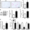
Effect of hyperhomocysteinemia (HHcy) on lipid accumulation and the expression of soluble epoxide hydrolase (sEH) in primary hepatocytes. Murine primary hepatocytes were cultured and treated with Hcy (100 μM) for 24 h. A: Oil-red O staining of representative images (left) and quantification (right) of intracellular lipid deposition. Images were taken at ×200 magnification. B: Western blot analysis (top) and quantification (bottom) of sEH protein level. β-Actin was an internal control. C: mRNA level of sEH in Hcy-treated hepatocytes. D: sEH activity was evaluated by the metabolic rate of 11,12-dihydroxyeicosatrienoic acid (DHET) to 11,12-epoxyeicosatrienoic acid (EET) by liquid chromatography with tandem mass spectrometry (LC-MS/MS). E: LC-MS/MS analysis of each regioisomer of EET level in hepatocytes. Data are means ± SE of 3 independent experiments by unpaired t-test. *P < 0.05; **P < 0.01 vs. Ctrl.
Fig. 3.
Fig. 3.
Inhibition of soluble epoxide hydrolase (sEH) prevented hyperhomocysteinemia (HHcy)-induced hepatic steatosis. Male C57BL/6 mice were fed a chow diet or high-methionine diet (HMD) with or without 1-trifluoromethoxyphenyl-3-(1-propionylpiperidin-4-yl) urea (TPPU) in drinking water (0.8 mg·kg−1·day−1) for 4 wk. A: representative Oil-red O staining for lipid deposition and hematoxylin and eosin staining for morphological changes in liver. Images were taken at ×200 magnification. B and C: hepatic triglyceride (TG) and cholesterol (Chol) content. D: Western blot analysis (top) and quantification (bottom) of hepatic sEH protein level in mice with 4-wk treatment. E: quantiative PCR analysis of the mRNA level of sEH. F: sEH activity was evaluated by the metabolic rate of 11,12-dihydroxyeicosatrienoic acid (DHET) to 11,12-epoxyeicosatrienoic acid (EET) by liquid chromatography with tandem mass spectrometry (LC-MS/MS). G: LC-MS/MS analysis of each regioisomer of EETs level in the liver. TG. Data are means ± SE by one-way ANOVA; n = 5 for chow diet; n = 7 for HMD; and n = 8 for HMD + TPPU. *P < 0.05, **P < 0.01.
Fig. 4.
Fig. 4.
Inhibition of soluble epoxide hydrolase (sEH) enhanced hepatic fatty acid β-Oxidation. C57BL/6 mice were fed a chow diet or high-methionine diet (HMD) for 8 wk with or without 1-trifluoromethoxyphenyl-3-(1-propionylpiperidin-4-yl) urea (TPPU) in drinking water (0.8 mg·kg−1·day−1) for 4 wk. A and C: quantitative PCR analysis of mRNA levels of genes involved in hepatic fatty acid uptake and fatty acid oxidation. C: Western blot analysis (left) and quantification (right) of peroxisome proliferator-activated receptor-α (PPAR-α) and target genes protein content in the liver. β-Actin was an internal control. D: plasma level of β-hydroxybutyrate in mice. Cpt1a, carnitine palmitoyl transferase 1a; Acox1, acyl-coenzyme A oxidase 1; Scad, Mcad, and Lcad, short-, medium- and long-chain acyl-coenzyme A dehydrogenase; Fatp, fatty acid transporter protein; Fabp, fatty acid binding protein. Data are means ± SE by one-way ANOVA. n = 5 for chow diet; n = 7 for HMD; and n = 8 for HMD + TPPU. *P < 0.05, **P < 0.01.
Fig. 5.
Fig. 5.
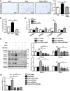
Inhibition of soluble epoxide hydrolase (sEH) activated peroxisome proliferator-activated receptor-α (PPAR-α) in hepatocytes. AC: murine primary hepatocytes were incubated with Hcy with or without 1-trifluoromethoxyphenyl-3-(1-propionylpiperidin-4-yl) urea (TPPU); 1 μM) for 24 h. A: Oil-red O staining of representative images (left) and quantification (right) of intracellular lipid deposition. Images were taken at ×200 magnification. B: sEH activity was evaluated by the metabolic rate of 11,12-dihydroxyeicosatrienoic acid (DHET) to 11,12-epoxyeicosatrienoic acid (EET) by liquid chromatography with tandem mass spectrometry (LC-MS/MS). C: LC-MS/MS analysis of each regioisomer of EET level in hepatocytes. D and E: hepatocytes were treated with Hcy with or without TPPU for 24 h after siRNA transfection of PPAR-α (siPPAR-α) or negative control (siNC) for 36 h. D: Western blot analysis (left) and quantification (right) of PPAR-α and protein levels of target genes. β-Actin was an internal control. E: relative PPAR-response element (PPRE) luciferase activity. Cpt1a, carnitine palmitoyl transferase 1a; Acox1, acyl-coenzyme A oxidase 1;, Mcad, medium-chain acyl-coenzyme A dehydrogenase. Data are means ± SE of 3 independent experiments by one-way ANOVA. *P < 0.05; **P < 0.01.
Fig. 6.
Fig. 6.
Activation of peroxisome proliferator-activated receptor-α (PPAR-α) depends on 11,12-epoxyeicosatrienoic acid (EET). A: primary hepatocytes were transfected with PPAR-response element (PPRE)-luciferase reporter for 36 h, then treated with each regioisomer of EETs (100 nM) for 24 h. Data are means ± SE of 3 independent experiments by unpaired t-test. *P < 0.05 vs. Ctrl. B: proposed mechanism of improvement of hyperhomocysteinemia (HHcy)-induced hepatic steatosis by sEH inhibitor in liver. High levels of Hcy induced fatty acid uptake by upregulating of CD36. Inhibition of soluble epoxide hydrolase (sEH) prevented EETs degradation from HHcy. As an endogenous activator of PPAR-α, EETs promotes the binding of PPAR-α to target genes, which facilitates the β-oxidation of fatty acids and lipid removal from the liver. DHET, 11,12-dihydroxyeicosatrienoic acid; TPPU, 1-trifluoromethoxyphenyl-3-(1-propionylpiperidin-4-yl) urea.

Similar articles

Cited by

References

    1. Abdelmegeed MA, Yoo SH, Henderson LE, Gonzalez FJ, Woodcroft KJ, Song BJ. PPARalpha expression protects male mice from high fat-induced nonalcoholic fatty liver. J Nutr 141: 603–610, 2011. doi:10.3945/jn.110.135210. - DOI - PMC - PubMed
    1. Angulo P. Nonalcoholic fatty liver disease. N Engl J Med 346: 1221–1231, 2002. doi:10.1056/NEJMra011775. - DOI - PubMed
    1. Audelin MC, Genest J Jr. Homocysteine and cardiovascular disease in diabetes mellitus. Atherosclerosis 159: 497–511, 2001. doi:10.1016/S0021-9150(01)00531-7. - DOI - PubMed
    1. Barbier O, Torra IP, Duguay Y, Blanquart C, Fruchart JC, Glineur C, Staels B. Pleiotropic actions of peroxisome proliferator-activated receptors in lipid metabolism and atherosclerosis. Arterioscler Thromb Vasc Biol 22: 717–726, 2002. doi:10.1161/01.ATV.0000015598.86369.04. - DOI - PubMed
    1. Bocher V, Pineda-Torra I, Fruchart JC, Staels B. PPARs: transcription factors controlling lipid and lipoprotein metabolism. Ann N Y Acad Sci 967: 7–18, 2002. doi:10.1111/j.1749-6632.2002.tb04258.x. - DOI - PubMed

Publication types

LinkOut - more resources