. 2019 Jun;15(3):448-455.
doi: 10.1007/s12015-019-09880-9.

Wnt Signaling Mediates the Aging-Induced Differentiation Impairment of Intestinal Stem Cells

Affiliations

Wnt Signaling Mediates the Aging-Induced Differentiation Impairment of Intestinal Stem Cells

Hui Cui et al. Stem Cell Rev Rep. 2019 Jun.

Abstract

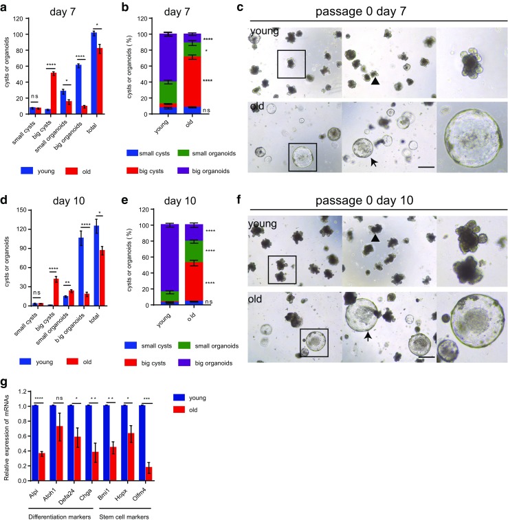
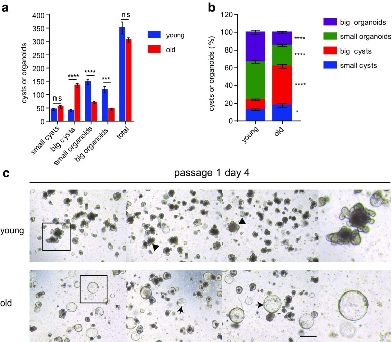
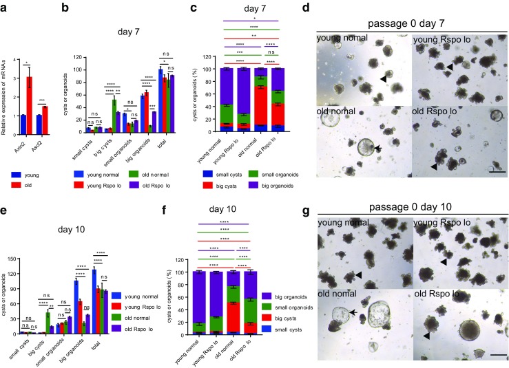
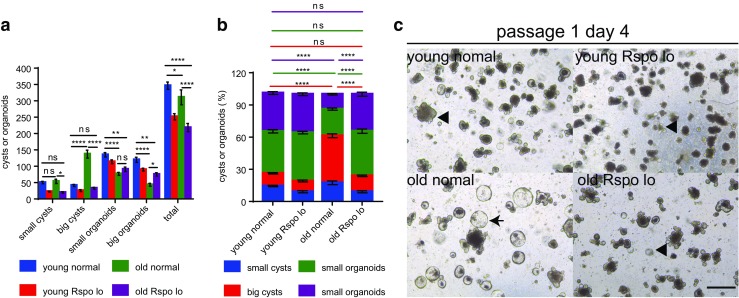
Stem cell aging underlies aging-associated disorders, such as steeply increased incidences of tumors and impaired regeneration capacity upon stress. However, whether and how the intestinal stem cells age remains largely unknown. Here we show that intestinal stem cells derived from 24-month-old mice hardly form typical organoids with crypt-villus structures, but rather mainly form big, rounded cysts devoid of differentiated cell types, which mimics the culturing of heterozygous APC-deficient cells from the APCmin mouse line. Further analysis showed that cultured crypts derived from aged mice exhibited reduced expression levels of differentiation genes and higher expression of Wnt target genes. Lowering the concentration of R-spondin-1 in the culture system significantly reduced formation of rounded cysts, accompanied by an increased formation of organoids from crypts derived from old mice. We are the first to uncover that intestinal stem cells derived from old mice harbor significant deficiency in differentiation that can be partially rescued through a reduction in R-spondin-1 exposure. This could be highly relevant to intestinal tumor development and the reduced regeneration potential observed in the aged population. Our study provides the first experimental evidence that an over-responsiveness to Wnt/beta-catenin signaling of aged intestinal stem cells mediates the aging-induced deficiency in differentiation, and could serve as a potential target to ameliorate aging-associated intestinal pathologies.

Keywords: Aging; Differentiation; Intestinal stem cells; R-spondin-1; Wnt signaling.


Conflict of interest statement

The authors declare no potential conflicts of interest.

Figures

Fig. 1
Fig. 1
ISCs from aged mice exhibit significant deficiency in differentiation in primary culture. Freshly isolated crypts from 2 months old (young) and 24 months old (old) mice were plated at a density of 200 crypts per well (results show data from one representative experiment out of 2 independent experiments; n = 3 mice per group). (a, b) Absolute number (a) and percentage (b) of grown out structures on day 7 after plating. c Representative pictures of indicated groups on day 7 after plating. d,e Absolute number (d) and percentage (e) of grown out structures on day 10 after plating. f Representative pictures of indicated groups on day 10 after plating. Small cysts: diameters ≤70 μm; big cysts: diameters >70 μm; small organoids: with crypt-villus architectures, budding number ≤ 3 (for a and b) and budding number ≤ 5 (for d and e); big organoids: with crypt-villus architectures, budding number > 3 (for a and b) and budding number > 5 (for d and e). Arrow heads indicate typical organoids; arrows indicate typical big cysts. g mRNA expression of indicated genes in crypts cultured for 7 days (n = 3 independent experiments). mRNA expression of genes was normalized to beta-actin with the expression level of each gene in young crypts set to 1. Data are displayed as mean ± SEM. *, P < 0.05; **, P < 0.01; ****, P < 0.0001. ns, not significant. Unpaired two tailed Student’s t test was used. Scale bar: 200 μm
Fig. 2
Fig. 2
ISCs from aged mice exhibit significant deficiency in differentiation in subculture. a-c Cultured crypts were dissociated and passaged on day 14 after primary plating. a,b Absolute number (a) and percentage (b) of grown out structures. c Representative pictures of indicated groups on day 4 after secondary plating. Small cysts: diameters ≤70 μm; big cysts: diameters >70 μm; small organoids: with crypt-villus architectures, budding number ≤ 3; big organoids: with crypt-villus architectures, budding number > 3. Arrow heads indicate typical organoids; arrows indicate typical big cysts. Data are displayed as mean ± SEM. *, P < 0.05; ***, P < 0.001; ****, P < 0.0001. ns, not significant. Unpaired two tailed Student’s t test was used. Scale bar: 200 μm
Fig. 3
Fig. 3
Reduction in R-spondin-1 exposure during primary culture ameliorates the aging-induced deficiency in differentiation of ISCs. a mRNA expression of Wnt target genes in crypts derived from 2 months old (young) and 24 months old (old) mice after culturing for 7 days. mRNA expression of genes was normalized to beta-actin with the expression level of each gene in young crypts set to 1 (n = 3 independent experiments). b-g Freshly isolated crypts from 2 months old (young) and 24 months old (old) mice were plated at a density of 200 crypts per well. Concentration of R-spondin-1was reduced to 1/3 of the normal level (330 ng/ml) in half of the wells (results show data from one representative experiment out of 2 independent experiments; n = 3 mice per group). b,c Absolute number (b) and percentage (c) of grown out structures on day 7 after primary plating. d Representative pictures of indicated groups on day 7 after plating. e,f Absolute number (e) and percentage (f) of grown out structures on day 10 after plating. g Representative pictures of indicated groups on day 10 after plating. Young normal: ISCs derived from young mice culturing in normal concentration of R-spondin-1; young Rspo lo: ISCs derived from young mice culturing in 1/3 concentration of R-spondin-1; old normal: ISCs derived from old mice culturing in normal concentration of R-spondin-1; old Rspo lo: ISCs derived from old mice culturing in 1/3 concentration of R-spondin-1;Small cysts: diametres≤70 μm; big cysts: diameters>70 μm; small organoids: with crypt-villus architectures, budding number ≤ 3 (for b and c) and budding number ≤ 5 (for e and f); big organoids: with crypt-villus architectures, budding number > 3 (for b and c) and budding number > 5 (for e and f). Arrow heads indicate typical organoids; arrows indicate typical big cysts. Data are displayed as mean ± SEM. *, P < 0.05; **, P < 0.01; ***, P < 0.001; ****, P < 0.0001. ns, not significant. Unpaired two-tailed Student’s t test was used for a. Two-way ANOVA analysis was used for b,c,e,f. Scale bar: 200 μm
Fig. 4
Fig. 4
Reduction in R-spondin-1 exposure during subculture ameliorates the aging-induced deficiency in differentiation of ISCs. a-c) Cultured crypts were dissociated and passaged on day 14 after primary plating. a,b Absolute number (a) and percentage (b) of grown out structures. c Representative pictures of indicated groups on day 4 after secondary plating. Young normal: ISCs derived from young mice culturing in normal concentration of R-spondin-1; young Rspo lo: ISCs derived from young mice culturing in 1/3 concentration of R-spondin-1; old normal: ISCs derived from old mice culturing in normal concentration of R-spondin-1; old Rspo lo: ISCs derived from old mice culturing in 1/3 concentration of R-spondin-1; Small cysts: diametres≤70 μm; big cysts: diameters>70 μm; small organoids: with crypt-villus architectures, budding number ≤ 3; big organoids: with crypt-villus architectures, budding number > 3. Arrow heads indicate typical organoids; arrows indicate typical big cysts. Data are displayed as mean ± SEM. *, P < 0.05; **, P < 0.01; ****, P < 0.0001. ns, not significant. Two-way ANOVA analysis was used. Scale bar: 200 μm

References

    1. Barker N, van Es JH, Kuipers J, et al. Identification of stem cells in small intestine and colon by marker gene Lgr5. Nature. 2007;449:1003–1007. doi: 10.1038/nature06196. - DOI - PubMed
    1. Metcalfe C, Kljavin NM, Ybarra R, de Sauvage FJ. Lgr5+ stem cells are indispensable for radiation-induced intestinal regeneration. Cell Stem Cell. 2014;14:149–159. doi: 10.1016/j.stem.2013.11.008. - DOI - PubMed
    1. Gregorieff A, Liu Y, Inanlou MR, Khomchuk Y, Wrana JL. Yap-dependent reprogramming of Lgr5+ stem cells drives intestinal regeneration and cancer. Nature. 2015;526:715–718. doi: 10.1038/nature15382. - DOI - PubMed
    1. Barker N, Ridgway RA, van Es JH, et al. Crypt stem cells as the cells-of-origin of intestinal cancer. Nature. 2009;457:608–611. doi: 10.1038/nature07602. - DOI - PubMed
    1. He XC, Yin T, Grindley JC, et al. PTEN-deficient intestinal stem cells initiate intestinal polyposis. Nature Genetics. 2007;39:189–198. doi: 10.1038/ng1928. - DOI - PMC - PubMed

Publication types

Substances