Review
. 2019 Oct;25(5):622-633.
doi: 10.1007/s13365-019-00728-z. Epub 2019 Feb 21.

Pathogenesis of age-related HIV neurodegeneration

Affiliations
Review

Pathogenesis of age-related HIV neurodegeneration

Miroslaw Mack Mackiewicz et al. J Neurovirol. 2019 Oct.

Abstract

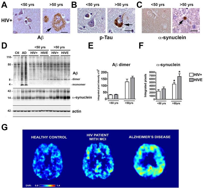
People over the age of 50 are the fastest growing segment of the HIV-infected population in the USA. Although antiretroviral therapy has remarkable success controlling the systemic HIV infection, HIV-associated neurocognitive disorder (HAND) prevalence has increased or remained the same among this group, and cognitive deficits appear more severe in aged patients with HIV. The mechanisms of HAND in the aged population are not completely understood; a leading hypothesis is that aged individuals with HIV might be at higher risk of developing Alzheimer's disease (AD) or one of the AD-related dementias (ADRD). There are a number of mechanisms through which chronic HIV disease alone or in combination with antiretroviral therapy and other comorbidities (e.g., drug use, hepatitis C virus (HCV)) might be contributing to HAND in individuals over the age of 50 years, including (1) overlapping pathogenic mechanisms between HIV and aging (e.g., decreased proteostasis, DNA damage, chronic inflammation, epigenetics, vascular), which could lead to accelerated cellular aging and neurodegeneration and/or (2) by promoting pathways involved in AD/ADRD neuropathogenesis (e.g., triggering amyloid β, Tau, or α-synuclein accumulation). In this manuscript, we will review some of the potential common mechanisms involved and evidence in favor and against a role of AD/ADRD in HAND.

Keywords: Aging; Alzheimer’s disease; HIV-associated cognitive impairment; Neurodegeneration.


Figures

Figure 1.
Figure 1.. Mechanisms of neurodegeneration in HIV associated neurocognitive disorders (HAND) in the pre and highly active antiretroviral (HAART) eras.
(A) Schematic representation of mechanisms leading to HIV encephalitis and neurodegeneration initiating with trafficking of HIV infected macrophages through an injured blood-brain barrier followed by activation and infection of microglial and astroglial cells resulting in neurotoxicity and inflammation with synapto-dendritic damage and selective neuronal damage. (B) In the pre-HAART era, HIV CNS pathology was characterized by the presence of microglial nodules, multinucleated giant cells, severe astrogliosis, myelin loss and neurodegenerative pathology accompanied by opportunistic infections. Modern treatments with HAART results in HIV suppression and immune recovery; the pathology has shifted to a more subtle, chronic neurodegenerative process with lower or undetectable HIV in the CNS and diffuse astrogliosis, some microglial nodules, white matter alterations and vascular changes accompanied by co-morbid conditions associated with aging.
Figure 2.
Figure 2.. Potential mechanisms of neurodegeneration in HIV and aging.
Common pathways involved in aging and chronic HIV disease in the CNS might interact leading to neurodegeneration. Such mechanisms include defective proteostasis (eg: altered proteasome, proteolysis, autophagy), mitochondrial abnormalities, epigenetic alterations, DNA damage, cell senescence, neuroinflammation, oxidative stress and stem cell defects. Together, these alterations might lead to abnormal protein accumulation (Aβ, Tau, α-synuclein) and hyperactivation of signaling pathways (eg: CDK5, JNK) involved in neuronal damage.
Figure 3.
Figure 3.. Brain Proteinopathy in aging and HIV.
With aging in an individual with chronic HIV disease protein aggregates involved in AD/ADRD might abnormally accumulate. (A) Representative images of the frontal cortex immunostained with an antibody against Aβ (4G8) from an HIV+ case under 50 yrs of age showing no amyloid deposition while an older case shows plaque-like lesion; (B) frontal cortex immunostained with an antibody against p-Tau (PHF-1) from HIV+ case under 50 yrs of age showing no neuronal alterations while an older case shows tangle-like lesion; (C) temporal cortex immunostained with an antibody against alpha-synuclein (SYN1) from HIV+ case under 50 yrs of age showing neuropil immunoreactivity while an older case shows Lewy-like dystrophic neurites (Bar=10 μm) . (D) Representative immunoblot analysis with the frontal cortex fractioned from control, AD and HIV+ cases under and over the age of 50 years. Samples from the frontal cortex were fractioned by ultracentrifugation and the membrane fractions ran in SDS-PAGE gels followed by blotting onto nitrocellulose membranes. Compared to controls in AD cases there was a considerable accumulation of monomers and multimers at various molecular weights, in older HIV cases Aβ dimers, as well as α-synuclein monomer and trimers appear to accumulate. (E, F) Image analysis corrected to actin for levels of Aβ dimers and α-synuclein (n=3 cases per group). (G) Representative FDDNP parametric PET images from normal control, HIV+ case with mild cognitive impairment (MCI) or Alzheimer’s disease. The analysis of FDDNP scans was performed for frames between 15 and 85 min using Logan graphical analysis with cerebellum as reference region. The resulting distribution volume ratios (DVR) were used to generate the DVR parametric images. The DVR values have been color-coded in such a way that all values below DVR 0.8 are shown as dark blue and all values above DVR 1.4 are shown as dark red. The DVR values between are shown in colors of a rainbow as shown on the color bar at the bottom of the image. The control shown is a 43-year-old female with MMSE score 30. The HIV patient with MCI is a 45-year-old male. The AD patient is an 81-year-old female with MMSE score 15. The images shown are transaxial cuts slightly above the basal ganglia so that posterior cingulate gyrus is visible. The frontal lobe is at the top and parietal lobe is at the bottom. The AD case shows high levels of FDDNP binding (high DVR values) throughout the neocortex, most prominently in parietal and frontal cortices and in posterior cingulate. The control case has a uniformly low level of FDDNP binding (low DVR) throughout the cortex. The HIV cases show a limited number of isolated areas with medium to high level of FDDNP binding in prefrontal areas, in posterior and anterior cingulate gyri, and asymmetrically in the parietal lobe.

References

    1. (2005). HIV/AIDS surveillance report. 1–63.
    1. (2007). HIV/AIDS surveillance report. 1–54.
    1. Achim CL, Adame A, Dumaop W, Everall IP, Masliah E, Neurobehavioral Research C (2009). Increased accumulation of intraneuronal amyloid beta in HIV-infected patients. J Neuroimmune Pharmacol 4: 190–9. - PMC - PubMed
    1. Alakkas A, Ellis RJ, Watson CW, Umlauf A, Heaton RK, Letendre S, Collier A, Marra C, Clifford DB, Gelman B, Sacktor N, Morgello S, Simpson D, McCutchan JA, Kallianpur A, Gianella S, Marcotte T, Grant I, Fennema-Notestine C, Group C (2018). White matter damage, neuroinflammation, and neuronal integrity in HAND. Journal of neurovirology: 1–10. - PMC - PubMed
    1. Alirezaei M, Kiosses WB, Flynn CT, Brady NR, Fox HS (2008a). Disruption of neuronal autophagy by infected microglia results in neurodegeneration. PloS one 3: e2906. - PMC - PubMed

Publication types

MeSH terms