. 2019 Aug:185:107585.
doi: 10.1016/j.exer.2019.02.001. Epub 2019 Feb 18.

BFSP1 C-terminal domains released by post-translational processing events can alter significantly the calcium regulation of AQP0 water permeability

Affiliations

BFSP1 C-terminal domains released by post-translational processing events can alter significantly the calcium regulation of AQP0 water permeability

Antal Tapodi et al. Exp Eye Res. 2019 Aug.

Abstract

BFSP1 (beaded filament structural protein 1, filensin) is a cytoskeletal protein expressed in the eye lens. It binds AQP0 in vitro and its C-terminal sequences have been suggested to regulate the water channel activity of AQP0. A myristoylated fragment from the C-terminus of BFSP1 was found in AQP0 enriched fractions. Here we identify BFSP1 as a substrate for caspase-mediated cleavage at several C-terminal sites including D433. Cleavage at D433 exposes a cryptic myristoylation sequence (434-440). We confirm that this sequence is an excellent substrate for both NMT1 and 2 (N-myristoyl transferase). Thus caspase cleavage may promote formation of myristoylated fragments derived from the BFSP1 C-terminus (G434-S665). Myristoylation at G434 is not required for membrane association. Biochemical fractionation and immunogold labeling confirmed that C-terminal BFSP1 fragments containing the myristoylation sequence colocalized with AQP0 in the same plasma membrane compartments of lens fibre cells. To determine the functional significance of the association of BFSP1 G434-S665 sequences with AQP0, we measured AQP0 water permeability in Xenopus oocytes co-transfected with transcripts expressing both AQP0 and various C-terminal domain fragments of BFSP1 generated by caspase cleavage. We found that different fragments dramatically alter the response of AQP0 to different concentrations of Ca2+. The complete C-terminal fragment (G434-S665) eliminates calcium regulation altogether. Shorter fragments can enhance regulation by elevated calcium or reverse the response, indicative of the regulatory potential of BFSP1 with respect to AQP0. In particular, elimination of the myristoylation site by the mutation G434A reverses the order of water permeability sensitivity to different Ca2+ concentrations.


Figures

Fig. 1.
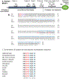
Fig. 1.. Bioinformatic analysis of the C-terminal sequences of BFSP1.
A. The main structural features of human BFSP1 and the location of predicted caspase cleavage sites. The residue numbers for potential cleavage sites are included. B. Sequence alignment of human, rat and bovine BFSP1 using CLUSTALW to identify regions of homology. Caspase sites as predicted for BFSP1 are indicated (red). The caspase site adjacent to the cryptic myristoylation motif (mauve) is in a region of homology between human, rat and cow. The construct used to study the potential functional interaction of rat BFSP1 with AQP0 is written in blue script. The BFSP1 residue identified by crosslinking with AQP0 is indicated (green). Notice the relative positions of the myristoylation sequence and the predicted caspase sites and the variability in the location of these between the three species. C. The caspase site and adjacent cryptic myristoylation sequence in BFSP1 is conserved over a wide range of animals from whale shark to human.
Fig. 2.
Fig. 2.. Characterization of the caspase sensitivity of BFSP1.
A. In vitro cleavage of BFSP1 by purified caspases 1–10. Full length recombinant BFSP1 containing a C-terminal polyhistidine tag is indicated (chevron). Using the polyclonal antibody 3241 that was raised against the bovine 53 kDa fragment of BFSP1, two major fragments were detected (arrows). One corresponded to the 53 kDa fragment and like the slower migrating band of the two, both were undetected by the His-tag directed antibodies. Therefore both these fragments likely lack different proportions of the C-terminal sequences. Using the anti-His tag antibodies, two prominent bands were detected in the caspase 2 treated BFSP1 sample (arrowheads). Neither of these bands comigrated with bands detected by the 3241 polyclonal antibodies. B. To evidence cleavage of BFSP1 by caspase 2 at the D433 and D549 sites, two BFSP1 mutants were produced, D433A and D549A. Comparing WT, D433A and D549A BFSP1 sensitivity to caspase 2 cleavage resulted in the absence of the characteristic 53 kDa fragment (arrowhead) in the G433A BFSP1 as detected by the 3241 polyclonal antibodies. Using the anti-His tag antibodies, two characteristic fragments (arrowheads) were detected in the wild type and D549A BFSP1 samples, but only the faster migrating band in the D433A BFSP1 sample. Similarly the bands indicated (Asterisks) were present in the WT and D433A BFSP1 samples but missing from the D549A BFSP1 sample. This indicates that the mutations have abolished one site at position D433 and one at D549 for caspase 2.
Fig. 3.
Fig. 3.. Caspase sensitivity of transfected BFSP1 in MCF7 and FHL124 cells.
MCF7 and FHL124 cells were transiently transfected with constructs encoding eGFP alone, or as a N-terminal fusion with human BFSP1, and the mutants G433A (MCF7 and FHL124) and G434A BFSP1 (MCF7 only). Transfected BFSP1 was detected using the polyclonal antibodies 3241. A. MCF7 cells were either untreated or treated with the generic caspase inhibitor (zVAD) or with 0.15 M H202 in order to induce effector caspases. Full length eGFP-BFSP1 is indicated (chevron). A 53 kDa fragment (arrow) was detected by the 3241 antibodies. Note the absence of the 53 kDa signal in cells transfected with D433A, but not the G434A BFSP1 construct. The caspase inhibitor also prevented the formation of this fragment. B. The D433A mutation blocks the caspase sensitivity of BFSP1 constructs transiently transfected into the human lens epithelial cell line FHL124. The BFSP1 sequence 1–460 was fused to the N-terminus of eGFP and transiently transfected into FHL124 cells resulting in an 80 kDa product (chevrons). Notice the major breakdown product (53 kDa) for the construct containing the wild type D433 residue and the very significant reduction when this was replaced by A433. Slower migrating products (arrowheads) could arise by caspase cleavage either within the BFSP1 or eGFP parts of the fusion protein. Marker proteins (m) are indicated (•) and correspond to 250, 130, 100, 70, 55, 35, 25 and 15 kDa as per PageRuler prestained standards.
Fig. 4.
Fig. 4.. In vivo Myristoylation assay of truncated Tail fragment of BFSP1:
A: Truncated Tail fragment of BFSP1 was co-expressed with CaNMT in E. coli in the presence of azido-myristate. CLICK-chemistry was used to detect myristolyated proteins and detected in the gel after fluorescently labelled with Cy3-TAMRA. PfARF1 (Plasmodium falciparum ADP-ribosylation factor-1) was included as a positive control of myristoylation. Track1 and 2, Pre- and post-induction of PfARF expression in E.coli; Tracks 3 and 4, Pre- and post-induction of PfARF with C.albicans NMT expression in E.coli; Tracks 5 and 6: Pre- and post-induction of G434-P548 BFSP1 expression in E.coli; Tracks 7 and 8, Pre- and post-induction of BFSP1 G434-P548 with C.albicans NMT expression in E.coli; Tracks 9 and 10, Pre- and post-induction of PfARF with C.albicans NMT expression in E.coli fed with azido-myristate followed by CLICK chemistry detection; Tracks 11 and 12, Pre- and post-induction of BFSP1 G434-P548 with C.albicans NMT expression in E.coli fed with azidomyristate followed by CLICK chemistry detection. Notice the positive bands in tracks 10 and 12 only. B. Km and Vmax determination for recombinant human NMT1 and 2 for the BFSP1 sequence G434- F441. C. Characterization of the polyclonal antibodies NP-Tail and NP53 using human lens samples. The soluble protein fraction (S1), the plasma membrane cytoskeleton complex (PMCC) and plasma membrane fraction (P3) were isolated from the human cortex and separated by SDS PAGE and the 10% (w/v) polyacrylamide gel prior to either staining with Coomassie Blue or immunoblotting and probing with the polyclonal antibodies 3241, NP-53 and NP-Tail. Notice that the major immunoreactive band detected in the P3 sample by the NP-tail antibodies would correspond to a C-terminal fragment of less than 25 kDa. Marker proteins (m) are indicated (•) and correspond to 250, 130, 100, 70, 55, 35, 25 and 15 kDa as per PageRuler prestained standards.
Fig. 5.
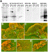
Fig. 5.. Cofractionation and colocalization of BFSP1 and AQP0 to the plasma membrane compartment of lens fibre cells.
A. Plasma membranes were prepared from the nucleus (N) and cortical (C) parts of the lens from a 51 year old male donor. The plasma membranes were extracted with buffer containing 8 M urea (UREA) and then with 0.1 M NaOH (NaOH) and samples of the pelleted material taken and separated by SDS PAGE. Coomassie blue staining of the proteins in these fractions (CB Stain) revealed a few prominent low molecular weight proteins between the 25 and 15 kDa markers. Immunoblotting of these plasma membrane fractions with the polyclonal antibodies NP-53 identified BFSP1 N-terminal-derived fragments only in the urea extracted membranes. These were removed after alkali extraction. In contrast, the NP Tail antibodies identified bands in both urea and alkali extracted membrane fractions, although the fragments retained in the alkali stripped membrane fraction were those with faster electrophoretic mobility equivalent to approx. 25 kDa. Probing these fractions with AQP0 antibodies demonstrated the enrichment of AQP0 signal in the alkali stripped plasma membrane fractions of both the nuclear and cortical parts of the lens. Marker proteins (m) are indicated (•) and correspond to 250, 130, 100, 70, 55, 35, 25 and 15 kDa as per PageRuler prestained standards. B, C. Co-immuno-gold labeling of alkali stripped plasma membranes from the nucleus (B) and cortex (C) of human lenses using the rabbit polyclonal NP Tail antibodies to detect BFSP1 and mouse monoclonal antibodies to detect AQP0. To visualize the antibodies in the SEM, 5 nm and 10 nm gold labelled secondary antibodies were used to detect AQP0 (arrowheads) and BFSP1(arrows) respectively. D, E and F. Immunogold labeling of alkali stripped membranes from human lens nuclei and labelled with either the preimmune serum for the NP Tail antibodies (D) or the polyclonal antibodies NP-53 (E). These two evidence the background labeling achievable either with preimmune (D) or closely related polyclonal rabbit antibodies (E). The NP53 detectable BFSP1 fragments have been removed by the alkali extraction of these membrane preparations (A). The signal detected by the polyclonal NP Tail antibodies (F) is very clearly above background levels.
Fig. 6.
Fig. 6.. Regulation of AQP0 by BFSP1 C-terminal domains.
A. Xenopus oocytes were injected with AQP0 alone or in combination with the indicated BFSP1 constructs. C-terminal constructs could be divided into two classes: ones which left the calcium response essentially identical to wild type, that is Pf was low in 1.8 mM Ca2+ and high in 0 mM Ca2+; or ones in which Pf did not change with Ca2+ concentration. Results were analyzed by omnibus ANOVA followed by pair wise t-tests for individual 0 mM Ca2+ - 1.8 mM Ca2+ comparisons. In the cases where Pf increased in response to no calcium, pairwise p values were less than 0.05 (*). In cases where Pf did not respond to changes in calcium, p values were greater than 0.3 (ns). Conditional p values for 1.8 mM Ca2+ vs no Ca2+: AQP0 alone = 0.0232, +434–665 = 0.0328, +434–548 = 0.6823, +A434–548 = 0.0128, B. Immunoblots showing that BFSP1 was expressed in Xenopus oocytes when appropriate constructs were injected. Two major immunoreactive bands were detected (arrows), the faster migrating band a fragment derived by proteolytic cleavage because the D433E mutation in BFSP1 altered the pattern achieved. Notice too that the presence of AQP0 altered the banding pattern achieved with the D433A mutant (*). BFSP1 immunoreactive bands are found in the pellet fraction prepared from injected oocytes. C. Full length BFSP1 and full length BFSP1 with mutations at putative caspase sites eliminated Pf regulation by calcium. Conditional p values for 1.8 mM Ca2+ vs no Ca2+: AQP0 alone = 0.001, +BFSP1 = 0.617, +BFSP1 D433A = 0.3913, +BFSP1 D549A = 0.6445.
Fig. 7.
Fig. 7.. AQP0 activity is differentially modulated by BFSP1 and its post-translationally modified fragments.
Full length human BFSP1 is cleaved by caspases at sites 433 and 549. The 433 site reveals a cryptic myristoylation site. The lens expresses both NMT1 and NMT2. The 434–549 and 434–665 fragments, once myristoylated, can effectively eliminate the regulation of AQP0 Pf by Ca2+. Preventing myristoylation restores the Ca2+ regulation of AQP0 Pf (right hand side of the figure).

Similar articles

Cited by

References

    1. Backes C, Kuentzer J, Lenhof HP, Comtesse N, Meese E, 2005. GraBCas: a bioinformatics tool for score-based prediction of Caspase- and Granzyme B-cleavage sites in protein sequences. Nucleic Acids Res. 33, W208–W213. - PMC - PubMed
    1. Bassnett S, Wilmarth PA, David LL, 2009. The membrane proteome of the mouse lens fiber cell. Mol. Vis 15, 2448–2463. - PMC - PubMed
    1. Block J, Breitsprecher D, Kuhn S, Winterhoff M, Kage F, Geffers R, Duwe P, Rohn JL, Baum B, Brakebusch C, Geyer M, Stradal TE, Faix J, Rottner K, 2012. FMNL2 drives actin-based protrusion and migration downstream of Cdc42. Curr. Biol. : CB 22, 1005–1012. - PMC - PubMed
    1. Bologna G, Yvon C, Duvaud S, Veuthey AL, 2004. N-Terminal myristoylation predictions by ensembles of neural networks. Proteomics 4, 1626–1632. - PubMed
    1. Brunkener M, Georgatos SD, 1992. Membrane-binding properties of filensin, a cytoskeletal protein of the lens fiber cells. J. Cell Sci 103 (Pt 3), 709–718. - PubMed

Publication types

MeSH terms