

Postoperative Endothelial Cell Density Is Associated with Late Endothelial Graft Failure after Descemet Stripping Automated Endothelial Keratoplasty
- PMID: 30790587
- PMCID: PMC6646077
- DOI: 10.1016/j.ophtha.2019.02.011
Postoperative Endothelial Cell Density Is Associated with Late Endothelial Graft Failure after Descemet Stripping Automated Endothelial Keratoplasty
Abstract
Purpose: To determine whether preoperative endothelial cell density (ECD) and postoperative ECD after Descemet stripping automated endothelial keratoplasty (DSAEK) are associated with late endothelial graft failure (LEGF) in the Cornea Preservation Time Study (CPTS).
Design: Cohort study within a multicenter, randomized clinical trial.
Participants: A total of 1007 individuals (1223 study eyes), mean age 70 years, undergoing DSAEK for Fuchs' dystrophy (94% of eyes) or pseudophakic or aphakic corneal edema (PACE) (6% of eyes) and followed for up to 5 years.
Methods: Central ECD was determined by a central image analysis reading center. Preoperative ECD was determined for 1209 eyes that did not fail and 14 eyes that experienced LEGF. The ECD at 6 and 12 months after DSAEK, the change in ECD from preoperative to 6 and 12 months, surgeon-reported operative complications, and postoperative graft dislocation were investigated for an association with LEGFs unrelated to other postoperative events. Univariable and multivariable Cox proportional hazards regression models were used to assess associations.
Main outcome measures: Late endothelial graft failure and its associations with pre- and postoperative ECD and operative complications.
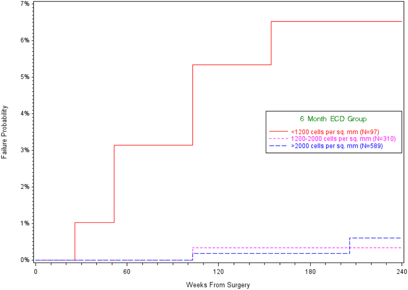
Results: The cumulative probability of LEGF was 1.3% (95% confidence interval [CI], 0.8%-2.4%). Median (interquartile range [IQR]) preoperative ECDs were similar for eyes with LEGF (2523; 2367-3161) cells/mm2) and eyes without failure (2727; 2508-2973) cells/mm2) (P = 0.34). The ECD at 6 months was associated with LEGF (P < 0.001) in time-to-event analyses, whereas preoperative ECD was not (P = 0.55). The cumulative incidence (95% CI) of LEGF was 6.5% (3.0%, 14.0%) for 97 grafts with a 6-month ECD less than 1200 cells/mm2, 0.3% (0.0%, 2.4%) for 310 grafts with a 6-month ECD between 1200 and 2000 cells/mm2, and 0.6% (0.1%, 2.7%) for 589 grafts with a 6-month ECD greater than 2000 cells/mm2. In multivariable analyses, ECD at 6 months and operative complications were both associated with LEGF (P = 0.002 and P = 0.01, respectively), whereas graft dislocation was not (P = 0.61).
Conclusions: In eyes undergoing DSAEK, preoperative ECD is unrelated to LEGF, whereas lower ECD at 6 months is associated with LEGF. Early endothelial cell loss after DSAEK and intraoperative complications should be minimized to improve graft survival.
Copyright © 2019 American Academy of Ophthalmology. All rights reserved.
Conflict of interest statement
Figures
References
Publication types
MeSH terms
Grants and funding
LinkOut - more resources
Full Text Sources
Other Literature Sources
