Comparative Study
. 2019 Mar 28:298:186-193.
doi: 10.1016/j.jconrel.2019.02.017. Epub 2019 Feb 18.

Poly(ethylene glycol)-block-poly(d,l-lactic acid) micelles containing oligo(lactic acid)8-paclitaxel prodrug: In Vivo conversion and antitumor efficacy

Affiliations
Comparative Study

Poly(ethylene glycol)-block-poly(d,l-lactic acid) micelles containing oligo(lactic acid)8-paclitaxel prodrug: In Vivo conversion and antitumor efficacy

Yu Tong Tam et al. J Control Release. .

Abstract

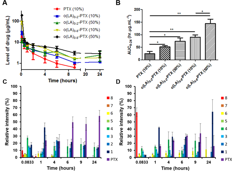
Poly(ethylene glycol)-block-poly(d,l-lactic acid) (PEG-b-PLA) micelles affect drug solubilization, and a paclitaxel (PTX) loaded-PEG-b-PLA micelle (PTX-PM) is approved for cancer treatment due to injection safety and dose escalation (Genexol-PM®) compared to Taxol®. However, PTX-PM is unstable in blood, has rapid clearance, and causes dose-limiting toxicity. We have synthesized a prodrug for PTX (7-OH), using oligo(lactic acid) as a novel pro-moiety (o(LA)8-PTX) specifically for PEG-b-PLA micelles, gaining higher loading and slower release of o(LA)8-PTX over PTX. Notably, o(LA)8-PTX prodrug converts into PTX by a backbiting reaction in vitro, without requiring esterases. We hypothesize that o(LA)8-PTX-loaded PEG-b-PLA micelles (o(LA)8-PTX-PM) has a lower Cmax and higher plasma AUC than PTX-PM for improved therapeutic effectiveness. In Sprague-Dawley rats at 10 mg/kg, compared to o(LA)8-PTX-PM (10% w/w loading) and PTX-PM (10%), o(LA)8-PTX-PM (50% w/w loading) produces a 2- and 3-fold higher plasma AUC0-24 of PTX, lactic acid-PTX, and o(LA)2-PTX (o(LA)0-2-PTX), respectively. For o(LA)8-PTX-PM at 10 and 50% w/w loading, PTX and lactic acid-PTX are major bioactive metabolites, respectively. Fast prodrug conversion of o(LA)8-PTX in vivo versus in vitro (by backbiting) suggests that o(LA)8 is a good substrate for esterases. At 60 mg/kg (qwx3), o(LA)8-PTX-PM (50%) has higher antitumor activity than o(LA)8-PTX-PM (10%) and PTX-PM (10%) in a syngeneic 4T1-luc breast tumor model based on measurements of tumor volume, 4T1-luc breast tumor bioluminescence, and survival. Importantly, intravenous administration of o(LA)8-PTX-PM is well tolerated by BALB/c mice. In summary, oligo(lactic acid)8-PTX is more compatible than PTX with PEG-b-PLA micelles, more stable, and may expand the role of PEG-b-PLA micelles from "solubilizer" into "nanocarrier" for PTX as a next-generation taxane for cancer.

Keywords: Block copolymer; Oligo(lactic acid); PEG; Polymeric micelle; Prodrug; Taxanes.


Figures

Figure 1.
Figure 1.
An o(LA)8-PTX prodrug-loaded PEG-b-PLA micelle (o(LA)8-PTX-PM). Relative to a PTX-loaded PEG-b-PLA micelle (PTX-PM), o(LA)8-PTX-PM may be more stable in blood. After release, o(LA)8-PTX converts into PTX, lactic acid-PTX, and o(LA)2-PTX (o(LA)0–2-PTX) as metabolites for a lower Cmax and higher plasma AUC relative to PTX-PM, aiming for reduced toxicity and higher antitumor efficacy.
Figure 2.
Figure 2.
(A) Images of PTX-PM (10%), o(LA)8-PTX-PM (10%), and o(LA)8-PTX-PM (50%) at 2 mg/mL drug level at 25 °C after 24 hours, (B) Particle size analysis by dynamic light scattering (DLS) of PTX-PM (10%), o(LA)8-PTX-PM (10%), and o(LA)8-PTX-PM (50%), (C) Representative AFM image of o(LA)8-PTX-PM (50%), and (D) In vitro drug release profiles of PTX from PTX-PM (10%) (drug precipitation < 4 hours), o(LA)8-PTX from o(LA)8-PTX-PM (10%), and o(LA)8-PTX-PM (50%) in 10 mM PBS at 37 °C and pH 7.4 (mean ± SD, n=3).
Figure 3.
Figure 3.
In vitro rat plasma stability of (A) o(LA)8-PTX, (B) o(LA)8-PTX-PM (10%), and (C) o(LA)8-PTX-PM (50%) at a final concentration of 10 μM at 37 °C (mean ± SEM, n=3).
Figure 4.
Figure 4.
In vitro cytotoxicity of PTX, o(LA)2-PTX, o(LA)8-PTX, PTX-PM (10%), o(LA)8-PTX-PM (10%), and o(LA)8-PTX-PM (50%) for 4T1-luc cells (mean ± SEM, n=4) (**. p < 0.01; *, p < 0.05).
Figure 5.
Figure 5.
(A) Concentration-time profile in rat plasma and (B) area under the concentration-time curve (AUC0–24) of PTX from PTX-PM (10%) at 10 mg/kg, o(LA)0–2-PTX and o(LA)0–8-PTX from o(LA)8-PTX-PM (10%) at 10 mg/kg PTX-eq and o(LA)0–2-PTX and o(LA)0–8-PTX from o(LA)8-PTX-PM (50%) at 10 mg/kg PTX-eq in male Sprague-Dawley rats. (mean ± SEM, n=4; ***, p < 0.001; **, p < 0.01; *, p < 0.05). Relative intensity of o(LA)0–8-PTX over time in rat plasma after IV injection of (C) o(LA)8-PTX-PM (10%) at 10 mg/kg PTX-eq, and (D) o(LA)8-PTX-PM (50%) at 10 mg/kg PTX-eq in male Sprague-Dawley rats (Digits in Figures 5C and 5D represent the number of lactic acid oligomers (n) of o(LA)n-PTX; mean ± SEM, n=4).
Figure 6.
Figure 6.
In vivo antitumor efficacy in BALB/c mice bearing s.c. 4T1-luc breast tumors. Treatments were initiated 14 days post tumor implantation: PTX-PM (10%), o(LA)8-PTX-PM (10%), and o(LA)8-PTX-PM (50%) IV injection at 60 mg/kg PTX-eq (qwx3), indicated by arrows. (A) Whole-body bioluminescence images of 4T1-luc mice, (B) Relative tumor volume (mean ± SEM, n=3–4; *: p < 0.05 (o(LA)8-PTX-PM (50%) vs. o(LA)8-PTX-PM (10%), PTX-PM (10%), or saline control), (C) Relative body weight change, (D) % bioluminescence intensity of region of interest (ROI) (mean ± SEM, n=3; *: p < 0.05 (o(LA)8-PTX-PM (50%) vs. o(LA)8-PTX-PM (10%), PTX-PM (10%), or saline control), and (E) Kaplan-Meier analysis for survival (> 1200 mm3 endpoint tumor volume; **: p < 0.01 (o(LA)8-PTX-PM (50%) vs. PTX-PM (10%)).
Figure 7.
Figure 7.
(A) Relative body weight of female BALB/c mice over time after single tail-vein injection: PTX-PM (10%) at 100 mg/kg, o(LA)8-PTX-PM (10%) at 100 mg/kg PTX-eq, and o(LA)8-PTX-PM (50%) at 100, 150, and 200 mg/kg PTX-eq, respectively (mean ± SEM, n=3). Two mice suffered from loss of consciousness, flushed skin, and dyspnea were euthanized on day 0 and day 1 for PTX-PM (10%) at 100 mg/kg. (B) Relative body weight of female BALB/c mice over time after daily injection on days 0, 1 and 2 of PTX-PM (10%) at 50 mg/kg, o(LA)8-PTX-PM (10%) at 50 mg/kg PTX-eq, and o(LA)8-PTX-PM (50%) at 50 mg/kg PTX-eq (mean ± SEM, n=5; *: p < 0.05 (o(LA)8-PTX-PM (50%) vs. PTX-PM (10%)); **: p < 0.01 (saline vs. PTX-PM (10%)).

Similar articles

Cited by

References

    1. McGuire WP, Hoskins WJ, Brady MF, Kucera PR, Partridge EE, Look KY, Clarke-Pearson DL, Davidson M, Cyclophosphamide and cisplatin compared with paclitaxel and cisplatin in patients with stage III and stage IV ovarian cancer, N. Engl. J. Med 334 (1996) 1–6. - PubMed
    1. Sparano JA, Wang M, Martino S, Jones V, Perez EA, Saphner T, Wolff AC, Sledge GW Jr, Wood WC, Davidson NE, Weekly paclitaxel in the adjuvant treatment of breast cancer, N. Engl. J. Med 358 (2008) 1663–1671. - PMC - PubMed
    1. Murphy WK, Fossella FV, Winn RJ, Shin DM, Hynes HE, Gross HM, Davilla E, Leimert J, Dhingra H, Raber MN, Krakoff IH, Phase II study of taxol in patients with untreated advanced non-small-cell lung cancer, J. Natl. Cancer Inst 85 (1993) 384–388. - PubMed
    1. Silvestris N, Galetta D, Colucci G, Successful treatment with three-weekly paclitaxel of an anthracycline-refractory classical Kaposi’s sarcoma, Anticancer Res. 29 (2009) 675–676. - PubMed
    1. Arnal I, Wade RH, How does taxol stabilize microtubules?, Curr. Biol 5 (1995) 900–908. - PubMed

Publication types

MeSH terms