. 2019 Jun;69(6):2546-2561.
doi: 10.1002/hep.30571. Epub 2019 Apr 11.

The Antiresection Activity of the X Protein Encoded by Hepatitis Virus B

Affiliations

The Antiresection Activity of the X Protein Encoded by Hepatitis Virus B

Laifeng Ren et al. Hepatology. 2019 Jun.

Abstract

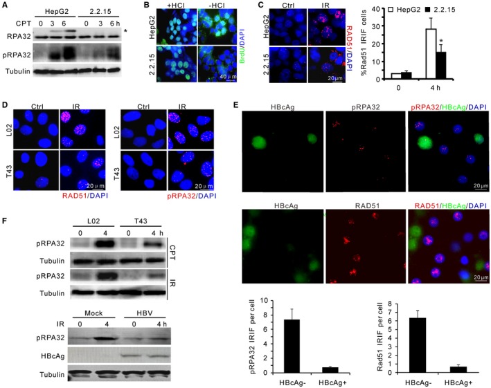
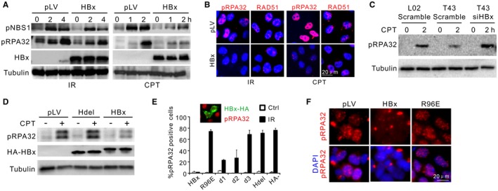
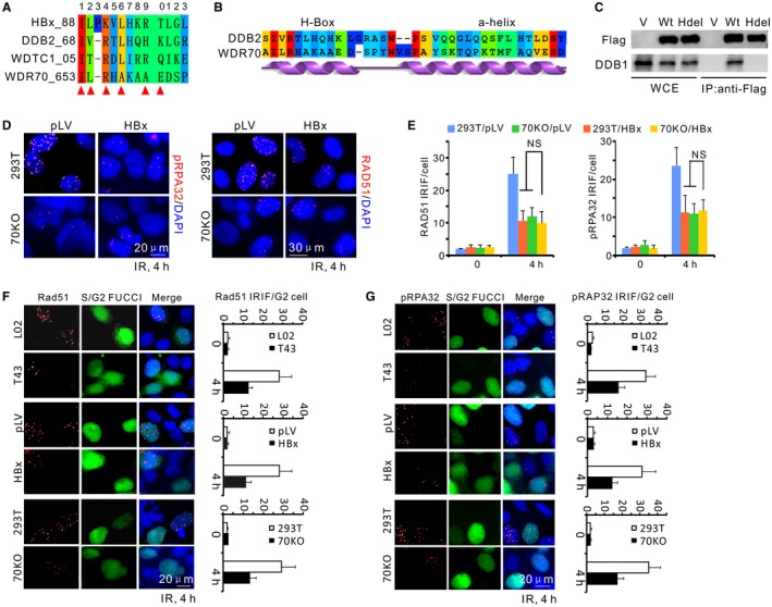
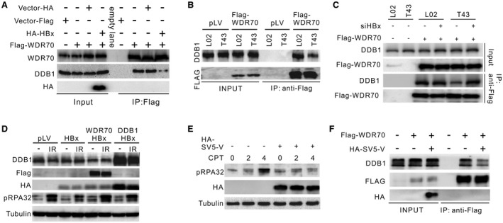
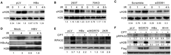
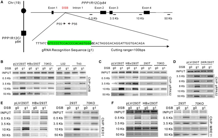
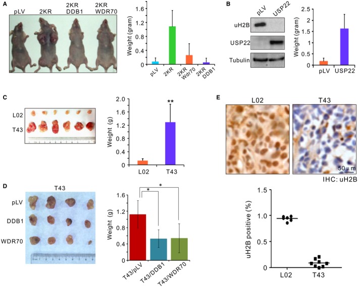
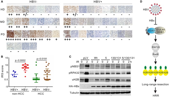
Chronic infection of hepatitis B virus (HBV) is associated with an increased incidence of hepatocellular carcinoma (HCC). HBV encodes an oncoprotein, hepatitis B x protein (HBx), that is crucial for viral replication and interferes with multiple cellular activities including gene expression, histone modifications, and genomic stability. To date, it remains unclear how disruption of these activities contributes to hepatocarcinogenesis. Here, we report that HBV exhibits antiresection activity by disrupting DNA end resection, thus impairing the initial steps of homologous recombination (HR). This antiresection activity occurs in primary human hepatocytes undergoing a natural viral infection-replication cycle as well as in cells with integrated HBV genomes. Among the seven HBV-encoded proteins, we identified HBx as the sole viral factor that inhibits resection. By disrupting an evolutionarily conserved Cullin4A-damage-specific DNA binding protein 1-RING type of E3 ligase, CRL4WDR70 , through its H-box, we show that HBx inhibits H2B monoubiquitylation at lysine 120 at double-strand breaks, thus reducing the efficiency of long-range resection. We further show that directly impairing H2B monoubiquitylation elicited tumorigenesis upon engraftment of deficient cells in athymic mice, confirming that the impairment of CRL4WDR70 function by HBx is sufficient to promote carcinogenesis. Finally, we demonstrate that lack of H2B monoubiquitylation is manifest in human HBV-associated HCC when compared with HBV-free HCC, implying corresponding defects of epigenetic regulation and end resection. Conclusion: The antiresection activity of HBx induces an HR defect and genomic instability and contributes to tumorigenesis of host hepatocytes.


Figures

Figure 1
Figure 1
HBV attenuates DNA end resection upon genotoxic insults. (A) Immunoblotting for RPA32 phosphorylation in HepG2 and HepG2.2.15 cells upon CPT treatment. pRPA32 indicates phosphorylated serine 33 of RPA32. *Band shift of phosphorylated RPA32 detected by α‐RPA32 serum. (B) Non‐(−HCl) and denaturing‐(+HCl) indirect immunofluorescence showing ssDNA at resected DSBs and equal incorporation of BrdU after CPT treatment, respectively. Scale bar, 40 μm. (C) Representative image and percentage of RAD51 IRIF‐positive cells after 5‐Gy irradiation in HepG2 and HepG2.2.15 cells. (D) Representative images for IRIF of RAD51 and phosphorylated RPA32 in L02 and T43 cells after IR treatment. (E) Images (top) and quantification (bottom) of indicated IRIF in mock or HBV‐infected PHHs at day 7. Cells were fixed for immunostaining 4 hours after 1‐Gy irradiation. HBcAg (green) marks HBV‐infected PHHs. (F) Immunoblotting for RPA32 phosphorylation in L02/T43 cells or HBV‐infected PHHs at indicated times. Genotoxic treatments: 2 μM CPT or 5‐Gy irradiation. Abbreviation: DAPI, 4′,6‐diamidino‐2‐phenylindole.
Figure 2
Figure 2
HBx impairs DNA end resection. (A) Immunoblotting for IR‐induced or CPT‐induced RPA32 and NBS1 phosphorylation in the presence or not of HBx. L02 cells were preinfected with 2 multiplicities of infection/cell of pLV (mock infection) or pLV‐HA‐HBx lentivirus. (B) pLV‐transfected or pLV‐HBx‐transfected L02 cells were treated as in (A), followed by immunostaining of RAD51 and pRPA32 foci. (C) Monitoring of RPA32 phosphorylation in indicated cells treated or not with siHBx. (D) Immunoblotting for pRPA32 in CPT‐treated L02 cells transfected with indicated expression plasmids. HBx and Hdel (HBx with H‐box deleted: amino acids 88‐98) were monitored by α‐HA. (E) Percentage of pRPA32 IRIF‐positive L02 cells pretransfected with different HA‐HBx constructs. pRPA32 foci were monitored 4 hours after irradiation. Cells with double‐positive staining of HA and phosphorylated serine 33 were judged as positive except in “HBx” cells (no HA‐HBx transfection) where pRPA32 were counted in random cells. Inset: Costaining of HA‐HBx (green) and phosphorylated serine 33 (red). n = 3 biological repeats. Error bars = SD. (F) Immunofluorescence for pRPA32 in L02 cells infected with HBx wild‐type or R96E lentivirus (2 multiplicities of infection/cell) followed by IR treatment. Abbreviations: DAPI, 4′,6‐diamidino‐2‐phenylindole; HA, hemagglutinin; LV, lentivirus.
Figure 3
Figure 3
Disruption of CRL4WDR70 complex by HBx results in a resection defect. (A) Sequence alignment for the H‐boxes of cellular DCAF proteins and HBx (WDTC1, alias of DCAF9). Red triangle, binding hotspots serving as key DDB1‐contacting residues. (B) By sequence alignment and secondary structure prediction, the WDR70 C‐terminal sequence possesses a similar H‐box sequence and an adjunct α‐helical structure to that of DDB2, indicating their similar docking mode to DDB1. (C) Immunoprecipitation of endogenous DDB1 in 293T cells by Flag‐tagged WDR70 with or without H‐boxes (amino acids 653‐662). (D) Immunofluorescent staining for phosphorylated RPA32 or RAD51 IRIF in parental and WDR70 knockout 293T cells with or without HBx expression. IRIF defects in 70KO cells were not exacerbated by the presence of HBx. (E) Quantification for IRIF as in (D). n = 3 biological repeats. Error bars = SD. (F,G) Representative images and quantification for RAD51 (F) and pRPA32 (G) foci in IR‐treated cells (4 Gy). Geminin‐green fluorescent protein represents S/G2 cells (S/G2‐FUCCI). Abbreviations: DAPI, 4′,6‐diamidino‐2‐phenylindole; FUCCI, fluorescent ubiquitylation‐based cell cycle indicator; Hdel, H‐box deletion mutant of WDR70; IP, immunoprecipitation; 70KO, WDR70 knockout; LV, lentivirus; NS, no significant difference; WCE, whole cell extracts; V, empty vector.
Figure 4
Figure 4
HBx influences CRL4WDR70‐dependent end resection through its H‐box. (A) Immunoprecipitation of endogenous DDB1 by Flag‐WDR70 in the presence or not of overexpressed HBx. Flag‐WDR70 plasmid was transfected into L02 cells 24 hours before infection by pLV‐HA‐HBx lentivirus. Cells were lysed for coimmunoprecipitation analysis 48 hours after infection. α‐WDR70 and α‐DDB1 were used to detected individual proteins. (B) Immunoprecipitation of endogenous DDB1 by Flag‐WDR70 in L02 and T43 cells. (C) Rescue of DDB1–WDR70 interaction in T43 cells by introducing siHBx. (D) Restoration of IR‐induced pRPA32 by forced expression of DDB1 or Flag‐WDR70 in HBx‐expressing L02 cells. Note that the protein level of HBx is elevated upon DDB1 overexpression, possibly due to a stabilizing effect of their interaction.29 (E,F) Decreased RPA32 phosphorylation (E) and interaction between endogenous Flag‐WDR70 and DDB1 (F) in L02 cells in the presence of HA‐tagged SV5‐V serving as an H‐box surrogate for HBx. Abbreviations: HA, hemagglutinin; IP, immunoprecipitation.
Figure 5
Figure 5
HBx inhibits damage‐induced H2B monoubiquitylation. (A‐D) Evaluation of damage‐induced H2B monoubiquitylation in HBx‐expressing L02 cells (A), WDR70‐depleted (B) or DDB1‐depleted (C) 293T cells, and PHHs with HBV replication (D). The densitometry of uH2B was determined relative to corresponding loading controls. H2B, loading control. (E) Immunoblotting for chromatin fractions of RPA32 probed with α‐RAP32 serum in L02 cells with indicated treatments. Cells were treated with 2 μM CPT for 2 hours. (F) Partial restoration of H2B monoubiquitylation levels by overexpression of pLV‐Flag‐WDR70 in HBx‐expressing L02 (E). Abbreviations: H3, histone H3 level as loading control; W+H, coexpression of Flag‐WDR70 with HBx.
Figure 6
Figure 6
HBx impairs CRL4WDR70‐mediated extensive monoubiquitylation of H2B in the vicinity of DSBs. (A) Schematic representation for CRISPR‐gRNA‐DSB at the human PPP1R12C/p84 locus of chromosome 19. The gRNA targeting sequence in intron 1 is highlighted in green. Upon transfection, CRISPR‐RNA complexes digest DNA within a range of 100 bp downstream of the recognition site. Arrows, primer sets across cutting site; black bars, PCR amplicons for ChIP assays. The sizes of individual amplicons are 171 bp (0.5 kb), 169 bp (3.5 kb), 168 bp (10 kb), and 162 bp (50 kb). See also Supporting Fig. S7A,B. (B) Enrichment of H2B monoubiquitylation assayed by ChIP in HBx‐expressing and WDR70 knockout 293 cells or in L02 and T43 cells. Distances from PPP1R12C/p84‐specific CRISPR‐gRNA (g1)–induced DSBs are indicated at left. g0 is the control gRNA plasmid expressing no targeting sequence. (C,D) DSB loading of RPA32 assayed by ChIP in indicated cells and distance from breakpoint. (E,F) Chromatin loading of EXO1 (E) and MRE11 (F) assayed by ChIP in HBx‐expressing and WDR70 knockout 293T cells.
Figure 7
Figure 7
Loss of H2B monoubiquitylation promotes tumorigenesis. (A) Subcutaneous xenograft of L02 cells preinfected by indicated lentivirus. Note the suppression of tumorigenesis of 2KR by coexpression of DDB1 or WDR70. (B) Immunoblotting for H2B monoubiquitylation in L02 cells by overexpression of human USP22 (left). Cells expressing USP22 were inoculated in nude mice and measured for tumor formation (right). (C) Xenograft tumor formation assay in nude mice. Individual tumor samples were dissected from nude nice 10 days after subcutaneous injection of L02 or T43 cells (left), and average tumor weights (right) were calculated. n = 9 dissected samples. Error bars, SD. **P < 0.01. (D) As in (C), tumor formation assays for T43 cells preinfected with pLV‐vector, pLV‐DDB1, or pLV‐WDR70 lentivirus. (E) IHC images (inset) and quantification of uH2B positivity for paraffin‐embedded xenografts. Nuclei were counterstained by hematoxylin (blue). n = 10 dissected samples. Abbreviation: LV, lentivirus.
Figure 8
Figure 8
Implication of H2B monoubiquitylation and resection defects in human HBVHCC. (A) Comparison of H2B monoubiquitylation by IHC in hepatocarcinoma with or without HBV infection. HBV carriers were diagnosed according to enzyme‐linked immunosorbent assay for serological positivity of surface antigen. (B) Quantitative analysis by internal reflection spectroscopy for IHC staining of uH2B in HCC and non‐HCC samples, see also Supporting Information. P = 0.0057. (C) Impact on RPA32 phosphorylation and H2B monoubiquitylation in L02 cells by wild type and three clinically prevalent mutants of HBx at residuals 5, 130, and 131. None of the single, double, or triple combinations of mutations significantly diminishes the inhibitory effects of HBx on these cellular activities. (D) Model for the antiresection activity of HBx on HR through CRL4WDR70‐dependent chromatin modification. Mirroring the deletion of yeast Wdr70, HBx dissembles the functional CRL4WDR70 complex and results in decreased H2B monoubiquitylation, leading to an aberrant chromatin landscape. HBx impairs long‐range resection. The resultant loss of homologous recombination promotes genome instability and thus provides a driver for HBV‐associated carcinogenesis. Abbreviations: HA, hemagglutinin; HD, highly differentiated HCC; HRR, homologous recombinational repair; IRS, immunoreactive score; MD, moderately differentiated HCC; PD, poorly differentiated HCC.

Similar articles

Cited by

References

    1. El‐Serag HB. Hepatocellular carcinoma. N Engl J Med 2011;365:1118‐1127. - PubMed
    1. Farazi PA, DePinho RA. Hepatocellular carcinoma pathogenesis: from genes to environment. Nat Rev Cancer 2006;6:674‐687. - PubMed
    1. Beasley RP, Hwang LY, Lin CC, Chien CS. Hepatocellular carcinoma and hepatitis B virus. A prospective study of 22,707 men in Taiwan. Lancet 1981;2:1129‐1133. - PubMed
    1. Brechot C, Pourcel C, Louise A, Rain B, Tiollais P. Presence of integrated hepatitis B virus DNA sequences in cellular DNA of human hepatocellular carcinoma. Nature 1980;286:533‐535. - PubMed
    1. Seeger C, Mason WS. Molecular biology of hepatitis B virus infection. Virology 2015;479‐480:672‐686. - PMC - PubMed

Publication types

MeSH terms