

Massively parallel sequencing analysis of benign melanocytic naevi
- PMID: 30791119
- PMCID: PMC6752735
- DOI: 10.1111/his.13843
Massively parallel sequencing analysis of benign melanocytic naevi
Abstract
Aims: Melanocytic naevi are benign lesions of the skin or mucosa that may constitute non-obligate precursors of malignant melanoma, particularly when they show lentiginous and dysplastic features. The aim of this study was to investigate the repertoire of somatic genetic alterations in melanocytic naevi.
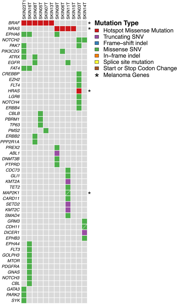
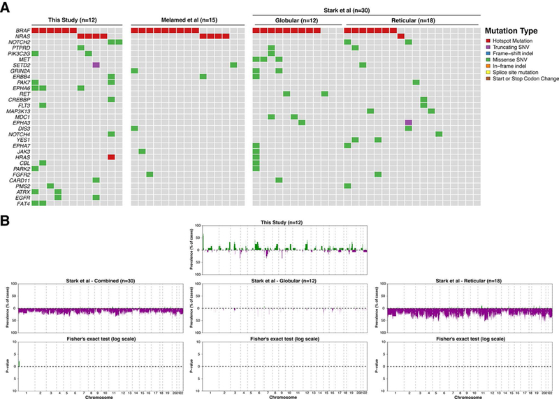
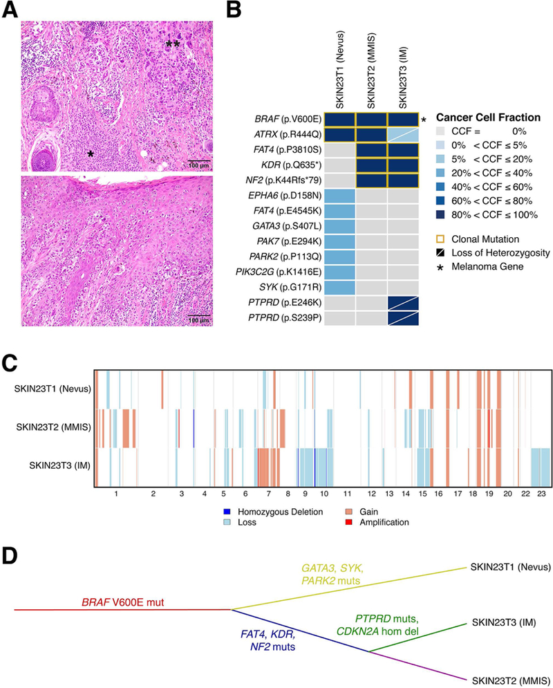
Methods and results: DNA extracted from 12 melanocytic naevi and DNA from matching normal tissue were separately microdissected and subjected to targeted massively parallel sequencing of ≥300 cancer genes. A median of 5.5 (range 1-12) non-synonymous somatic mutations were detected, with 10 cases harbouring mutually exclusive BRAF V600E (6/12) or NRAS (4/12) clonal hotspot mutations. One of the two cases lacking BRAF and NRAS mutations was a dysplastic naevus harbouring an HRAS Q61L hotspot mutation. Analysis of the laser-capture microdissected components of a naevus synchronously diagnosed with in-situ and invasive malignant melanoma revealed a truncal, clonal BRAF V600E mutation, and the acquisition of a CDKN2A homozygous deletion in the invasive component, in conjunction with additional clonal mutations affecting NF2, FAT4 and KDR in both in-situ and invasive malignant components.
Conclusion: Melanocytic naevi harbour recurrent BRAF V600E or NRAS hotspot mutations with low mutational burdens. Our findings also show that progression from naevi to malignant melanoma may be driven by the acquisition of additional genetic alterations, including CDKN2A homozygous deletions.
Keywords: melanocytic naevi; melanoma.
© 2019 John Wiley & Sons Ltd.
Conflict of interest statement
CONFLICTS OF INTEREST
All other authors declare no conflicts of interest relevant to this study.
Figures

Similar articles
-
The BRAF and NRAS mutation prevalence in dermoscopic subtypes of acquired naevi reveals constitutive mitogen-activated protein kinase pathway activation.Br J Dermatol. 2018 Jan;178(1):191-197. doi: 10.1111/bjd.15809. Epub 2017 Dec 18. Br J Dermatol. 2018. PMID: 28714107
-
BRAF, NRAS, and GNAQ Mutations in Conjunctival Melanocytic Nevi.Invest Ophthalmol Vis Sci. 2018 Jan 1;59(1):117-121. doi: 10.1167/iovs.17-22517. Invest Ophthalmol Vis Sci. 2018. PMID: 29332123 Free PMC article.
-
High BRAF mutation frequency does not characterize all melanocytic tumor types.Int J Cancer. 2004 Sep 20;111(5):705-10. doi: 10.1002/ijc.20325. Int J Cancer. 2004. PMID: 15252839
-
Defining the Molecular Genetics of Dermoscopic Naevus Patterns.Dermatology. 2019;235(1):19-34. doi: 10.1159/000493892. Epub 2018 Oct 17. Dermatology. 2019. PMID: 30332666 Review.
-
Genomic aberrations in spitzoid melanocytic tumours and their implications for diagnosis, prognosis and therapy.Pathology. 2016 Feb;48(2):113-31. doi: 10.1016/j.pathol.2015.12.007. Epub 2016 Jan 18. Pathology. 2016. PMID: 27020384 Free PMC article. Review.
Cited by
-
Malignant melanoma: evolving practice management in an era of increasingly effective systemic therapies.Curr Probl Surg. 2022 Jan;59(1):101030. doi: 10.1016/j.cpsurg.2021.101030. Epub 2021 Jul 7. Curr Probl Surg. 2022. PMID: 35033317 Free PMC article. Review. No abstract available.
-
Early contribution of germline and nevi genetic alterations to a rapidly-progressing cutaneous melanoma patient: a case report.BMC Med Genomics. 2023 Jan 5;16(1):1. doi: 10.1186/s12920-022-01426-2. BMC Med Genomics. 2023. PMID: 36604730 Free PMC article.
-
UV-Induced Somatic Mutations Driving Clonal Evolution in Healthy Skin, Nevus, and Cutaneous Melanoma.Life (Basel). 2022 Aug 29;12(9):1339. doi: 10.3390/life12091339. Life (Basel). 2022. PMID: 36143375 Free PMC article. Review.
-
Imaging Mass Spectrometry-Based Proteomic Analysis to Differentiate Melanocytic Nevi and Malignant Melanoma.Cancers (Basel). 2021 Jun 26;13(13):3197. doi: 10.3390/cancers13133197. Cancers (Basel). 2021. PMID: 34206844 Free PMC article.
References
-
- Happle R What is a nevus? A proposed definition of a common medical term. Dermatology 1995;191;1–5. - PubMed
-
- Lentigenes Weedon D., nevi and melanomas. Skin Pathology. Edinburgh: Churchill Livingstone, 2002;803–858.
-
- Shain AH, Yeh I, Kovalyshyn I et al. The Genetic Evolution of Melanoma from Precursor Lesions. New England Journal of Medicine 2015;373;1926–1936. - PubMed
-
- Smolle J, Kaddu S, Kerl H. Non-random spatial association of melanoma and naevi - a morphometric analysis. Melanoma Research 1999;9;407–412. - PubMed
-
- Elder DE. Dysplastic naevi: an update. Histopathology 2010;56;112–120. - PubMed
MeSH terms
Substances
Grants and funding
LinkOut - more resources
Full Text Sources
Medical
Research Materials
Miscellaneous
