. 2019 May;43(5):516-527.
doi: 10.1002/cbin.11117. Epub 2019 Mar 12.

Cellular responses to ionizing radiation change quickly over time during early development in zebrafish

Affiliations

Cellular responses to ionizing radiation change quickly over time during early development in zebrafish

Yasuko Honjo et al. Cell Biol Int. 2019 May.

Abstract

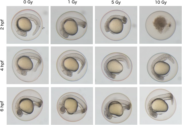
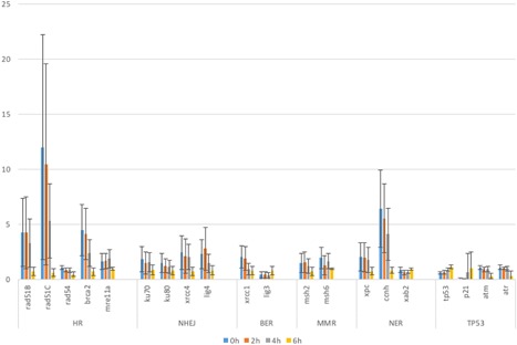
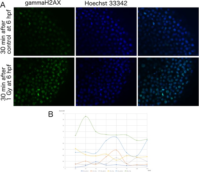
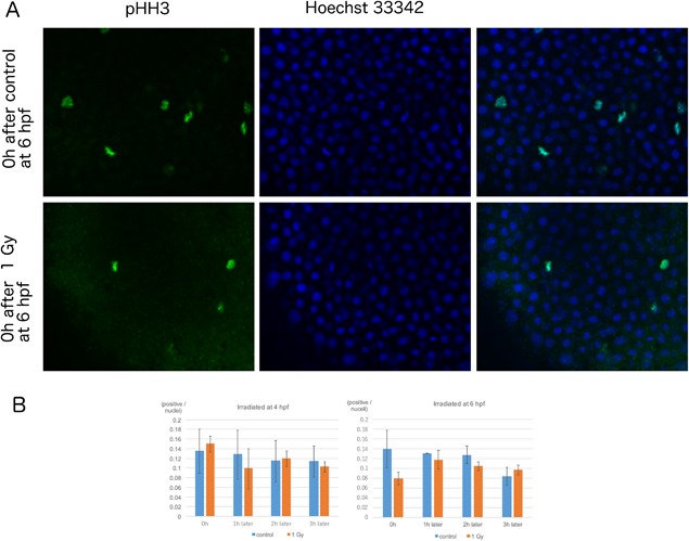
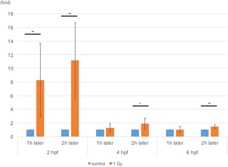
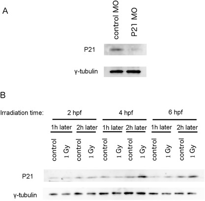
Animal cells constantly receive information about and respond to environmental factors, including ionizing radiation. Although it is crucial for a cell to repair radiation-induced DNA damage to ensure survival, cellular responses to radiation exposure during early embryonic development remain unclear. In this study, we analyzed the effects of ionizing radiation in zebrafish embryos and found that radiation-induced γH2AX foci formation and cell cycle arrest did not occur until the gastrula stage, despite the presence of major DNA repair-related gene transcripts, passed on as maternal factors. Interestingly, P21/WAF1 accumulation began ∼6 h post-fertilization, although p21 mRNA was upregulated by irradiation at 2 or 4 h post-fertilization. These results suggest that the cellular responses of zebrafish embryos at 2 or 4 h post-fertilization to radiation failed to overcome P21 protein accumulation and further signaling. Regulation of P21/WAF1 protein stabilization appears to be a key factor in the response to genotoxins during early embryogenesis.

Keywords: early development; radiation; zebrafish.


Figures

Figure 1
Figure 1
Differential effect of ionizing radiation on phenotypes of zebrafish embryos according to irradiated time‐points during early developmental stages. Zebrafish embryos at 24 hpf after irradiation at 2, 4, or 6 hpf with doses of 0, 1, 5, and 10 Gy of radiation. Non‐irradiated control embryos were shown in the left end panels (0 Gy). Whole embryos irradiated at 2 hpf (64‐cell stage) showed the most severe phenotype. At a dose of 10 Gy, embryos irradiated at 2 hpf were dead, whereas those at 4 hpf (blastula stage) or 6 hpf (gastrula stage) showed normal body shape.
Figure 2
Figure 2
Expression levels of DNA repair‐related genes in zebrafish embryos during early developmental stages. mRNA expression levels of various DNA repair genes in zebrafish embryos were quantified by real‐time RT‐PCR and their relative‐fold expression at 0 hpf (blue bars), 2 hpf (orange bars), 4 hpf (gray bars), and 6 hpf (bright yellow bars) are shown in Y‐axis compared with the expression level of reference gene. Types of genes denoted in X‐axis are categorized as below: homologous recombination (HR)‐related genes (rad51B, rad51C, rad54, brca2, mre11a), non‐homologous end‐joining (NHEJ) repair‐related genes (ku70, ku80, xrcc4, lig4)base excision repair (BER)‐related genes (xrcc1, lig3), nucleotide excision repair (NER)‐related genes (xpc, ccnh, xab2), and p53 signaling (TP53)‐related genes (tp53, p21, atm, atr). The columns and error bars represent the mean and SD, respectively.
Figure 3
Figure 3
Formation of gamma‐H2AX foci in zebrafish embryos after irradiation at early developmental stages. (A) Fluorescein immunostaining images of anti‐γH2AX antibody (green, left panels), Hoechst 33342 (blue, middle panels) and merged images (right panels) in control embryos (upper panels) and 1 Gy‐irradiated embryos (lower panels) at 6 hpf. Irradiated samples showed γH2AX foci within nuclei, whereas controls have almost no γH2AX foci. (B) Embryos irradiated with 1 Gy at 2, 4, or 6 hpf were fixed at 15‐min intervals and stained with an anti‐γH2AX antibody. The numbers of foci per nuclei at each timepoint were plotted as: 2 hpf control (light blue) and 1 Gy irradiated (orange), 4 hpf control (yellow), and 1 Gy irradiated (blue) or 6 hpf control (dark blue) and 1 Gy irradiated (brown). Only the 6 hpf embryos irradiated at 1 Gy showed γH2AX foci formation. Foci numbers peaked at 30 min after irradiation.
Figure 4
Figure 4
Effect of radiation exposure on M‐phase cells in zebrafish embryos at early developmental stages. (A) Immunostaining images of anti‐pHH3 antibody (green, left panels), Hoechst 33342 (blue, middle panels) and merged images (right panels) from embryos of 0 h after control (upper panels) or 1 Gy irradiation (lower panels) at 6 hpf. (B) Irradiated embryos were fixed at 1‐h intervals. The M‐phase marker, phospho‐histone H3 (phospho‐HH3) was detected by immunostaining; the ratio of positive cells/nuclei at each timepoint was calculated. In embryos irradiated at 4 hpf, the number of cells in M‐phase did not change compared to those in the control embryos (blue bars). However, cells irradiated at 6 hpf (orange bars) showed a reduction in M‐phase cells immediately after irradiation (P = 0.06). The columns and error bars represent the mean and SD, respectively.
Figure 5
Figure 5
Levels of p21 mRNA expression in irradiated zebrafish embryos. RNA samples were extracted 1 or 2 h after irradiating the embryos at 2, 4, or 6 hpf. Levels of p21 mRNA were measured via qPCR and normalized to those in control embryos at each timepoint. At all timepoints, the levels of p21 mRNA were upregulated in irradiated embryos. At all timepoints, 1 and 2 h after 2 hpf (P = 0.008 and P = 0.001), 2 h after 4 hpf (P = 0.02), and 2 h after 6 hpf (P = 0.005) were significantly different compared to the control at each timepoint. Note that upregulation of p21 mRNA levels in embryos irradiated at 2 hpf were much higher than those irradiated at other time points. The columns and error bars represent the mean and SD, respectively.
Figure 6
Figure 6
Levels of P21 protein expression in irradiated zebrafish embryos. A) Newly synthesized antibody recognized a protein between 20 and 25 kD in size. The amount of this protein decreased in the P21 antisense morpholino oligo (MO)‐injected embryos compared to embryos injected with standard control MO. B) Protein samples were obtained 1 or 2 h after irradiating embryos at 2, 4, or 6 hpf. In embryos irradiated at 6 hpf, the levels of P21/WAF1 protein were upregulated at 1 h after irradiation, whereas in embryos irradiated at 4 hpf, P21/WAF1 protein levels were upregulated only after 2 h.

Similar articles

Cited by

References

    1. Adiga SK, Toyoshima M, Shimura T, Takeda J, Uematsu N, Niwa O (2007) Delayed and stage specific phosphorylation of H2AX during preimplantation development of γ‐irradiated mouse embryos. Reproduction 133(2): 415–22. - PubMed
    1. Anderson JA, Lewellyn AL, Maller JL (1997) Ionizing radiation induces apoptosis and elevates cyclin A1‐Cdk2 activity before but not after the midblastula transition in Xenopus. Mol Biol Cell 8(7): 1195–206. - PMC - PubMed
    1. Bladen CL, Lam WK, Dynan WS, Kozlowski DJ (2005) DNA damage response and Ku80 function in the vertebrate embryo. Nucleic Acids Res 33(9): 3002–10. - PMC - PubMed
    1. Bladen CL, Navarre S, Dynan WS, Kozlowski DJ (2007) Expression of the Ku70 subunit (XRCC6) and protection from low dose ionizing radiation during zebrafish embryogenesis. Neurosci Lett 422(2): 97–102. - PMC - PubMed
    1. Brownlie A, Donovan A, Pratt SJ, Paw BH, Oates AC, Brugnara C, Witkowska HE, Sassa S, Zon LI (1998) Positional cloning of the zebrafish sauternes gene: a model for congenital sideroblastic anaemia. Nat Genet 20(3): 244–50. - PubMed

MeSH terms

Substances

LinkOut - more resources