. 2019 Apr;8(4):1731-1743.
doi: 10.1002/cam4.2041. Epub 2019 Feb 21.

SPHK1-induced autophagy in peritoneal mesothelial cell enhances gastric cancer peritoneal dissemination

Affiliations

SPHK1-induced autophagy in peritoneal mesothelial cell enhances gastric cancer peritoneal dissemination

Songcheng Yin et al. Cancer Med. 2019 Apr.

Abstract

Gastric cancer peritoneal dissemination (GCPD) has been recognized as the most common form of metastasis in advanced gastric cancer (GC), and the survival is pessimistic. The injury of mesothelial cells plays an important role in GCPD. However, its molecular mechanism is not entirely clear. Here, we focused on the sphingosine kinase 1 (SPHK1) in human peritoneal mesothelial cells (HPMCs) which regulates HPMCs autophagy in GCPD progression. Initially, we analyzed SPHK1 expression immunohistochemically in 120 GC peritoneal tissues, and found high SPHK1 expression to be significantly associated with LC3B expression and peritoneal recurrence, leading to poor prognosis. Using a coculture system, we observed that GC cells promoted HPMCs autophagy and this process was inhibited by blocking TGF-β1 secreted from GC cells. Autophagic HPMCs induced adhesion and invasion of GC cells. We also confirmed that knockdown of SPHK1 expression in HPMCs inhibited TGF-β1-induced autophagy. In addition, SPHK1-driven autophagy of HPMCs accelerated GC cells occurrence of GCPD in vitro and in vivo. Moreover, we explored the relationship between autophagy and fibrosis in HPMCs, observing that overexpression of SPHK1 induced HPMCs fibrosis, while the inhibition of autophagy weakened HPMCs fibrosis. Taken together, our results provided new insights for understanding the mechanisms of GCPD and established SPHK1 as a novel target for GCPD.

Keywords: SPHK1; autophagy; gastric cancer peritoneal dissemination; mesothelial cell.


Conflict of interest statement

The authors declare no conflict of interests.

Figures

Figure 1
Figure 1
Upregulated SPHK1 in peritoneum is correlated with LC3B and poor survival in GC. (A) Representative immunohistochemistry (IHC) staining with SPHK1 and LC3B in GC peritoneum tissues. (B) Scatter plots showing the positive correlation between SPHK1 and LC3B IHC scores in peritoneum tissues. (C) Kaplan‐Meier survival curves based on SPHK1. (D) Kaplan‐Meier survival curves based on LC3B
Figure 2
Figure 2
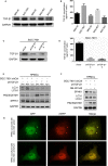
GC cell line SGC‐7901 upregulated HPMCs SPHK1 expression and induced HPMCs autophagy via TGF‐β1. (A) TGF‐β1 expression in five GC cell lines detected by western blotting. (B) TGF‐β1 levels in condition medium of five GC cell lines analyzed by ELISA. (C) Protein expression of TGF‐β1 after transfection with shTGF‐β1 lentivirus in SGC‐7901. (D) TGF‐β1 levels in condition medium of SGC‐7901 transfected with shRNA lentivirus. (E) Western blot showing the expression of LC3B, P62/SQSTM1, and SPHK1 in HPMCs cocultured with SGC‐7901 after 24, 48, and 72 hour. (F) The effect of TGF‐β1 receptor inhibitor SB431542 on LC3B, P62/SQSTM1, and SPHK1 expression in HPMCs. (G) Immunofluorescent micrographs demonstrating mRFPGFPLC3 fusion protein in HPMCs cocultured with SGC‐7901 after 48 hour. P < 0.05
Figure 3
Figure 3
Autophagy of HPMCs stimulated the adhesion and invasion of GC cells. (A) HPMCs were cocultured with SGC‐7901 cells (shCtrl or shTGF‐β1) in the absence/presence of the autophagy inhibitor 3‐MA (5 mmol L−1). Western blotting analysis revealed that 3‐MA abrogated the HPMCs’ autophagic effect of TGF‐β1 from SGC‐7901 cells. Adhesion (B) and invasion (C) of SGC‐7901 and MGC‐803 cells to the HPMCs cocultured with SGC‐7901 (shCtrl or shTGF‐β1) cells in the absence/presence of 3‐MA. P < 0.05
Figure 4
Figure 4
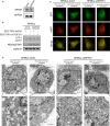
SPHK1 was required for TGF‐β1‐induced HPMC autophagy. (A) Western blotting detected SPHK1 expression after transfection with shSPHK1 lentivirus in HPMCs. (B‐D) HPMCs were transfected with shCtrl or shSPHK1 and cocultured with SGC‐7901 cells with existence or inhibition of TGF‐β1. (B) Western blotting analyzed the LC3B and P62/SQSTM1 expression in HPMCs. (C) Immunofluorescent micrographs and (D) transmission electron microscopy showed autophagosomes in HPMCs
Figure 5
Figure 5
SPHK1 expression in HPMCs regulated GC cells adhesion, invasion, and GCPD. (A, B) HPMCs were transfected with shCtrl or shSPHK1 and cocultured with SGC‐7901 cells with existence or inhibition of TGF‐β1. Adhesion (A) and invasion (B) of SGC‐7901 and MGC‐803 cells to the HPMCs. (C) Representative tumor nodules in the abdominal cavity of nude mice that were intraperitoneally injected with SGC‐7901 cells admixed with shSPHK1 and shCtrl HPMCs, respectively. P < 0.05
Figure 6
Figure 6
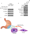
SPHK1 regulated HPMC fibrosis by promoting autophagy. (A) HPMCs were transfected with shCtrl or shSPHK1 and cocultured with SGC‐7901 cells with existence or inhibition of TGF‐β1. Western blotting analyzed MMT‐related proteins E‐cadherin, N‐cadherin, and α‐SMA expression in HPMCs. (B) HPMCs were transfected with vector or Flag‐SPHK1 plasmids and treated with 3‐MA (5 mmol L−1). Autophagy‐related proteins and MMT‐related proteins expression was detected by western blot. (C) Schematic diagram of the current study

References

    1. Bray F, Ferlay J, Soerjomataram I, Siegel RL, Torre LA, Jemal A. Global cancer statistics 2018: GLOBOCAN estimates of incidence and mortality worldwide for 36 cancers in 185 countries. CA Cancer J Clin. 68:394-424. - PubMed
    1. Van Cutsem E, Sagaert X, Topal B, Haustermans K, Prenen H. Gastric cancer. Lancet. 2016;388:2654‐2664. - PubMed
    1. Thomassen I, van Gestel YR, van Ramshorst B, et al. Peritoneal carcinomatosis of gastric origin: a population‐based study on incidence, survival and risk factors. Int J Cancer. 2014;134:622‐628. - PubMed
    1. Nashimoto A, Akazawa K, Isobe Y, et al. Gastric cancer treated in 2002 in Japan: 2009 annual report of the JGCA nationwide registry. Gastric Cancer. 2013;16:1731‐27. - PMC - PubMed
    1. Lv ZD, Zhao WJ, Jin LY, et al. Blocking TGF‐beta1 by P17 peptides attenuates gastric cancer cell induced peritoneal fibrosis and prevents peritoneal dissemination in vitro and in vivo. Biomed Pharmacother. 2017;88:27‐33. - PubMed

Publication types

MeSH terms

Substances