. 2019 Feb 19;7(1):5.
doi: 10.3390/jdb7010005.

Distinct Activities of Gli1 and Gli2 in the Absence of Ift88 and the Primary Cilia

Affiliations

Distinct Activities of Gli1 and Gli2 in the Absence of Ift88 and the Primary Cilia

Yuan Wang et al. J Dev Biol. .

Abstract

The primary cilia play essential roles in Hh-dependent Gli2 activation and Gli3 proteolytic processing in mammals. However, the roles of the cilia in Gli1 activation remain unresolved due to the loss of Gli1 transcription in cilia mutant embryos, and the inability to address this question by overexpression in cultured cells. Here, we address the roles of the cilia in Gli1 activation by expressing Gli1 from the Gli2 locus in mouse embryos. We find that the maximal activation of Gli1 depends on the cilia, but partial activation of Gli1 by Smo-mediated Hh signaling exists in the absence of the cilia. Combined with reduced Gli3 repressors, this partial activation of Gli1 leads to dorsal expansion of V3 interneuron and motor neuron domains in the absence of the cilia. Moreover, expressing Gli1 from the Gli2 locus in the presence of reduced Sufu has no recognizable impact on neural tube patterning, suggesting an imbalance between the dosages of Gli and Sufu does not explain the extra Gli1 activity. Finally, a non-ciliary Gli2 variant present at a higher level than Gli1 when expressed from the Gli2 locus fails to activate Hh pathway ectopically in the absence of the cilia, suggesting that increased protein level is unlikely the major factor underlying the ectopic activation of Hh signaling by Gli1 in the absence of the cilia.

Keywords: Gli3; Hh signaling; Shh; Smo; Sufu; intraflagellar transport; mouse; neural tube; patterning.


Conflict of interest statement

The authors declare no conflict of interest. The funders had no role in the design of the study; in the collection, analyses, or interpretation of data; in the writing of the manuscript, or in the decision to publish the results.

Figures

Figure 1
Figure 1
Expression of Gli1 from the Gli2 locus was independent of the primary cilia. RNA in situ hybridization images of transverse sections through the E10.5 neural tubes. (A) Gli1 was expressed in a ventral-to-dorsal gradient in the wild type (wt) neural tube. (B) Widespread Gli1 expression was found in Sufu−/− mutant embryos. (C) Gli1 expression was greatly reduced in Ift88−/− mutant embryos. (D) Gli1 expression was present in all progenitor cells in the Gli21ki/+;Ift88−/− double mutant neural tubes. Dashed lines outline the neural tubes. n = 3 embryos for each genotype. Gli: Glioma-associated oncogene homolog.
Figure 2
Figure 2
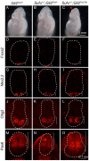
Partial activation of Gli1 in the absence of cilia. (AA”’) Lateral views of E10.5 embryos. Wild type (A) and Gli21ki/+ (A’) embryos look similar. Ift88−/− (A”) and Gli21ki/+;Ift88−/− (A”’) embryos exhibit exencephaly (arrowheads) and twisted body axes. (BG”’) Transverse sections of E10.5 embryos processed for immunofluorescence analyses. (BB”’) Foxa2 was expressed in the floor plates of the wild type (B) and Gli21ki/+ (B’) neural tubes. It was absent in the Ift88 (B”) and Gli21ki/+;Ift88−/− (B”’) neural tubes. (CC”’) Shh protein was present in the notochords and ventral neural tubes of wild type (C) and Gli21ki/+ (C’) embryos. In Ift88−/− (C”) and Gli21ki/+;Ift88−/− (C”’) embryos, Shh was present in the notochords, but not in the neural tubes. (D-D”’) Nkx2.2 labels V3 interneurons in the wild type (D) and Gli21ki/+ (D’) neural tubes. It was absent in the Ift88−/− (D”) neural tube. The Nkx2.2 expression domain was expanded both ventrally and dorsally in the Gli21ki/+;Ift88−/− (D”’) neural tube. (E–E”’) Olig2 labels motor neuron progenitors in the wild type (E) and Gli21ki/+ (E’) neural tubes. It was expanded ventrally in the Ift88−/− (E”) neural tube. The Olig2 expression domain was expanded both ventrally and dorsally in the Gli21ki/+;Ift88−/− (E”’) neural tube. (FF”’) Nkx6.1 labels progenitors of V1-3 interneurons, motor neurons and the floor plate in the wild type (F) and Gli21ki/+ (F’) neural tubes. It appears normal in the Ift88−/− (F”) neural tube. The Nkx6.1 expression domain was expanded dorsally in the Gli21ki/+;Ift88−/− (F”’) neural tube. (GG”’) Pax6 was expressed in the dorsal and lateral regions of the wild type (G) and Gli21ki/+ (G’) neural tubes. It was expanded ventrally in the Ift88−/− (G”) neural tube. Pax6 expression was restricted more dorsally in the Gli21ki/+;Ift88−/− (G”’) neural tube. Dashed lines outline the neural tubes and brackets mark the expression domains. n = 3 embryos for each genotype.
Figure 3
Figure 3
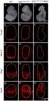
Gli3 antagonized Gli1 activity in the Gli21ki neural tube. (AE) Lateral views of E10.5 embryos. Wild type (A) and Gli21ki/+ (B) embryos looked similar. Gli3−/− embryo (C) exhibited reduced telencephalon (bracket, compare to the one in A). Gli21ki/+;Gli3+/− (D) and Gli21ki/+;Gli3−/− (E) embryos exhibit exencephaly (arrowheads). (FT) Transverse sections of E10.5 embryos processed for immunofluorescence analyses. Dashed lines outline the neural tubes and brackets mark the expression domains. (FJ) Foxa2 was expressed in the floor plates of the wild type (F), Gli21ki/+ (G), Gli3−/− (H), Gli21ki/+;Gli3+/− (I) and Gli21ki/+;Gli3−/− (J) neural tubes. (KO) Nkx2.2 labels V3 interneurons in the wild type (K), Gli21ki/+ (L), Gli3−/− (M), Gli21ki/+;Gli3+/− (N) and Gli21ki/+;Gli3−/− (O) neural tubes. (PT) Olig2 labels motor neuron progenitors in the wild type (P), Gli21ki/+ (Q) and Gli3−/− (R) neural tubes. The Olig2 expression domain was expanded dorsally in the Gli21ki/+;Gli3+/− (S) neural tube, and the expansion was more drastic in the Gli21ki/+;Gli3−/− (T) neural tube. n = 3 embryos for each genotype unless otherwise mentioned in the text.
Figure 4
Figure 4
Expression of Gli1 from the Gli2 locus did not change neural tube patterning in the absence of Smo. Transverse sections of E10.5 embryos processed for immunofluorescence analyses. (AC) Foxa2 was expressed in the floor plates of the wild type (A) and Gli21ki/1ki (B), but not in the Gli21ki/1ki;Smo−/− (C) neural tubes. (DF) Nkx2.2 labeled V3 interneurons in the wild type (D) and Gli21ki/1ki (E), but was absent in Gli21ki/1ki;Smo−/− (F) neural tubes. (GI) Olig2 labels motor neuron progenitors in the wild type (G) and Gli21ki/1ki (H), but not in Gli21ki/1ki;Smo−/− (I) neural tubes. (JL) Nkx6.1 was expressed in the wild type (J) and Gli21ki/1ki(K), but not in Gli21ki/1ki;Smo−/− (L) neural tubes. (MO) Pax7 was expressed in the dorsal region of the wild type (M) and Gli21ki/1ki(N), and was expanded ventrally in the Gli21ki/1ki;Smo−/− (O) neural tubes. Dashed lines outline the neural tubes and brackets mark the expression domains. Note the Gli21ki/1ki;Smo−/− images are of higher magnification to show the details of the neural tubes that are significantly smaller than those of their littermates. n = 3 embryos for each genotype.
Figure 5
Figure 5
Reducing the dosage of Sufu did not alter the patterning in the Gli21ki neural tube. (AC) Lateral views of E10.5 embryos. Gli21ki/+ (A) Gli21ki/+;Sufu+/− (B) embryos looked similar. One out of 4 Gli21ki/1ki;Sufu+/− embryos (C) exhibited midbrain exencephaly (arrowheads). (DO) Transverse sections of E10.5 embryos processed for immunofluorescence analyses. (DF) Foxa2 was expressed in the floor plates of the Gli21ki/+ (D), Gli21ki/+; Sufu+/− (E) and Gli21ki/1ki;Sufu+/− (F) neural tubes. (GI) Nkx2.2 labeled V3 interneurons in the Gli21ki/+ (G), Gli21ki/+; Sufu+/− (H) and Gli21ki/1ki;Sufu+/− (I) neural tubes. (JL) Olig2 labeled motor neuron progenitors in the Gli21ki/+ (J), Gli21ki/+; Sufu+/− (K) and Gli21ki/1ki;Sufu+/− (L) neural tubes. (MO) Pax6 was expressed in the dorsal and lateral regions of the Gli21ki/+ (M), Gli21ki/+; Sufu+/− (N) and Gli21ki/1ki;Sufu+/− (O) neural tubes. Dashed lines outline the neural tubes and brackets mark the expression domains. n = 3 embryos for each genotype unless otherwise mentioned in the text.
Figure 6
Figure 6
Gli2∆CLR failed to activate ectopic Hh signaling in the absence of cilia. (AC) Lateral views of E10.5 embryos. Wild type (A) and Gli2∆CLRki/∆CLRki (B) embryos looked similar. Gli2∆CLRki/∆CLRki;Ift88−/− embryo (C) exhibited exencephaly and twisted body. (DO)Immunofluorescent images of transverse sections through the E10.5 neural tubes. (DF) Foxa2 labeled floor plates in the wild type (D) neural tube. It was absent in the Gli2∆CLRki/∆CLRki (E) and Gli2∆CLRki/∆CLRki;Ift88−/− (F) mutant neural tubes. (GI) Nkx2.2 was expressed in the V3 interneurons of the wild type neural tube (G) but was absent in the Gli2∆CLRki/∆CLRki (H) and Gli2∆CLRki/∆CLRki;Ift88−/− (I) mutant neural tubes. (JL) Olig2 was expressed in motor neuron progenitors in the wild type neural tube (J). It was expanded ventrally in the Gli2∆CLRki/∆CLRki (K) and Gli2∆CLRki/∆CLRki;Ift88−/− (L) mutant neural tubes. (MO) Pax6 was excluded from the ventral-most regions of the wild type (M), Gli2∆CLRki/∆CLRki (N) and Gli2∆CLRki/∆CLRki;Ift88−/− (O) mutant neural tubes. Dashed lines outline the neural tubes. Brackets show the expression domains. n = 3 for each genotype.
Figure 7
Figure 7
The level of Gli1 in Gli21ki/+ embryos was lower than that of Gli2CLR in Gli2∆CLRki/+ embryos. Immunoblots of E10.5 whole embryo lysates with antibodies against FLAG and β-tubulin. **: p = 0.0018 in two-tailed student t-test.
Figure 8
Figure 8
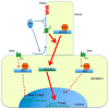
Cilia-dependent and independent activation of Gli proteins. Both Gli1 and Gli2 are activated by Smo-mediated Sonic hedgehog (Shh) pathway inside the cilia, leading to maximal activation (thick arrows) of target gene transcription (Foxa2). Outside of the cilia, Smo partially activates Gli1 and lower levels of transcriptional response (arrows with dashed lines, Nkx2.2 and Olig2). There is no direct evidence for cilia-independent Gli2 activation, but this possibility cannot be completely ruled out yet.

Similar articles

Cited by

References

    1. Briscoe J., Therond P.P. The mechanisms of Hedgehog signalling and its roles in development and disease. Nat. Re. Mol. Cell Biol. 2013;14:416–429. doi: 10.1038/nrm3598. - DOI - PubMed
    1. Nozawa Y.I., Lin C., Chuang P.T. Hedgehog signaling from the primary cilium to the nucleus: an emerging picture of ciliary localization, trafficking and transduction. Curr. Opin. Genet. Dev. 2013;23:429–437. doi: 10.1016/j.gde.2013.04.008. - DOI - PMC - PubMed
    1. Matise M.P., Joyner A.L. Gli genes in development and cancer. Oncogene. 1999;18:7852–7859. doi: 10.1038/sj.onc.1203243. - DOI - PubMed
    1. Haycraft C.J., Banizs B., Aydin-Son Y., Zhang Q., Michaud E.J., Yoder B.K. Gli2 and Gli3 localize to cilia and require the intraflagellar transport protein polaris for processing and function. PLoS Genet. 2005;1:e53. doi: 10.1371/journal.pgen.0010053. - DOI - PMC - PubMed
    1. Chiang C., Litingtung Y., Lee E., Young K.E., Corden J.L., Westphal H., Beachy P.A. Cyclopia and defective axial patterning in mice lacking Sonic hedgehog gene function. Nature. 1996;383:407–413. doi: 10.1038/383407a0. - DOI - PubMed

LinkOut - more resources