Review
. 2019 Feb 20;12(4):624.
doi: 10.3390/ma12040624.

Overview of Carbon Nanotubes for Biomedical Applications

Affiliations
Review

Overview of Carbon Nanotubes for Biomedical Applications

Juliette Simon et al. Materials (Basel). .

Abstract

The unique combination of mechanical, optical and electrical properties offered by carbon nanotubes has fostered research for their use in many kinds of applications, including the biomedical field. However, due to persisting outstanding questions regarding their potential toxicity when considered as free particles, the research is now focusing on their immobilization on substrates for interface tuning or as biosensors, as load in nanocomposite materials where they improve both mechanical and electrical properties or even for direct use as scaffolds for tissue engineering. After a brief introduction to carbon nanotubes in general and their proposed applications in the biomedical field, this review will focus on nanocomposite materials with hydrogel-based matrices and especially their potential future use for diagnostics, tissue engineering or targeted drug delivery. The toxicity issue will also be briefly described in order to justify the safe(r)-by-design approach offered by carbon nanotubes-based hydrogels.

Keywords: diagnostic; drug delivery; hydrogels; nanocomposites; tissue engineering.


Conflict of interest statement

The authors declare no conflict of interest.

Figures

Figure 1
Figure 1
Scheme of the topics addressed in this review: Carbon nanotubes (CNTs) are good materials for various biomedical applications but they raise several question about toxicity. Their utilization as component in nanocomposites like CNTs-based hydrogels could limit those concerns.
Figure 2
Figure 2
In vivo mouse brain imaging with SWNT–IRDye800 in different Near InfraRed (NIR) sub regions. (a) A C57Bl/6 mouse head with hair removed. (bd), Fluorescence images of the same mouse head in the NIR-I, NIR-II and NIR-IIa regions. In (d), the inferior cerebral vein, superior sagittal sinus and transverse sinus are labelled 1, 2 and 3, respectively. Reprinted with permission [26].
Figure 3
Figure 3
Neuro2a cells culture on a SiO2/CNT micropatterned surface. (a) SEM images of neuro2a cells cultured on patterned surfaces after 2 days of differentiation. Arrows point to neurites developed on CNT patterns. The letter A indicates a specific cell body on a CNT feature and the letter B points out a specific cell body outside of a CNT feature (on a SiO2 feature). (b) Optical fluorescence image of neural cells grown on CNT patterns after phalloidin staining. Note that neurites follow the CNT lines turning at an angle of 90°. Reprinted with permission [37].
Figure 4
Figure 4
(a) Schematic demonstration of the spinal cord in the human body. (b) Conductive nerve conduits for spinal cord injury treatment. (c) Structure of the conductive OPF-CNTpega hydrogel. Reproduced from [88] with permission from the Centre National de la Recherche Scientifique (CNRS) and The Royal Society of Chemistry.
Figure 5
Figure 5
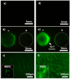
Visualization of skin electroporation. Representative pictures of mouse skin after electropermeabilisation with CTRL-AGsummuri and DWCNT-AG (1 wt. % DWCNT) containing 4 kDa FITC-dextran at 1 mm. Images (a,d) show CTRL-AG and DWCNT-AG without electrical stimulation under identical conditions at the same intensity level (×0.57). Images in (b,e) are with electrical stimulation: The anode is on the left-hand side and the cathode on the right-hand side (magnification ×0.57). (c,f) Magnifications (×4) of the anode area of (b,e), respectively. The frame on the left side of (c,f) pictures shows a numerical magnification ×400. Reprinted with permission [104].
Figure 6
Figure 6
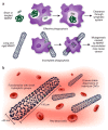
(a) The effect of CNT structure on phagocytosis by macrophages and clearing from tissues. Whereas macrophages can engulf MWNTs with a low aspect ratio (ratio of length to width) before their clearance by draining lymph vessels, MWNTs with a high aspect ratio cannot be cleared and accumulate in tissues, where they promote carcinogenesis. (b) In addition to their dimensions, other considerations relevant to the safety of CNT include increasing their solubility and preventing their aggregation, to facilitate urinary excretion and thereby prevent tissue accumulation. Reprinted with permission [123].

References

    1. Auffan M., Rose J., Bottero J.-Y., Lowry G.V., Jolivet J.-P., Wiesner M.R. Towards a Definition of Inorganic Nanoparticles from an Environmental, Health and Safety Perspective. Nat. Nanotechnol. 2009;4:634–641. doi: 10.1038/nnano.2009.242. - DOI - PubMed
    1. Wick P., Louw-Gaume A., Kucki M., Krug H., Kostarelos K., Fadeel B., Dawson K., Salvati A., Vazquez E., Ballerini L., et al. Classification Framework for Graphene-Based Materials. Angew. Chem. Int. Ed. 2014;53:149–158. doi: 10.1002/anie.201403335. - DOI - PubMed
    1. Neves V., Heister E., Costa S., Tîlmaciu C., Flahaut E., Soula B., Coley H.M., McFadden J., Silva S.R.P. Design of Double-Walled Carbon Nanotubes for Biomedical Applications. Nanotechnology. 2012;23:365102. doi: 10.1088/0957-4484/23/36/365102. - DOI - PubMed
    1. Monthioux M., Serp P., Caussat B., Flahaut E., Razafinimanana M., Valensi F., Laurent C., Peigney A., Mesguich D., Weibel A., et al. Carbon Nanotubes. In: Bhushan B., editor. Springer Handbook of Nanotechnology. Springer Handbooks; Berlin, Germany: 2017. pp. 193–247.
    1. Nel A.E., Mädler L., Velegol D., Xia T., Hoek E.M.V., Somasundaran P., Klaessig F., Castranova V., Thompson M. Understanding Biophysicochemical Interactions at the Nano–Bio Interface. Nat. Mater. 2009;8:543–557. doi: 10.1038/nmat2442. - DOI - PubMed

LinkOut - more resources