Review
. 2019 Feb 20;8(2):183.
doi: 10.3390/cells8020183.

Autophagy as a Therapeutic Target to Enhance Aged Muscle Regeneration

Affiliations
Review

Autophagy as a Therapeutic Target to Enhance Aged Muscle Regeneration

David E Lee et al. Cells. .

Abstract

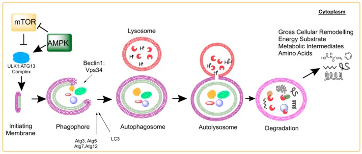
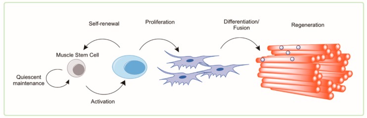
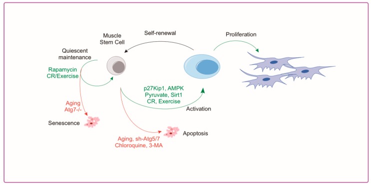
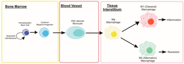
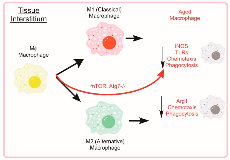
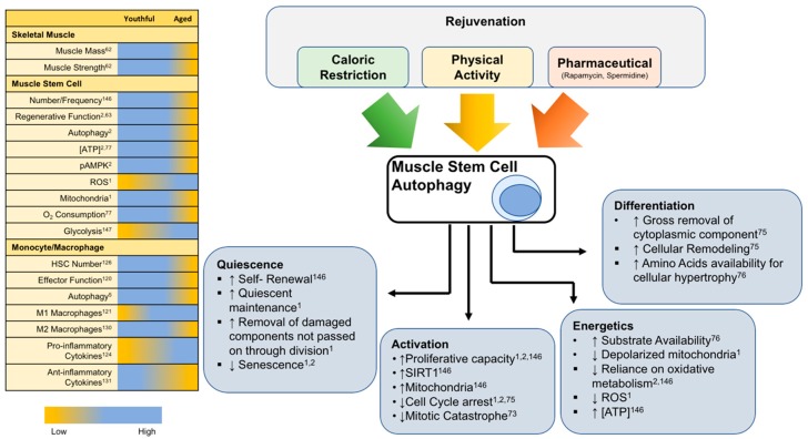
Skeletal muscle has remarkable regenerative capacity, relying on precise coordination between resident muscle stem cells (satellite cells) and the immune system. The age-related decline in skeletal muscle regenerative capacity contributes to the onset of sarcopenia, prolonged hospitalization, and loss of autonomy. Although several age-sensitive pathways have been identified, further investigation is needed to define targets of cellular dysfunction. Autophagy, a process of cellular catabolism, is emerging as a key regulator of muscle regeneration affecting stem cell, immune cell, and myofiber function. Muscle stem cell senescence is associated with a suppression of autophagy during key phases of the regenerative program. Macrophages, a key immune cell involved in muscle repair, also rely on autophagy to aid in tissue repair. This review will focus on the role of autophagy in various aspects of the regenerative program, including adult skeletal muscle stem cells, monocytes/macrophages, and corresponding age-associated dysfunction. Furthermore, we will highlight rejuvenation strategies that alter autophagy to improve muscle regenerative function.

Keywords: aging; caloric restriction; exercise; immune; macrophage; muscle regeneration; senescence; stem cell.


Conflict of interest statement

The authors declare no conflict of interest.

Figures

Figure 1
Figure 1
Molecular Events of Autophagy and related Signaling Pathways. Autophagy is a highly-conserved cellular process across eukaryotes from yeast to human. The initiation membrane matures and develops into a phagophore around cytoplasmic compartments containing a variety of macromolecules, organelles, and other cytoplasmic contents. Once fully enclosed, the autophagosome will fuse with the lysosome exposing the contents of the autophagosome to an acidic pH and various digestive enzymes of the lysosome. Following degradation of the contents of the autolysosome, the resulting molecules become available for cytoplasmic utilization (including amino acids, carbon fuel substrates, nucleotides, and reducing cofactors). This process simultaneously allows the cell to undergo drastic and rapid remodeling. Previous research has specifically shown the interaction of mTOR and AMPK in the initial steps of the autophagy process through phosphorylation interaction with the ULK1:Atg13:FIP200 complex.
Figure 2
Figure 2
Activation of the Muscle Stem Cell. Adult, resident skeletal muscle stem cells—termed satellite cells—were originally identified in the hind limb muscle of frogs and rats by electron microscopy by 1961 [55]. These cells lie in a quiescent state; dormant with low metabolic flux and little cytoplasm (low energy demand/low energy storage). Following damage by mechanical or chemical stimuli, an activated satellite cell will hypertrophy to accommodate cytosolic machinery and organelle requirements for proliferation. This key functional step leaves the (recently quiescent and still relatively small) satellite cell with little in the way of energy stores or metabolic machinery (i.e., mitochondria) but an extremely high demand for energy substrates, metabolic intermediates, and amino acids. This state of high energy demand/low energy storage in accompanied by a simultaneous remodeling of the cell architecture. By using autophagy as a process to remove aged, unnecessary, or damaged components, the young/healthy activated satellite cell is capable of recycling those components into the building blocks needed to further proliferate. The proliferated satellite cells will asymmetrically divide allowing the self-renewal of the activated stem cell into the quiescent state; likewise requiring significant cytoplasmic reorganization and remodeling. The satellite cell progeny will undergo subsequent differentiation into mature myogenic cells that are able to fuse with the damaged myofibers accomplishing successful and complete regeneration.
Figure 3
Figure 3
Aging and Autophagy Directly Impact Muscle Stem Cell Activation. Autophagy in the muscle stem cell can directly affect cell fate decisions including the maintenance of quiescence or entering into senescence. These can be controlled by aging, exercise, and caloric restriction or by direct manipulators of autophagy such as rapamycin and genetic ablation of the Atg system. Similarly, muscle stem cell activation requires sufficient energy substrate that can be limiting when autophagy is reduced (i.e., aging or with the autophagy inhibitiors chloroquine or 3-MA). Restoring the energetic signaling through AMPK activation (AICAR) or by supplementing the energy stores through pyruvate can help to replenish stem cell activation.
Figure 4
Figure 4
Development of Bone Marrow-derived Macrophages. Hematopoietic stem cells (HSCs) reside in their hypoxic niche within the bone marrow and maintain a quiescent state. When activated, the HSC can generate daughter cells of the common myeloid progenitor lineage and subsequently, into circulating monocytes. When stimulated by inflammatory cytokines (i.e., IL-6 or TNF, such as following muscle damage), monocytes undergo transcriptional changes that prevent apoptosis and enhance autophagy machinery during the differentiation into a macrophage. Mature macrophages (both classical and alterative) rely heavily on autophagy to aid in their function of clearing the damaged tissue and cellular debris that is the result of muscle injury.
Figure 5
Figure 5
Aging Negatively Effects Macrophage Autophagy and Associated Phenotype/Function. The mature macrophage uses autophagy as part of the cell’s inherent function to phagocytose and eliminate pathogens or debris. The aged macrophages tend to have impairments in the autophagic functions, phagocytosis, and chemotaxis. While aged individuals tend to have elevated circulating inflammatory markers, aged M1 and M2 macrophages show reductions in iNOS and Arg1, respectively. By limiting autophagy through Atg7 genetic ablation or constitutive mTOR activation, M2 macrophages show a shift towards M1 cytokine secretion.
Figure 6
Figure 6
Rejuvenation of Muscle Stem cell autophagy. Left: Chart summarizing various physiological and functional parameters involved in skeletal muscle regeneration and how they are susceptible to age-related alterations. Right: Conceptualization of the rejuvenation potential targeting stem cell autophagy and how various cellular functions are affected. Caloric restriction, physical activity, and certain pharmaceuticals are known to affect stem cell autophagy. The role of autophagy in the stem cell is multifaceted with implications in quiescence vs. senescence, activation/proliferation vs. apoptosis, and differentiation outcomes. A substantial contribution of autophagy in the function of a muscle stem cell is alterations in the cellular metabolic landscape or energetics.

Similar articles

Cited by

References

    1. Garcia-Prat L., Martinez-Vicente M., Perdiguero E., Ortet L., Rodriguez-Ubreva J., Rebollo E., Ruiz-Bonilla V., Gutarra S., Ballestar E., Serrano A.L., et al. Autophagy maintains stemness by preventing senescence. Nature. 2016;529:37–42. doi: 10.1038/nature16187. - DOI - PubMed
    1. White J.P., Billin A.N., Campbell M.E., Russell A.J., Huffman K.M., Kraus W.E. The AMPK/p27(Kip1) Axis Regulates Autophagy/Apoptosis Decisions in Aged Skeletal Muscle Stem Cells. Stem Cell Rep. 2018;11:425–439. doi: 10.1016/j.stemcr.2018.06.014. - DOI - PMC - PubMed
    1. Tekirdag K., Cuervo A.M. Chaperone-mediated autophagy and endosomal microautophagy: Joint by a chaperone. J. Biol. Chem. 2018;293:5414–5424. doi: 10.1074/jbc.R117.818237. - DOI - PMC - PubMed
    1. De Duve C. The lysosome. Sci. Am. 1963;208:64–72. doi: 10.1038/scientificamerican0563-64. - DOI - PubMed
    1. Takeshige K., Baba M., Tsuboi S., Noda T., Ohsumi Y. Autophagy in yeast demonstrated with proteinase-deficient mutants and conditions for its induction. J. Cell Biol. 1992;119:301–311. doi: 10.1083/jcb.119.2.301. - DOI - PMC - PubMed

Publication types

LinkOut - more resources