

State-of-the-Art of Profiling Immune Contexture in the Era of Multiplexed Staining and Digital Analysis to Study Paraffin Tumor Tissues
- PMID: 30791580
- PMCID: PMC6406364
- DOI: 10.3390/cancers11020247
State-of-the-Art of Profiling Immune Contexture in the Era of Multiplexed Staining and Digital Analysis to Study Paraffin Tumor Tissues
Abstract
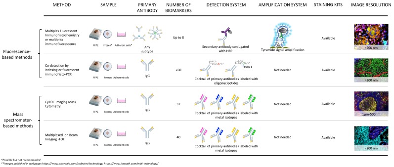
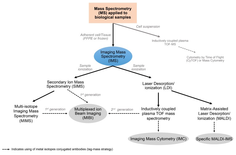
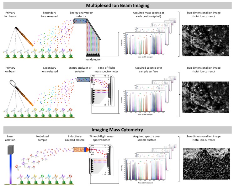
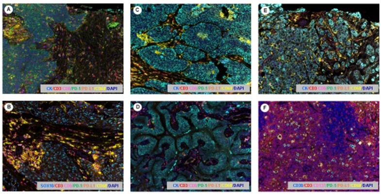
Multiplexed platforms for multiple epitope detection have emerged in the last years as very powerful tools to study tumor tissues. These revolutionary technologies provide important visual techniques for tumor examination in formalin-fixed paraffin-embedded specimens to improve the understanding of the tumor microenvironment, promote new treatment discoveries, aid in cancer prevention, as well as allowing translational studies to be carried out. The aim of this review is to highlight the more recent methodologies that use multiplexed staining to study simultaneous protein identification in formalin-fixed paraffin-embedded tumor tissues for immune profiling, clinical research, and potential translational analysis. New multiplexed methodologies, which permit the identification of several proteins at the same time in one single tissue section, have been developed in recent years with the ability to study different cell populations, cells by cells, and their spatial distribution in different tumor specimens including whole sections, core needle biopsies, and tissue microarrays. Multiplexed technologies associated with image analysis software can be performed with a high-quality throughput assay to study cancer specimens and are important tools for new discoveries. The different multiplexed technologies described in this review have shown their utility in the study of cancer tissues and their advantages for translational research studies and application in cancer prevention and treatments.
Keywords: cancer tissues; image analysis; immune profiling; multiplexed methodologies; spatial analysis.
Conflict of interest statement
The authors declare no conflict of interest.
Figures
References
-
- Steiner C., Ducret A., Tille J.C., Thomas M., McKee T.A., Rubbia-Brandt L., Scherl A., Lescuyer P., Cutler P. Applications of mass spectrometry for quantitative protein analysis in formalin-fixed paraffin-embedded tissues. Proteomics. 2014;14:441–451. doi: 10.1002/pmic.201300311. - DOI - PMC - PubMed
-
- Stauber J., MacAleese L., Franck J., Claude E., Snel M., Kaletas B.K., Wiel I.M., Wisztorski M., Fournier I., Heeren R.M. On-tissue protein identification and imaging by MALDI-ion mobility mass spectrometry. J. Am. Soc. Mass Spectrom. 2010;21:338–347. doi: 10.1016/j.jasms.2009.09.016. - DOI - PubMed
-
- Sood A., Miller A.M., Brogi E., Sui Y., Armenia J., McDonough E., Santamaria-Pang A., Carlin S., Stamper A., Campos C., et al. Multiplexed immunofluorescence delineates proteomic cancer cell states associated with metabolism. JCI Insight. 2016;1:e87030. doi: 10.1172/jci.insight.87030. - DOI - PMC - PubMed
-
- Gorris M.A.J., Halilovic A., Rabold K., van Duffelen A., Wickramasinghe I.N., Verweij D., Wortel I.M.N., Textor J.C., de Vries I.J.M., Figdor C.G. Eight-Color Multiplex Immunohistochemistry for Simultaneous Detection of Multiple Immune Checkpoint Molecules within the Tumor Microenvironment. J. Immunol. 2017;200:347–354. doi: 10.4049/jimmunol.1701262. - DOI - PubMed
Publication types
LinkOut - more resources
Full Text Sources
Other Literature Sources
