. 2019 Feb 20;11(2):176.
doi: 10.3390/v11020176.

Identification of Broad-Spectrum Antiviral Compounds by Targeting Viral Entry

Affiliations

Identification of Broad-Spectrum Antiviral Compounds by Targeting Viral Entry

Michela Mazzon et al. Viruses. .

Abstract

Viruses are a major threat to human health and economic well-being. In recent years Ebola, Zika, influenza, and chikungunya virus epidemics have raised awareness that infections can spread rapidly before vaccines or specific antagonists can be made available. Broad-spectrum antivirals are drugs with the potential to inhibit infection by viruses from different groups or families, which may be deployed during outbreaks when specific diagnostics, vaccines or directly acting antivirals are not available. While pathogen-directed approaches are generally effective against a few closely related viruses, targeting cellular pathways used by multiple viral agents can have broad-spectrum efficacy. Virus entry, particularly clathrin-mediated endocytosis, constitutes an attractive target as it is used by many viruses. Using a phenotypic screening strategy where the inhibitory activity of small molecules was sequentially tested against different viruses, we identified 12 compounds with broad-spectrum activity, and found a subset blocking viral internalisation and/or fusion. Importantly, we show that compounds identified with this approach can reduce viral replication in a mouse model of Zika infection. This work provides proof of concept that it is possible to identify broad-spectrum inhibitors by iterative phenotypic screenings, and that inhibition of host-pathways critical for viral life cycles can be an effective antiviral strategy.

Keywords: Semliki Forest virus; Zika; alphaviruses; broad-spectrum antivirals; dengue; endocytosis; flaviviruses; host-targeted antivirals; phenotypic screening; virus entry.


Conflict of interest statement

The authors declare no conflict of interest.

Figures

Figure 1
Figure 1
Strategy for the identification of broad-spectrum antiviral (BSA) compounds inhibiting Semliki Forest virus (SFV) and dengue virus serotype 2 (DENV-2) infection. (A) Schematic of the screening procedure. Cells in 96 well plates were pre-treated with 10 μM compounds for 45 min and then infected in the presence of compounds for a time sufficient to detect expression of viral proteins (7h for SFV and 24h for DENV-2). Uninfected cells, and infected cells treated with DMSO or with Monensin were included as controls. After fixation, plates were stained and images acquired using a PE Opera LX high-throughput confocal microscope, which images multiple fields in each well, allowing calculation of the percentage of infected cells. (B) Schematic of the screening strategy. In order to identify compounds with BSA activity, all 43 compounds displaying inhibitory activity against SFV (complete list in Table S1) were tested for their ability to block infection by DENV-2. Of the 22 compounds blocking both viruses (complete list in Table S2), the 12 least toxic, as determined by MTT assay, were selected for further studies (Table S3).
Figure 2
Figure 2
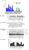
Analysis of compounds modes of action in the virus entry pathway. (A) Percentage of VSV_Blam (blue) or EBOV_Blam (green) pseudotyped VLP fusion events upon compound treatment, measured as cytosolic release of β-lactamase. HeLa Kyoto cells were treated with the indicated compounds for 1 h before infection with pseudotyped Blam VLPs. After 1 h at 37 °C, compounds were removed, the β-lactamase substrate added, and the percentage of cells in which fusion had occurred was quantified by flow cytometry. Data are normalised to Blam signal in DMSO-treated control cells (100%, dotted line). Monensin was used as a positive control. Statistics: one-way analysis of variance (Anova), Fisher’s least significant difference (LSD) test. * = p < 0.05; ** = 0.01 > p > 0.005; *** = p < 0.005. (B) Western blots showing the amount of SFV E1/E2 proteins that remains bound to the surface of infected HeLa Kyoto cells after 1 h compound treatment at 37 °C, and 1 h SFV infection on ice, in the presence of compounds. Untreated samples were included as controls. A Western blot for tubulin was used as a loading control. (C) Western blots showing SFV E1/E2 protein after subtilisin treatment. HeLa Kyoto cells were treated with the indicated compounds for 1 h at 37 °C, and SFV bound for 1 h on ice in the presence of compounds. Next, virus was allowed to internalise at 37 °C for 20 min, before subtilisin treatment on ice to remove surface-bound virus Ice-treated samples (where the virus was not internalised) treated or not with subtilisin, as well as untreated samples incubated at 37 °C (where the virus was internalised) were included as controls. (D) Western blot showing SFV E1/E2 proteins and low pH-induced E1 trimers. HeLa Kyoto cells were treated with the indicated compounds for 1 h at 37 °C, SFV bound 1 h on ice in the presence of compounds, and then internalised at 37 °C for 40 min, before cell lysis. A fraction of each lysate was treated trypsin to verify the identity of the trypsin-resistant E1 trimer (top panel). Monensin and Chloroquine (100 μM), known inhibitors of endosomal acidification were used as positive controls. Untreated samples were included as negative controls. (E) Percentage of DID-labelled SFV hemifusion/fusion events normalised to DMSO treated cells (100%, dashed line). HeLa Kyoto cells were pre-treated with compounds for 1 h at 37 °C before adding DID-SFV for an additional hour on ice. Unbound virus was then washed away and infection left to proceed for 40 min at 37 °C to allow virus internalisation and fusion. Bafilomycin (100 nM), a known inhibitor of viral fusion, was used as positive control. Hemifusion/fusion events were quantified on a PE Opera LX. Averages from three independent experiments are shown. Statistics: one-way Anova, Fisher’s LSD test. * = p < 0.05; ** = 0.01> p > 0.005; *** = p < 0.005.
Figure 3
Figure 3
Analysis of compounds mode of action after fusion. (A) A Renilla expressing plasmid was transfected in HeLa Kyoto cells. 24 h later, cells were treated with compounds for 8 h and then lysed to measure Renilla activity. Data are normalised to a DMSO treated control (100%). Emetine (10 μM), a known inhibitor of protein synthesis, was used as a control. Averages of three independent experiments are shown. Statistics: one-way Anova, Fisher’s LSD test. * = p < 0.05; ** = 0.01> p >0.005; *** = p < 0.005. (B) Percentage of inhibition of SFV infection following administration of each compound 60 min before infection, or at 30 and 60 min after infection. Cells (HeLa Kyoto) were fixed at 7 hpi and percentages of infection quantified following images acquisition on a PE Opera LX. Data are normalised to pre-treated controls samples (100% inhibition). Averages of two independent experiments are shown. Statistical significance is shown in Table S4, as determined with a two-way Anova with Dunnett’s test for multiple comparisons.
Figure 4
Figure 4
Niclosamide and Tyrphostin A9 impact on endocytosis. (A) Western blot showing SFV E1/E2 protein after subtilisin treatment. HeLa Kyoto cells were treated with 10 μM Niclosamide or Tyrphostin A9 for 1 h at 37 °C, and infected with SFV for 1 h on ice in the presence of compounds. Virus was left to internalise at 37 °C for 20, 40, or 60 min, before subtilisin treatment to remove surface-bound virus. Untreated cells not exposed to subtilisin, were included as controls. (B) HeLa Kyoto cells were treated with 10 μM Niclosamide or Tyrphostin A9 for 1 h before internalisation of Alexa 488-conjugated transferrin for 10, 20, and 30 min. DMSO-treated cells were used as controls. One representative of three independent experiments is shown. Statistics: two-way Anova with Dunnett’s test for multiple comparisons. *** = p < 0.005.
Figure 5
Figure 5
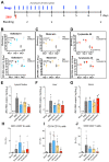
In vivo activity of Tyrphostin A9, Monensin, and Sofosbuvir. (A) Schematic of the drug treatment and infection regime in AG129 mice. Mice were treated with 5.7 mg/kg of Sofosbuvir, 10 mg/kg of Monensin, or 1mg/kg of Tyrphostin A9 before and after infection with 105 pfu of ZIKV, at the indicated intervals. Administration of Tyrphostin A9 was suspended at day 3 due to toxicity. Focus-forming units (ffu, top panels) and ZIKV RNA copies (bottom panels) per ml of serum at day 1, 3, and 7 p.i. upon treatment with Sofosbuvir (B), Monensin (C), or Tyrphostin A9 (D). N.D. (not detected), indicates that no infectious units were recovered. ZIKV RNA copies/ng at day 7 p.i. in the lymph nodes (E), liver (F), and brain (G) upon indicated treatment. Percentages of CD14+CD11b+ macrophages (H), CD14+CD11c+ DC (I), and CD3+CD4+ T cells (J) infected with ZIKV at day 7 p.i. upon indicated treatment. P values from unpaired T tests are displayed for statistically significant comparisons.

Similar articles

Cited by

References

    1. Coltart C.E.M., Lindsey B., Ghinai I., Johnson A.M., Heymann D.L. The ebola outbreak, 2013–2016: Old lessons for new epidemics. Philos. Trans. R. Soc. Lond. B Biol. Sci. 2017;372 doi: 10.1098/rstb.2016.0297. - DOI - PMC - PubMed
    1. Lowe R., Barcellos C., Brasil P., Cruz O.G., Honório N.A., Kuper H., Carvalho M.S. The zika virus epidemic in brazil: From discovery to future implications. Int. J. Environ. Res. Public Health. 2018;15:96. doi: 10.3390/ijerph15010096. - DOI - PMC - PubMed
    1. Paules C.I., Fauci A.S. Yellow fever—Once again on the radar screen in the Americas. N. Engl. J. Med. 2017;376:1397–1399. doi: 10.1056/NEJMp1702172. - DOI - PubMed
    1. Ziegler T., Mamahit A., Cox N.J. 65 years of influenza surveillance by a world health organization-coordinated global network. Influenza Other Respi. Viruses. 2018;12:558–565. doi: 10.1111/irv.12570. - DOI - PMC - PubMed
    1. Wahid B., Ali A., Rafique S., Idrees M. Global expansion of chikungunya virus: Mapping the 64-year history. Int. J. Infect. Dis. 2017;58:69–76. doi: 10.1016/j.ijid.2017.03.006. - DOI - PubMed

Publication types