Review
. 2019 Feb 20;7(1):14.
doi: 10.3390/biomedicines7010014.

Mechanisms of Neurodegeneration and Axonal Dysfunction in Progressive Multiple Sclerosis

Affiliations
Review

Mechanisms of Neurodegeneration and Axonal Dysfunction in Progressive Multiple Sclerosis

Jorge Correale et al. Biomedicines. .

Abstract

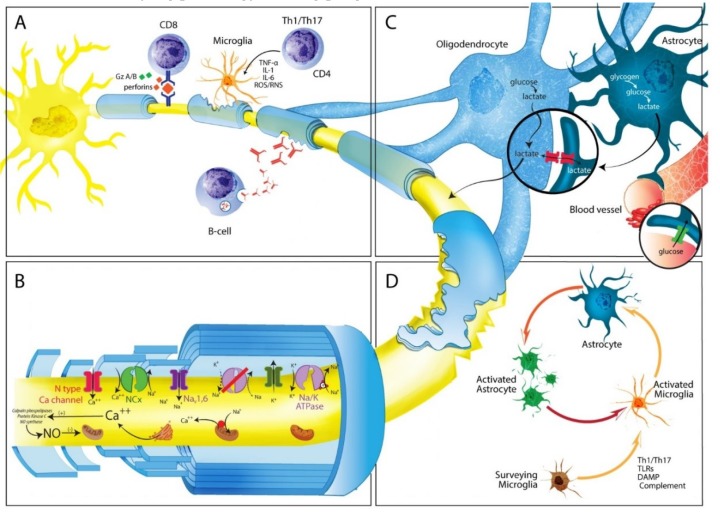
Multiple Sclerosis (MS) is a major cause of neurological disability, which increases predominantly during disease progression as a result of cortical and grey matter structures involvement. The gradual accumulation of disability characteristic of the disease seems to also result from a different set of mechanisms, including in particular immune reactions confined to the Central Nervous System such as: (a) B-cell dysregulation, (b) CD8⁺ T cells causing demyelination or axonal/neuronal damage, and (c) microglial cell activation associated with neuritic transection found in cortical demyelinating lesions. Other potential drivers of neurodegeneration are generation of oxygen and nitrogen reactive species, and mitochondrial damage, inducing impaired energy production, and intra-axonal accumulation of Ca2+, which in turn activates a variety of catabolic enzymes ultimately leading to progressive proteolytic degradation of cytoskeleton proteins. Loss of axon energy provided by oligodendrocytes determines further axonal degeneration and neuronal loss. Clearly, these different mechanisms are not mutually exclusive and could act in combination. Given the multifactorial pathophysiology of progressive MS, many potential therapeutic targets could be investigated in the future. This remains however, an objective that has yet to be undertaken.

Keywords: autoimmunity; axon; cortex; demyelination; mitochondria; multiple sclerosis; myelin; neurodegeneration; oligodendrocyte; progressive multiple sclerosis.


Conflict of interest statement

J.C. is a board member of Merck-Serono Argentina, Biogen-Idec LATAM, Merck-Serono LATAM, and Genzyme global. Jorge Correale has received reimbursement for developing educational presentations for Merck-Serono Argentina, Merck-Serono LATAM, Biogen-Idec Argentina, Genzyme Argentina, and TEVA Argentina as well as professional travel/accommodations stipends. M.M. has nothing to disclose. M.C.Y. has received reimbursement for developing educational presentations and for travel/accommodations stipends from Merck-Serono Argentina, Biogen-Idec Argentina, Genzyme Argentina, Bayer Inc, Novartis Argentina and TEVA-Tuteur Argentina.

Figures

Figure 1
Figure 1
(A) Three-dimension sagittal T1-weighted. Hypointense cortical lesion (white arrow). (B) Three-dimension sagittal T2-Fluid Attenuated Inversion Recovery (FLAIR). Hyperintense leukocortical lesion (white arrow). (C) Axial FLAIR. Subcortical temporal demyelinating plaque and perithalamic internal capsule lesion (white arrow). (D) Post-contrast 3D sagittal FLAIR. Focal area of leptomeningeal enhancement (white arrow).
Figure 2
Figure 2
Possible mechanisms involved in MS progression. (A) In progressive MS the inflammatory phenomena eventually leading to axonal degeneration and loss are compartmentalized within the CNS. Cellular components are represented by cells that come from the periphery (T and B lymphocytes), as well as by resident CNS cells (microglia cells and astrocytes). B cells can form ectopic follicle-like structures resembling tertiary lymph nodes, producing antibodies against myelin and non-myelin antigens, shown to play an important role in axonal and neuronal damage through complement cascade activation. In turn, CD8+ lymphocytes can recognize specific axonal antigens and produce tissue damage through secretion of perforin or granzymes A and B. Autoreactive CD4+ Th1 and Th17 lymphocytes can activate microglial cells, which in turn produce pro-inflammatory cytokines (IL-1, IL-6, TNF-α) or oxygen or nitrogen free radicals (ROS/RNS) causing axonal damage and neuronal loss through a bystander mechanism. (B) Following demyelination, energy requirements increase due to disruption of paranodal myelin loops. Reduction in neuronal ATP production may lead to failure of the Na+/K+ pump failure, generating a sustained sodium current, which drives reverse sodium/calcium exchange and accumulation of intra-axonal calcium. This, in turn activates degradative enzymes, including proteases, phospholipases, and calpains, resulting in further neuronal and/or axonal damage as well as impaired ATP production. (C) Axonal damage could be cause by poor trophic support. Oligodendrocytes capture glucose from circulation, breaking it down glucose to form pyruvate or lactate, which can enter axons, and be imported by mitochondria for ATP synthesis. An alternative source of energy for axons comes from glycogen stored in astrocytes, which can be transformed into glucose and later into pyruvate or lactate, depending on oxygen availability. (D) Several mechanisms cause surveillance microglia activation including Th1 or Th17 T cells; presence of microbial pathogens (PAMPs) recognized by Toll-like receptors (TLRs) or leucin-rich repeat containing receptors (NLRs); release of intracellular components from necrotic or apoptotic cells; presence of heat shock proteins, misfolded proteins (DAMPs), or components of the complement cascade. Once activated they in induce activation and proliferation of astrocytes, leading to astrogliosis.

References

    1. Thomson A.J., Baranzini S.E., Geurts J., Hemmer B., Ciccarelli O. Multiple Sclerosis. Lancet. 2018;391:1622–1636. doi: 10.1016/S0140-6736(18)30481-1. - DOI - PubMed
    1. Lassmann H., Brück W., Lucchinetti C.F. The immunopathology of multiple sclerosis: An overview. Brain Pathol. 2007;17:210–218. doi: 10.1111/j.1750-3639.2007.00064.x. - DOI - PMC - PubMed
    1. Baecher-Allan C., Kaskow B.J., Weiner H.L. Multiple sclerosis: Mechanisms and immunotherapy. Neuron. 2018;97:742–768. doi: 10.1016/j.neuron.2018.01.021. - DOI - PubMed
    1. Lublin F.D., Reingold S.C. Defining the clinical course of multiple sclerosis: Results of an international survey. Neurology. 1996;46:907–911. doi: 10.1212/WNL.46.4.907. - DOI - PubMed
    1. Skulina C., Schmidt S., Dornmair K., Babbe H., Roers A., Rajewsky K., Wekerle H., Hohlfeld R., Goebels N. Multiple sclerosis: Brain-infiltrating CD8+ T cells persist as clonal expansions in the cerebrospinal fluid and blood. Proc. Natl. Acad. Sci. USA. 2004;101:2428–2433. doi: 10.1073/pnas.0308689100. - DOI - PMC - PubMed