

Human Embryonic Stem Cell-Derived Retinal Pigment Epithelium-Role in Dead Cell Clearance and Inflammation
- PMID: 30791639
- PMCID: PMC6412543
- DOI: 10.3390/ijms20040926
Human Embryonic Stem Cell-Derived Retinal Pigment Epithelium-Role in Dead Cell Clearance and Inflammation
Abstract
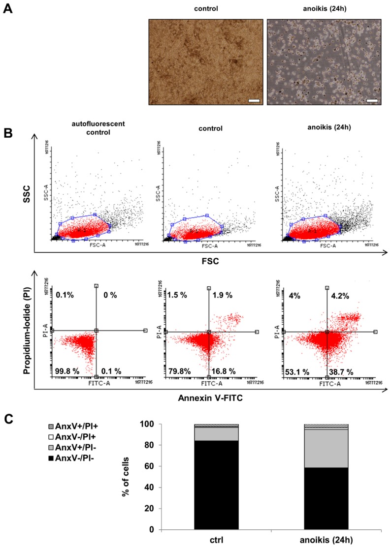
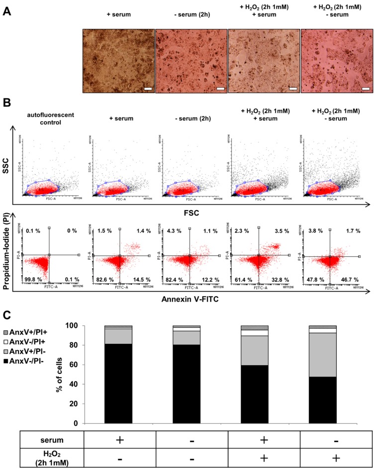
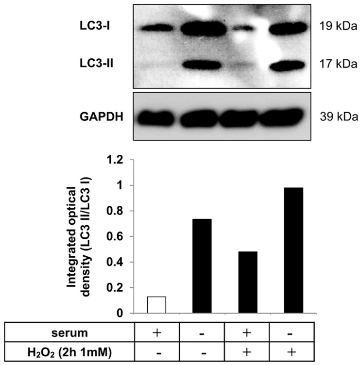
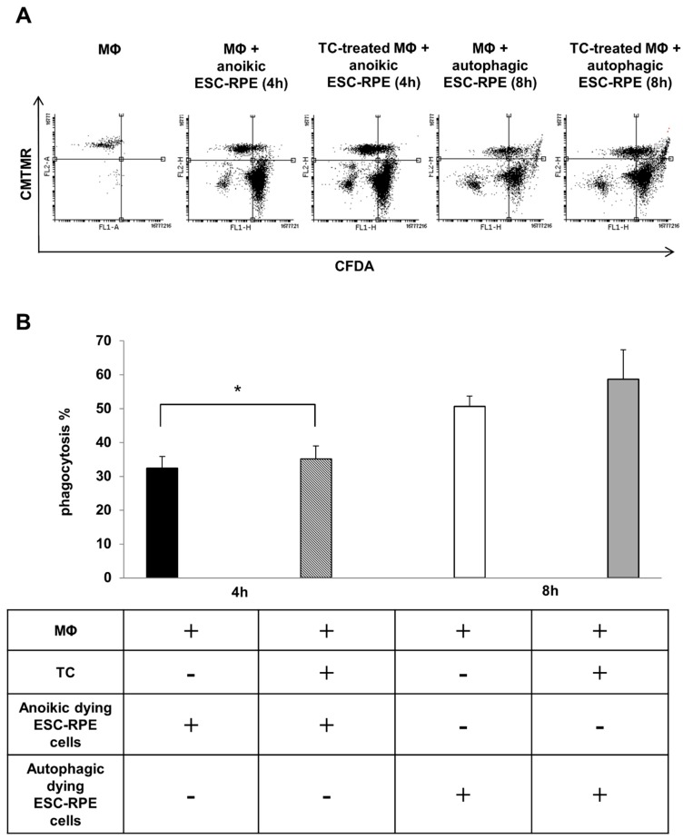
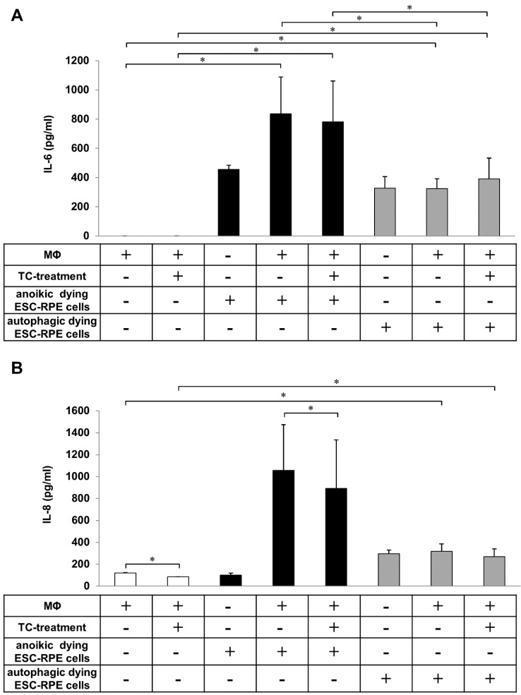
Inefficient removal of dying retinal pigment epithelial (RPE) cells by professional phagocytes can result in debris formation and development of age-related macular degeneration (AMD). Chronic oxidative stress and inflammation play an important role in AMD pathogenesis. Only a few well-established in vitro phagocytosis assay models exist. We propose human embryonic stem cell-derived-RPE cells as a new model for studying RPE cell removal by professional phagocytes. The characteristics of human embryonic stem cells-derived RPE (hESC-RPE) are similar to native RPEs based on their gene and protein expression profile, integrity, and barrier properties or regarding drug transport. However, no data exist about RPE death modalities and how efficiently dying hESC-RPEs are taken upby macrophages, and whether this process triggers an inflammatory responses. This study demonstrates hESC-RPEs can be induced to undergo anoikis or autophagy-associated cell death due to extracellular matrix detachment or serum deprivation and hydrogen-peroxide co-treatment, respectively, similar to primary human RPEs. Dying hESC-RPEs are efficiently engulfed by macrophages which results in high amounts of IL-6 and IL-8 cytokine release. These findings suggest that the clearance of anoikic and autophagy-associated dying hESC-RPEs can be used as a new model for investigating AMD pathogenesis or for testing the in vivo potential of these cells in stem cell therapy.
Keywords: age-related macular degeneration; anoikis; autophagy; hESC-RPE; inflammation; macrophages; phagocytosis; triamcinolone.
Conflict of interest statement
The authors declare no conflict of interest.
Figures
Similar articles
-
Clearance of autophagy-associated dying retinal pigment epithelial cells - a possible source for inflammation in age-related macular degeneration.Cell Death Dis. 2016 Sep 8;7(9):e2367. doi: 10.1038/cddis.2016.133. Cell Death Dis. 2016. PMID: 27607582 Free PMC article.
-
Stem Cell Derived Retinal Pigment Epithelium: The Role of Pigmentation as Maturation Marker and Gene Expression Profile Comparison with Human Endogenous Retinal Pigment Epithelium.Stem Cell Rev Rep. 2017 Oct;13(5):659-669. doi: 10.1007/s12015-017-9754-0. Stem Cell Rev Rep. 2017. PMID: 28730556 Free PMC article.
-
Effects of Cytokine Activation and Oxidative Stress on the Function of the Human Embryonic Stem Cell-Derived Retinal Pigment Epithelial Cells.Invest Ophthalmol Vis Sci. 2015 Oct;56(11):6265-74. doi: 10.1167/iovs.15-17333. Invest Ophthalmol Vis Sci. 2015. PMID: 26431480
-
Stem cell based therapies for age-related macular degeneration: The promises and the challenges.Prog Retin Eye Res. 2015 Sep;48:1-39. doi: 10.1016/j.preteyeres.2015.06.004. Epub 2015 Jun 23. Prog Retin Eye Res. 2015. PMID: 26113213 Review.
-
Human-induced pluripotent stem cells-derived retinal pigmented epithelium, a new horizon for cells-based therapies for age-related macular degeneration.Stem Cell Res Ther. 2022 May 26;13(1):217. doi: 10.1186/s13287-022-02894-0. Stem Cell Res Ther. 2022. PMID: 35619143 Free PMC article. Review.
Cited by
-
The Role of Autophagy in Eye Diseases.Life (Basel). 2021 Feb 27;11(3):189. doi: 10.3390/life11030189. Life (Basel). 2021. PMID: 33673657 Free PMC article. Review.
-
A Safe GDNF and GDNF/BDNF Controlled Delivery System Improves Migration in Human Retinal Pigment Epithelial Cells and Survival in Retinal Ganglion Cells: Potential Usefulness in Degenerative Retinal Pathologies.Pharmaceuticals (Basel). 2021 Jan 11;14(1):50. doi: 10.3390/ph14010050. Pharmaceuticals (Basel). 2021. PMID: 33440745 Free PMC article.
-
Cannabidiol-Loaded Retinal Organoid-Derived Extracellular Vesicles Protect Oxidatively Stressed ARPE-19 Cells.Biomedicines. 2025 May 10;13(5):1167. doi: 10.3390/biomedicines13051167. Biomedicines. 2025. PMID: 40426994 Free PMC article.
-
Autophagy in Age-Related Macular Degeneration: A Regulatory Mechanism of Oxidative Stress.Oxid Med Cell Longev. 2020 Aug 8;2020:2896036. doi: 10.1155/2020/2896036. eCollection 2020. Oxid Med Cell Longev. 2020. PMID: 32831993 Free PMC article. Review.
-
Anoikis in cell fate, physiopathology, and therapeutic interventions.MedComm (2020). 2024 Sep 15;5(10):e718. doi: 10.1002/mco2.718. eCollection 2024 Oct. MedComm (2020). 2024. PMID: 39286778 Free PMC article. Review.
References
MeSH terms
Substances
Grants and funding
- ÚNKP-17-4 New National Excellence Program of the Ministry of Human Capacities of Hungary, TÁMOP-4.2.2.A-11/1/KONV-2012-0023, TÁMOP-4.2.2. A-11/1/KONV-2012-0025 grant, the National Brain Research Program (KTIA_NAP_13-A_III/9), as well as the GINOP-2.3.2-15/ÚNKP-17-4 New National Excellence Program of the Ministry of Human Capacities of Hungary, TÁMOP-4.2.2.A-11/1/KONV-2012-0023, TÁMOP-4.2.2. A-11/1/KONV-2012-0025 grant, the National Brain Research Program (KTIA_NAP_13-A_III/9), as well as the GINOP-2.3.2-15
- grant numbers: 218050 and 133879/Academy of Finland
LinkOut - more resources
Full Text Sources
