. 2019 Jan-Dec:33:2058738418821837.
doi: 10.1177/2058738418821837.

Possible treatment for cutaneous lichen planus: An in vitro anti-inflammatory role of Angelica polysaccharide in human keratinocytes HaCaT

Affiliations

Possible treatment for cutaneous lichen planus: An in vitro anti-inflammatory role of Angelica polysaccharide in human keratinocytes HaCaT

Jun Wang et al. Int J Immunopathol Pharmacol. 2019 Jan-Dec.

Retraction in

Abstract

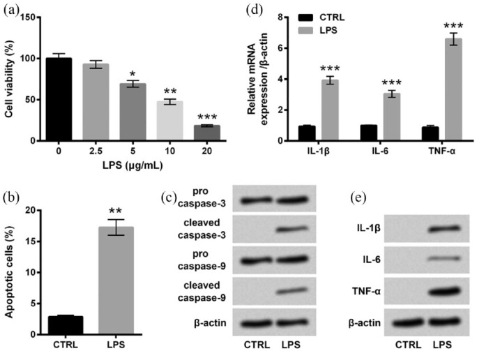
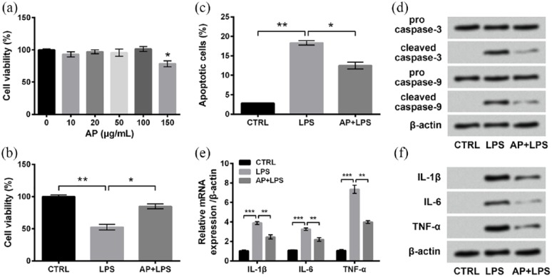
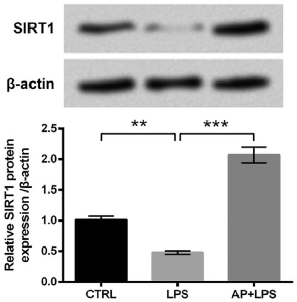
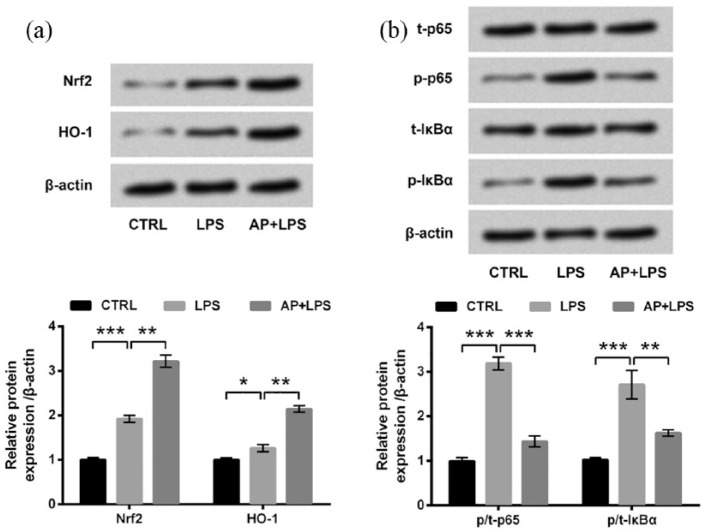
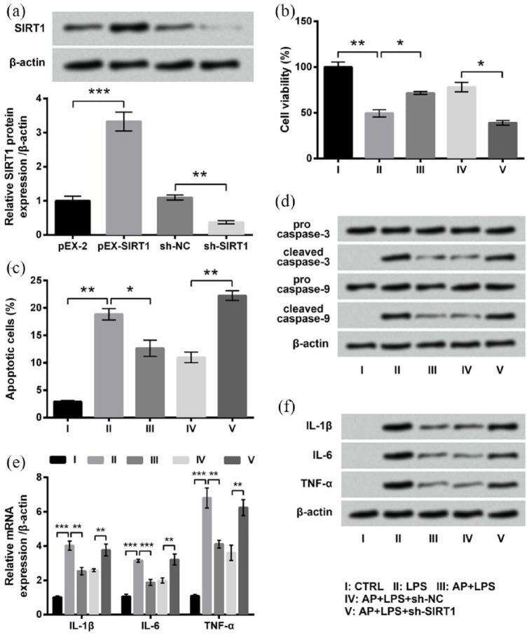
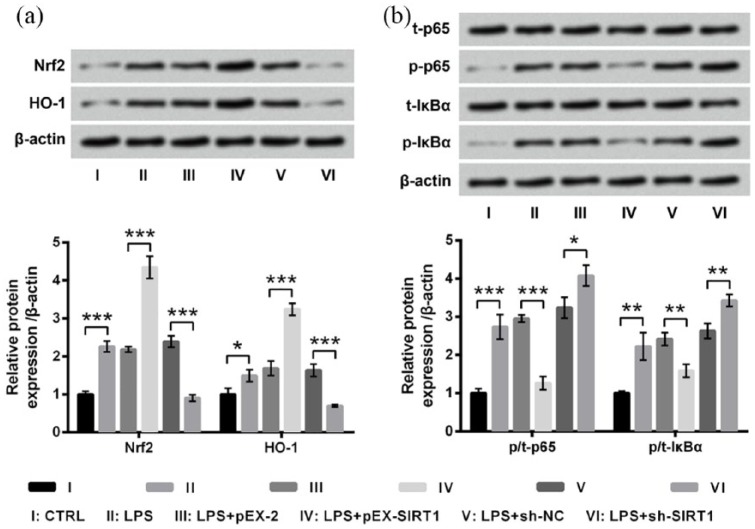
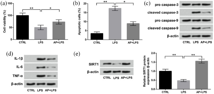
Cutaneous lichen planus (CLP) is an autoimmune disease. Angelica polysaccharide (AP) has been found to exert immunomodulation activity. In this study, we explored the roles of AP in lipopolysaccharide (LPS)-induced inflammatory injury of human keratinocytes (HaCaT cells), as well as the underlying mechanisms. LPS-induced cell injury was evaluated by alterations of cell viability, apoptosis, and expressions of proteins associated with apoptosis and inflammatory cytokines. Then, the protective effects of AP on LPS-induced cell injury were assessed. The protein expressions of sirtuin 1 (SIRT1) and key kinases in the Nrf2/HO-1 and nuclear factor κB (NF-κB) pathways were measured using western blotting. SIRT1 knockdown and overexpression were used to analyze whether AP affected HaCaT cells through regulating SIRT1. Finally, the possible inhibitory effects of AP on cell injury after LPS treatment were also evaluated. We found that LPS reduced HaCaT cell viability, enhanced apoptosis, and induced release of inflammatory cytokines. AP alleviated LPS-induced HaCaT cell inflammatory injury. The expression of SIRT1 was enhanced after AP treatment. AP activated Nrf2/HO-1 pathway while inhibited NF-κB pathway in HaCaT cells. The protective effects of AP on LPS-induced HaCaT cell injury were reversed by SIRT1 knockdown. Dysregulation of SIRT1 altered the activation of Nrf2/HO-1 and NF-κB pathways in LPS-treated HaCaT cells. Furthermore, AP also exerted inhibitory effects on HaCaT cell injury after LPS stimulation. In conclusion, AP could alleviate LPS-induced inflammatory injury of HaCaT cells through upregulating SIRT1 expression and then activating Nrf2/HO-1 pathway but inactivating NF-κB pathway. This study provided a possible therapeutic strategy for clinical CLP treatments.

Keywords: polysaccharide; NF-κB pathway; Nrf2/HO-1 pathway; cutaneous lichen planus; inflammatory injury; sirtuin 1.


Conflict of interest statement

Declaration of conflicting interests: The author(s) declared no potential conflicts of interest with respect to the research, authorship, and/or publication of this article.

Figures

Figure 1.
Figure 1.
LPS-induced inflammatory injury of HaCaT cells. (a) HaCaT cells were stimulated with 0–20 μg/mL LPS for 12 h, and cell viability was determined by CCK-8 assay. Cells were stimulated with 0 (control group) or 10 μg/mL LPS for 12 h. Then, (b) percentage of apoptotic cells, (c) expression of proteins associated with apoptosis, and (d) mRNA and (e) protein expressions of inflammatory cytokines were measured by flow cytometry assay, western blot analysis, RT-qPCR, and Western blot analysis, respectively. Data were presented as the mean ± SEM of three independent experiments. CTRL: control. *P < 0.05; **P < 0.01; ***P < 0.001.
Figure 2.
Figure 2.
LPS-induced inflammatory injury of HaCaT cells was alleviated by Angelica polysaccharide (AP) pre-treatments. (a) HaCaT cells were stimulated with 0–150 μg/mL AP for 48 h, and cell viability was determined by CCK-8 assay. Cells were stimulated with 10 μg/mL LPS or 100 μg/mL AP plus 10 μg/mL LPS. Non-treated cells acted as control. Then, (b) cell viability, (c) percentage of apoptotic cells, (d) expression of proteins associated with apoptosis, and (e) mRNA and (f) protein expressions of inflammatory cytokines were measured by CCK-8 assay, flow cytometry assay, western blot analysis, RT-qPCR, and Western blot analysis, respectively. Data were presented as the mean ± SEM of three independent experiments. CTRL: control. *P < 0.05; **P < 0.01; ***P < 0.001.
Figure 3.
Figure 3.
Angelica polysaccharide (AP) upregulated SIRT1 expression in HaCaT cells. HaCaT cells were stimulated with 10 μg/mL LPS or 100 μg/mL AP plus 10 μg/mL LPS. Non-treated cells acted as control. Then, SIRT1 protein expression was assessed by western blot analysis. Data were presented as the mean ± SEM of three independent experiments. CTRL: control. **P < 0.01; ***P < 0.001.
Figure 4.
Figure 4.
Angelica polysaccharide (AP) activated the Nrf2/HO-1 pathway while inhibited the NF-κB pathway in HaCaT cells. HaCaT cells were stimulated with 10 μg/mL LPS or 100 μg/mL AP plus 10 μg/mL LPS. Non-treated cells acted as control. Then, expressions of key kinases in (a) the Nrf2/HO-1 pathway and (b) the NF-κB pathway were evaluated by western blot analysis. Data were presented as the mean ± SEM of three independent experiments. CTRL: control; t-: total; p-: phospho-. *P < 0.05; **P < 0.01; ***P < 0.001.
Figure 5.
Figure 5.
Effects of Angelica polysaccharide (AP) pre-treatments on HaCaT cell were reversed by SIRT1 knockdown. (a) HaCaT cells were transfected with pEX-2, pEX-SIRT1, sh-NC, or sh-SIRT1 and expression of SIRT1 was measured by western blot analysis. Transfected and untransfected cells were stimulated with 10 μg/mL LPS or 100 μg/mL AP plus 10 μg/mL LPS. Non-treated cells acted as control. Then, (b) cell viability, (c) percentage of apoptotic cells, (d) expression of proteins associated with apoptosis, and (e) mRNA and (f) protein expressions of inflammatory cytokines were measured by CCK-8 assay, flow cytometry assay, western blot analysis, RT-qPCR, and western blot analysis, respectively. Data were presented as the mean ± SEM of three independent experiments. CTRL: control. *P < 0.05; **P < 0.01; ***P < 0.001.
Figure 6.
Figure 6.
Dysregulated SIRT1 affected the Nrf2/HO-1 and the NF-κB pathways in HaCaT cells. Transfected and untransfected HaCaT cells were stimulated with 10 μg/mL LPS for 12 h. Non-treated cells acted as control. Then, expressions of key kinases in (a) the Nrf2/HO-1 pathway and (b) the NF-κB pathway were evaluated by western blot analysis. Data were presented as the mean ± SEM of three independent experiments. CTRL: control; t-: total; p-: phospho-. *P < 0.05; **P < 0.01; ***P < 0.001.
Figure 7.
Figure 7.
AP post-treatment also attenuated the LPS-induced HaCaT cell inflammatory injury and upregulated the expression of SIRT1. HaCaT cells were stimulated with 10 μg/mL LPS for 12 h and then exposed to 100 μg/mL AP for 24 h. Non-treated cells acted as control. Then, (a) cell viability, (b) percentage of apoptotic cells, (c) expression of proteins associated with apoptosis, (d) expression of inflammatory cytokines, and (e) expression of SIRT1 were measured by CCK-8 assay, flow cytometry assay, and western blotting, respectively. Data were presented as the mean ± SEM of three independent experiments. CTRL: control. *P < 0.05; **P < 0.01.

References

    1. Canto AM, Muller H, Freitas RR, et al. (2010) Oral lichen planus (OLP): Clinical and complementary diagnosis. Anais Brasileiros de Dermatologia 85: 669–675. - PubMed
    1. Gorouhi F, Davari P, Fazel N. (2014) Cutaneous and mucosal lichen planus: A comprehensive review of clinical subtypes, risk factors, diagnosis, and prognosis. The Scientific World Journal 2014: 742826. - PMC - PubMed
    1. Arora SK, Chhabra S, Saikia UN, et al. (2014) Lichen planus: A clinical and immuno-histological analysis. Indian Journal of Dermatology 59: 257–261. - PMC - PubMed
    1. Sontheimer RD. (2009) Lichenoid tissue reaction/interface dermatitis: Clinical and histological perspectives. Journal of Investigative Dermatology 129: 1088–1099. - PubMed
    1. Chung PI, Hwang CY, Chen YJ, et al. (2015) Autoimmune comorbid diseases associated with lichen planus: A nationwide case-control study. Journal of the European Academy of Dermatology and Venereology 29: 1570–1575. - PubMed

Publication types

MeSH terms

LinkOut - more resources